Review
. 2014 Apr 28:5:54.
doi: 10.3389/fneur.2014.00054. eCollection 2014.

Cervical dystonia: a disorder of the midbrain network for covert attentional orienting

Affiliations
Review

Cervical dystonia: a disorder of the midbrain network for covert attentional orienting

Michael Hutchinson et al. Front Neurol. .

Abstract

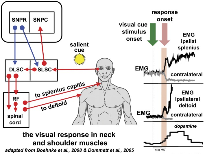
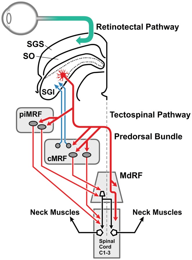
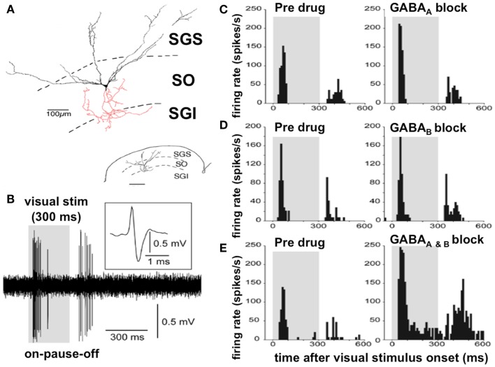
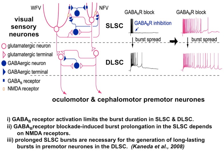
While the pathogenesis of cervical dystonia remains unknown, recent animal and clinical experimental studies have indicated its probable mechanisms. Abnormal temporal discrimination is a mediational endophenotype of cervical dystonia and informs new concepts of disease pathogenesis. Our hypothesis is that both abnormal temporal discrimination and cervical dystonia are due to a disorder of the midbrain network for covert attentional orienting caused by reduced gamma-aminobutyric acid (GABA) inhibition, resulting, in turn, from as yet undetermined, genetic mutations. Such disinhibition is (a) subclinically manifested by abnormal temporal discrimination due to prolonged duration firing of the visual sensory neurons in the superficial laminae of the superior colliculus and (b) clinically manifested by cervical dystonia due to disinhibited burst activity of the cephalomotor neurons of the intermediate and deep laminae of the superior colliculus. Abnormal temporal discrimination in unaffected first-degree relatives of patients with cervical dystonia represents a subclinical manifestation of defective GABA activity both within the superior colliculus and from the substantia nigra pars reticulata. A number of experiments are required to prove or disprove this hypothesis.

Keywords: GABA; cervical dystonia; covert attention; superior colliculus; temporal discrimination.


Figures

Figure 1
Figure 1
Reflexive covert orienting: the visual grasp reflex. This figure illustrates the postulated pathway for the visual grasp reflex in man. In a macaque, time-locked EMG responses recorded in the ipsilateral splenius capitis and deltoid, occur 80–90 ms following the sudden appearance of a cue in the visual field in a test of reflexive covert orienting (44). Responses are most marked when there is a gap of 200 ms between the fixation target and onset of the cue. Note that the EMG responses in the contralateral muscles diminish at the same time as the ipsilateral muscle activity increases. The onset of a saccade is not invariable and, if it does occur, the neck and shoulder muscle activity may precede saccade onset. The short latency phasic dopamine response is also included indicating the effect of salient events signaled to the substantia nigra pars compacta [adapted from Ref. (56)]. The postulated basal ganglia and brainstem pathway for this time-locked response is discussed in the text. Blue arrows indicated inhibition; red arrows indicate excitation. Figure adapted from Ref. (44) with permission. DLSC, intermediate and deep laminae of the superior colliculus; EMG, electromyographic; RF, reticular formation; ipsilat, ipsilateral; contralat, contralateral; SLSC, superficial laminae of superior colliculus; SNPR, substantia nigra pars reticulata; SNPC, substantia nigra pars compacta.
Figure 2
Figure 2
The visual head-turn pathway: the cephalomotor premotor pathway from the intermediate and deep lamina of the superior colliculus. This diagram [adapted from Ref. (76) with permission] illustrates the brainstem pathways involved in the head turn in response to a salient environmental change. Magnocellular retinal ganglion cells form the retinotectal pathway (green arrow) to visual sensory cells in the SGS of the superior colliculus. The direct connection between the superficial and the intermediate and deep laminae of the superior colliculus (not drawn) is illustrated in Figure 4. The cephalomotor premotor neurons, originating in the SGI of the superior colliculus, form the tectospinal and tecto-reticulospinal tracts to reach the motor neurons in the upper cervical cord by two routes: (a) direct pathway (thicker red arrow): the decussating predorsal bundle pathway (medial longitudinal fasciculus in man) can access the upper spinal cord directly or via the MdRF; (b) indirect pathway (thinner red arrows): via (i) the rostral portion of the MRF adjacent to the interstitial nucleus of Cajal (piMRF, vertical gaze), and (ii) the caudal portion of the MRF, the central mesencephalic reticular formation (cMRF, horizontal gaze). The cMRF also provides feedback to the superior colliculus (blue arrows) in relation to head and gaze position. MdRF, medullary reticular formation; MRF, mesencephalic reticular formation; cMRF, central mesencephalic reticular formation; piMRF, peri-interstitial nucleus of Cajal mesencephalic reticular formation; SGI, stratum griseum intermediale; SGS, stratum griseum superficiale; SO, stratum opticum.
Figure 3
Figure 3
Discharge properties of the wide field vertical cells in the superficial lamina of the superior colliculus: GABAA and GABAB receptors cooperatively shape transient “ON” responses. (A) A typical WFV cell is illustrated in the SGS and SO; both form part of the superficial laminae of the superior colliculus, contacting a premotor neuron (in red) in the SGI (part of the DLSC). Inset: small-scale drawing of same section. (B) Typical ON–PAUSE–OFF pattern response in a WFV cell to a static visual stimulus lasting 300 ms. Five traces are superimposed. The inset shows the expanded trace of a single spike with a positive–negative sequence due to close apposition of the recording electrode to the cell. Gray-shaded areas in the panels indicate duration of visual stimulus presentation. (C) Local application of the GABAA receptor antagonist significantly increased the peak-firing rate but did not affect “ON” response duration. (D) Local application of the GABAB receptor antagonist significantly prolonged the “ON” response duration, but did not affect the peak-firing rate. (E) Local application of both GABAA and GABAB receptor antagonists significantly increased the peak-firing rate and prolonged the “ON” response duration. Figure adapted from an original study kindly provided by Professor Tadashi Isa, from Ref. (80) with permission. GABA, gamma-aminobutyric acid; WFV, wide field vertical; SGS, stratum griseum superficiale; SGI, stratum griseum intermediale; SO, stratum opticum; DLSC, intermediate and deep laminae of the superior colliculus.
Figure 4
Figure 4
The microcircuit of the superior colliculus. Reduced GABAB inhibition causes pronged burst duration firing in both the sensory and premotor neurons. A postulated mechanism for both abnormal temporal discrimination and cervical dystonia. Diagram of the local circuit underlying GABAB receptor-mediated regulation of bursts in the superior colliculus [adapted from an original study kindly provided by Professor Tadashi Isa, from Ref. (82)]. Post-synaptic GABAB receptors expressed in both NFV and WFV cells and presynaptic GABAB receptors located on glutamatergic synaptic terminals in the SLSC are activated by synaptically released GABA during bursts of SLSC GABAergic neurons. GABAB inhibition normally curtails the discharge of both the sensory and premotor neurons. When GABAB receptors are blocked, burst duration in sensory neurons, the SLSC, may be prolonged in an NMDA receptor-dependent manner. Under conditions of reduced GABAB inhibition, this prolonged bursting may spread to the DLSC involving the oculomotor and cephalomotor premotor neurons. This prolonged bursting is postulated to result in excessive cephalomotor neuronal activity and result in cervical dystonia. DLSC, intermediate and deep laminae of the superior colliculus; GABA, gamma-aminobutyric acid; NMDA, n-methyl-D-aspartate; NFV, narrow field vertical cell; SLSC, superficial lamina of superior colliculus; WFV, wide field vertical.

Similar articles

Cited by

References

    1. Albanese A, Bhatia K, Bressman SB, Delong MR, Fahn S, Fung VS, et al. Phenomenology and classification of dystonia: a consensus update. Mov Disord (2013) 28:863–7310.1002/mds.25475 - DOI - PMC - PubMed
    1. Jinnah HA, Berardelli A, Comella C, Defazio G, Delong MR, Factor S, et al. The focal dystonias: current views and challenges for future research. Mov Disord (2013) 28:926–4310.1002/mds.25567 - DOI - PMC - PubMed
    1. Stamelou M, Edwards MJ, Hallett M, Bhatia KP. The non-motor syndrome of primary dystonia: clinical and pathophysiological implications. Brain (2012) 135:1668–8110.1093/brain/awr224 - DOI - PMC - PubMed
    1. Hutchinson M, Kimmich O, Molloy A, Whelan R, Molloy F, Lynch T, et al. The endophenotype and the phenotype: temporal discrimination and adult-onset dystonia. Mov Disord (2013) 28:1766–7410.1002/mds.25676 - DOI - PubMed
    1. Kimmich O, Molloy A, Whelan R, Williams L, Bradley D, Balsters J, et al. Temporal discrimination, a cervical dystonia endophenotype: penetrance and functional correlates. Mov Disord (2014).10.1002/mds.25822 - DOI - PubMed

LinkOut - more resources