Review
. 2014:2014:864058.
doi: 10.1155/2014/864058. Epub 2014 Apr 3.

The genetic architecture of multiple myeloma

Affiliations
Review

The genetic architecture of multiple myeloma

Steven M Prideaux et al. Adv Hematol. 2014.

Abstract

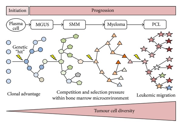
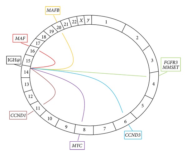
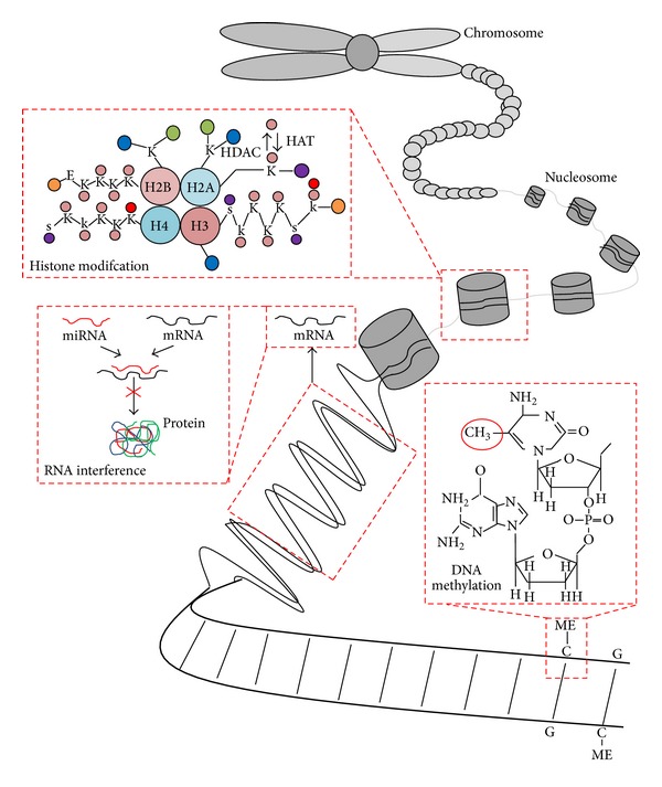
Multiple myeloma is a malignant proliferation of monoclonal plasma cells leading to clinical features that include hypercalcaemia, renal dysfunction, anaemia, and bone disease (frequently referred to by the acronym CRAB) which represent evidence of end organ failure. Recent evidence has revealed myeloma to be a highly heterogeneous disease composed of multiple molecularly-defined subtypes each with varying clinicopathological features and disease outcomes. The major division within myeloma is between hyperdiploid and nonhyperdiploid subtypes. In this division, hyperdiploid myeloma is characterised by trisomies of certain odd numbered chromosomes, namely, 3, 5, 7, 9, 11, 15, 19, and 21 whereas nonhyperdiploid myeloma is characterised by translocations of the immunoglobulin heavy chain alleles at chromosome 14q32 with various partner chromosomes, the most important of which being 4, 6, 11, 16, and 20. Hyperdiploid and nonhyperdiploid changes appear to represent early or even initiating mutagenic events that are subsequently followed by secondary aberrations including copy number abnormalities, additional translocations, mutations, and epigenetic modifications which lead to plasma cell immortalisation and disease progression. The following review provides a comprehensive coverage of the genetic and epigenetic events contributing to the initiation and progression of multiple myeloma and where possible these abnormalities have been linked to disease prognosis.


Figures

Figure 1
Figure 1
Initiation and progression of myeloma. A postgerminal centre B cell receives a genetic “hit” which immortalizes the cell and initiates transition to the indolent phase of monoclonal gammopathy of undetermined significance (MGUS). MGUS clones may then transition through the other disease phases of smouldering multiple myeloma (SMM), myeloma, and plasma cell leukemia (PCL) as genetic “hits”, which confer a survival advantage and are acquired over time. Clonal evolution develops through branching pathways whereby numerous ecosystems composed of multiple subclones exist at each disease phase, as represented by the differing shapes. At the end of this process, proliferative clones no longer become confined to the bone marrow and expand rapidly as a leukemic phase. At each disease phase, the precursor clones are present only at a low level as they have been outcompeted by more advantageous clones. It should be noted that the above figure represents an oversimplification of myeloma initiation and progression, as the process is highly complex with multiple pathways possible at any one time (adapted from Morgan et al., 2012 [8]).
Figure 2
Figure 2
Overexpression of cyclin D genes influence cell cycle progression at the G 1 /S transition point in myeloma. Increased cyclin D gene expression through hyperdiploid or nonhyperdiploid events in myeloma facilitates activation of a cyclin-dependent kinase (CDK 4 or 6). The respective CDK then phosphorylates Rb (retinoblastoma protein), which subsequently resides from its role inhibiting E2F transcription factors allowing these to facilitate cell cycle progression at the G1/S transition. G1: Gap-1 phase; S: synthesis phase; G2: Gap-2 phase; M: mitosis; P: phosphate group.
Figure 3
Figure 3
The key chromosomal translocations in myeloma. A Circos plot, with the chromosomes arranged in a clockwise direction, demonstrating the key translocations in myeloma. The translocations are represented as lines emerging from the immunoglobulin heavy chain (IGH@) locus on chromosome 14 to their respective partner chromosomes. The genes involved in each translocation are represented in boxes outside the plot. All translocations represent primary events except t(8; 14) involving MYC which is a secondary translocation.
Figure 4
Figure 4
Signalling pathways involved in myeloma pathogenesis. The various pathways involved in myeloma pathogenesis may be stimulated via exogenous factors, such as Wnt proteins, myeloma-stromal interactions, cytokines, growth and survival factors, and myeloma-extracellular matrix (ECM) interactions, or the pathways may be aberrantly activated endogenously through genetic abnormalities such as activating mutations in RAS, RAF, and NF-κB genes.
Figure 5
Figure 5
Mechanisms of epigenetic regulation. Three main forms of epigenetic modification include histone modification, RNA interference, and DNA methylation. Histone (chromatin) modification refers to the covalent posttranslational modifications to the N-terminal tails of the four core histone proteins; this modification is commonly acetylation/deacetylation changes at lysine residues mediated by histone acetyltransferases (HATs) and histone deacetylases (HDACs). RNA interference is predominantly mediated through microRNAs, which inhibit the translation of mRNA into protein. DNA methylation occurs at cytosine residues of CpG dinucleotides and acts to regulate gene expression. Pink circle = acetyl group, purple circle = phosphate group, red circle = methyl group, blue circle = carboxyl terminus, green circle = ubiquitin, orange circle = amino terminus, k = lysine, E = glutamic acid, S = serine. H2A, histone 2A; H2B, histone 2B; H3, histone 3; H4, histone 4.

References

    1. Kyle RA, Durie BG, Rajkumar SV, et al. Monoclonal gammopathy of undetermined significance (MGUS) and smoldering (asymptomatic) multiple myeloma: IMWG consensus perspectives risk factors for progression and guidelines for monitoring and management. Leukemia. 2010;24(6):1121–1127. - PMC - PubMed
    1. Kyle RA, Therneau TM, Rajkumar SV, et al. Prevalence of monoclonal gammopathy of undetermined significance. The New England Journal of Medicine. 2006;354(13):1362–1369. - PubMed
    1. Kyle RA, Therneau TM, Vincent Rajkumar S, et al. A long-term study of prognosis in monoclonal gammopathy of undetermined significance. The New England Journal of Medicine. 2002;346(8):564–569. - PubMed
    1. Kyle RA, Remstein ED, Therneau TM, et al. Clinical course and prognosis of smoldering (asymptomatic) multiple myeloma. The New England Journal of Medicine. 2007;356(25):2582–2590. - PubMed
    1. Chapman MA, Lawrence MS, Keats JJ, et al. Initial genome sequencing and analysis of multiple myeloma. Nature. 2011;471(7339):467–472. - PMC - PubMed