. 2014:2014:104248.
doi: 10.1155/2014/104248. Epub 2014 Apr 7.

Pharmacokinetic and Biodistribution Assessment of a Near Infrared-Labeled PSMA-Specific Small Molecule in Tumor-Bearing Mice

Affiliations

Pharmacokinetic and Biodistribution Assessment of a Near Infrared-Labeled PSMA-Specific Small Molecule in Tumor-Bearing Mice

Joy L Kovar et al. Prostate Cancer. 2014.

Abstract

Prostate cancer is the most frequently diagnosed cancer in men and often requires surgery. Use of near infrared (NIR) technologies to perform image-guided surgery may improve accurate delineation of tumor margins. To facilitate preclinical testing of such outcomes, here we developed and characterized a PSMA-targeted small molecule, YC-27. IRDye 800CW was conjugated to YC-27 or an anti-PSMA antibody used for reference. Human 22Rv1, PC3M-LN4, and/or LNCaP prostate tumor cells were exposed to the labeled compounds. In vivo targeting and clearance properties were determined in tumor-bearing mice. Organs and tumors were excised and imaged to assess probe localization. YC-27 exhibited a dose dependent increase in signal upon binding. Binding specificity and internalization were visualized by microscopy. In vitro and in vivo blocking studies confirmed YC-27 specificity. In vivo, YC-27 showed good tumor delineation and tissue contrast at doses as low as 0.25 nmole. YC-27 was cleared via the kidneys but bound the proximal tubules of the renal cortex and epididymis. Since PSMA is also broadly expressed on the neovasculature of most tumors, we expect YC-27 will have clinical utility for image-guided surgery and tumor resections.


Figures

Figure 1
Figure 1
Western blot and probe localization. Human A431, MCF7, U87 GM, PC3 M-LN4, and 22Rv1 cell lysates were prepared for western blot analysis (a) to confirm PSMA reactivity of anti-PSMA antibody candidate. PSMA 800CW distinguished between 22Rv1 (PSMA positive) and PC3 M-LN4 (PSMA negative) prostate tumor cell lysates, with no signal detected for cell lysates of nonprostate origin (A431, MCF-7, and U-87 MG). Microscopy images captured cell binding for PSMA 800CW ((b) and (c)) and YC-27 800CW ((d) and (e)) to 22Rv1 (PSMA positive, (b) and (d)) and PC3 M-LN4 (PSMA negative, (c) and (d)). Images were obtained by fluorescence microscopy using an Olympus IX81 Inverted Microscope.
Figure 2
Figure 2
Immunofluorescent cell-based comparison of binding and inhibition. (a) 22Rv1 and PC3 M-LN4 cells were incubated with YC-27 800CW (1 nM to 0.5 μM). Apparent K d = 8 nM. K d for PC3 M-LN4 could not be determined. (b) LNCaP cells were incubated with YC-27 800CW (0.5 nM to 0.5 μM, apparent K d = 36 nM). Competition of YC-27 800CW binding to LNCaP cells by unlabeled YC-27 ((c), IC50 = 1.7 μM) or inhibition by 2-PMPA preincubation ((d), IC50 = 0.1 μM).
Figure 3
Figure 3
Tumor imaging with YC-27 800CW. SCID Hairless mice implanted with 22Rv1 (right flank) and PC3 M-LN4 (left flank) cells received (a) 1X PBS, 100 μL; (b) 800CW carboxylate, 1 nmol; (c) IgG 800CW, 75 μg; (d) YC-27 800CW, 1 nmol (D/P = 1; SNR = 11.8); or (e) PSMA 800CW, 75 μg (D/P = 1; SNR = 8.9), which was allowed to clear for 24 h prior to imaging on Pearl Impulse Imaging System. Signal is presented in pseudocolor. All images were normalized to the same LUT. White arrows indicate the location of the 22Rv1 tumor. Inset panels show the white light image of the mouse.
Figure 4
Figure 4
Tissue distribution of fluorescence signal. Tissues were excised at endpoint from animals given YC-27 800CW (column (a)) or PSMA 800CW (column (b)). Representative whole organs (liver, kidney, and tumors) were imaged on the Pearl Impulse and reviewed for residual fluorescence of each probe 24 h after intravenous administration. (c) Tissue sections (8 μm; frozen) were prepared from muscle, brain, intestine, lung, heart, liver, kidney, spleen, testes, epididymis, and tumors, 22Rv1 and PC3 M-LN4. Sections were imaged on Odyssey CLx (21 μm) and 800 nm signal intensity per pixel was plotted for both probes.
Figure 5
Figure 5
Fluorescence signal retention in specific clearance organs. (a) Tissue sections from mice receiving 1X PBS, carboxylate 800CW, IgG 800CW, PSMA 800CW, and YC-27 800CW were scanned on Odyssey CLx (21 micron). In all images, green indicates signals captured at 800 nm and red represents autofluorescence at 700 nm. Colocalization of the signals is yellow. (b) Higher magnification image (40x) of kidney renal cortex from YC-27 800CW treated mouse.
Figure 6
Figure 6
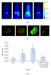
Specificity of YC-27 800CW for tumor imaging in vivo. SCID Hairless mice implanted with 22Rv1 (right flank, white arrows) and PC3 M-LN4 (left flank) cells received intravenous injections of 0.25, 0.5, or 1.0 nmol YC27-800CW or 2-PMPA (2 μg) plus YC-27 800CW (0.5 nmol). (a) Animals were imaged intact after 24 hours, using Pearl Impulse; (b) 22Rv1 tumor sections from each animal at endpoint were imaged on Odyssey CLx; (c) signal intensity of each tumor was quantified and normalized to unit area. Intact animal images are presented in pseudocolor. All images are normalized to the same LUT. Odyssey CLx images for tumor sections are shown in green (800 nm) and red (700 nm) and standardized to the same LUT.

References

    1. Siegel R, Naishadham D, Jemal A. Cancer statistics, 2012. CA Cancer Journal for Clinicians. 2012;62(1):10–29. - PubMed
    1. Imai K, Takaoka A. Comparing antibody and small-molecule therapies for cancer. Nature Reviews Cancer. 2006;6(9):714–727. - PubMed
    1. Israeli RS, Powell CT, Corr JG, Fair WR, Heston WDW. Expression of the prostate-specific membrane antigen. Cancer Research. 1994;54(7):1807–1811. - PubMed
    1. Silver DA, Pellicer I, Fair WR, Heston WDW, Cordon-Cardo C. Prostate-specific membrane antigen expression in normal and malignant human tissues. Clinical Cancer Research. 1997;3(1):81–85. - PubMed
    1. Futterer JJ, Barentsz JO. MRI-guided and robotic-assisted prostate biopsy. Current Opinion in Urology. 2012;22:316–319. - PubMed