Review
. 2014 May 1;3(5):390-399.
doi: 10.1089/wound.2013.0520.

The Role of Hypoxia-Inducible Factor in Wound Healing

Affiliations
Review

The Role of Hypoxia-Inducible Factor in Wound Healing

Wan Xing Hong et al. Adv Wound Care (New Rochelle). .

Abstract

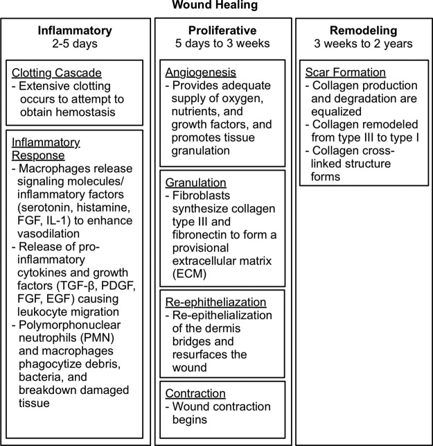
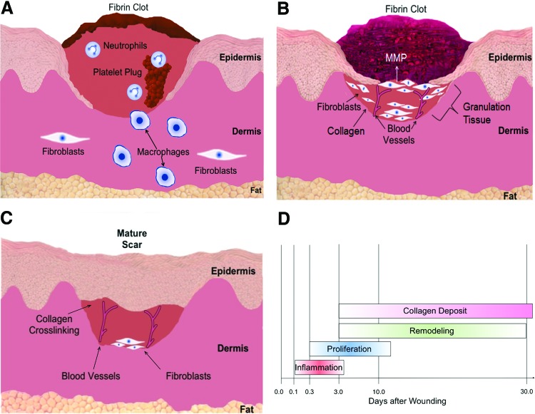
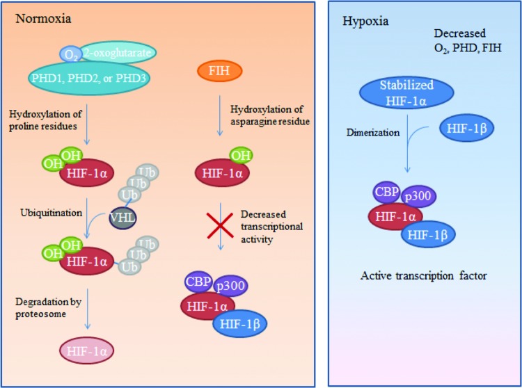
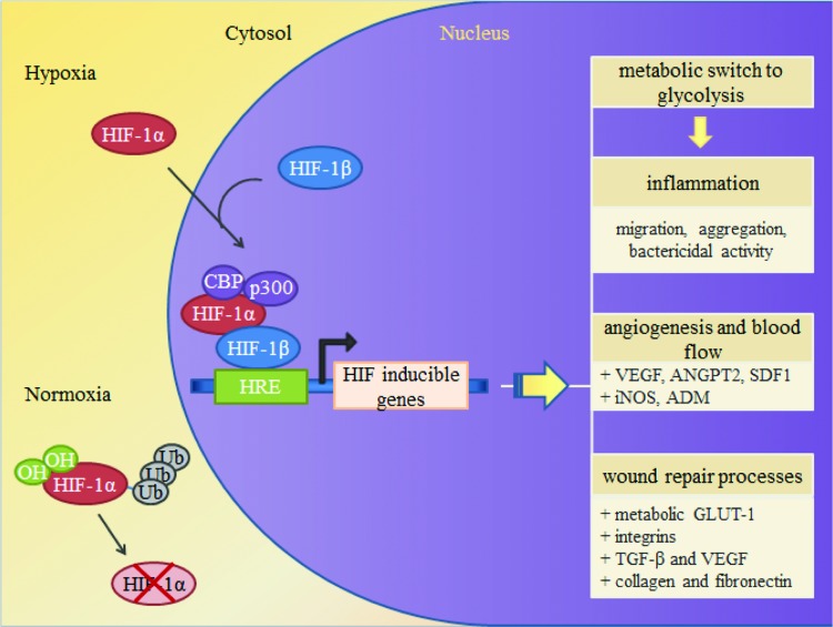
Significance: Poor wound healing remains a significant health issue for a large number of patients in the United States. The physiologic response to local wound hypoxia plays a critical role in determining the success of the normal healing process. Hypoxia-inducible factor-1 (HIF-1), as the master regulator of oxygen homeostasis, is an important determinant of healing outcomes. HIF-1 contributes to all stages of wound healing through its role in cell migration, cell survival under hypoxic conditions, cell division, growth factor release, and matrix synthesis throughout the healing process. Recent Advances: Positive regulators of HIF-1, such as prolyl-4-hydroxylase inhibitors, have been shown to be beneficial in enhancing diabetic ischemic wound closure and are currently undergoing clinical trials for treatment of several human-ischemia-based conditions. Critical Issues: HIF-1 deficiency and subsequent failure to respond to hypoxic stimuli leads to chronic hypoxia, which has been shown to contribute to the formation of nonhealing ulcers. In contrast, overexpression of HIF-1 has been implicated in fibrotic disease through its role in increasing myofibroblast differentiation leading to excessive matrix production and deposition. Both positive and negative regulators of HIF-1 therefore provide important therapeutic targets that can be used to manipulate HIF-1 expression where an excess or deficiency in HIF-1 is known to correlate with pathogenesis. Future Directions: Targeting HIF-1 during wound healing has many important clinical implications for tissue repair. Counteracting the detrimental effects of excessive or deficient HIF-1 signaling by modulating HIF-1 expression may improve future management of poorly healing wounds.


Figures

None
Michael T. Longaker, MD, MBA
<b>Figure 1.</b>
Figure 1.
Comparison of the major characteristics of the three phases of wound healing: inflammation, proliferation, and remodeling.
<b>Figure 2.</b>
Figure 2.
(A) Inflammation. Hemostasis and inflammation occur immediately following injury. Extravasation of blood leads to the formation of a clot. Numerous signaling factors are released leading to the recruitment of inflammatory cells, such as neutrophils and monocytes, to the wound. Monocytes then differentiate into mature macrophages at the wound site. Both neutrophils and macrophages are phagocytes and act to debride the wound while releasing additional factors to stimulate migration of fibroblasts to the wound site. (B) Proliferation and remodeling. During proliferation, fibroblasts secrete extracellular matrix (ECM) to form granulation tissue. Angiogenesis also occurs simultaneously as endothelial cells migrate to the area of the wound. Collagen secreted by the fibroblasts is concurrently degraded by matrix metalloproteinases (MMPs) as part of the remodeling process. (C) Maturation. During maturation, collagen production and degradation equalize. Disorganized collagen fibers are cross-linked and aligned along tension lines, leading to an increase in the tensile strength of the wound. (D) Time course of different processes that occur in the wound during healing. To see this illustration in color, the reader is referred to the web version of this article at www.liebertpub.com/wound
<b>Figure 3.</b>
Figure 3.
Hypoxia-inducible factor-1 (HIF-1) regulation during hypoxia and normoxia. During normoxia HIF-1α is degraded by a major pathway through PHD1, 2 or 3, and by a minor pathway through FIH. PHD hydroxylates select proline residues on HIF-1α causing a conformational change that allows ubiquitination by a VHL ubiquitin–protein ligase complex followed by proteosomal degradation. FIH hydroxylates asparagine residues within HIF-1α, preventing interaction between HIF-1α and its coactivators. During hypoxia, HIF-1α is stabilized due to reduced oxygen, which is required for PHD binding, and reduced PHD and FIH levels. The stabilized HIF-1α subunit binds to HIF-1β subunit to form an active transcription factor. Active HIF-1 then binds to coactivators CBP and p300 to promote expression of downstream target genes.To see this illustration in color, the reader is referred to the web version of this article at www.liebertpub.com/wound
<b>Figure 4.</b>
Figure 4.
Activation of HIF-1. Under hypoxia, HIF-1α is stabilized and binds to the HIF-1β subunit to form the active transcription factor HIF-1. HIF-1 translocates to the nucleus where it binds to hypoxia regulatory elements (HREs) within the promoter region of HIF-1-inducible genes along with coactivators p300 and CBP to promote expression of HIF-1 target genes, such as VEGF and SDF-1. To see this illustration in color, the reader is referred to the web version of this article at www.liebertpub.com/wound

References

    1. Botusan IR, Sunkari VG, Savo O, et al. : Stabilization of HIF-1alpha is critical to improve wound healing in diabetic mice. Proc Natl Acad Sci USA 2008; 105:19426. - PMC - PubMed
    1. Sen CK, Gordillo GM, Roy S, et al. : Human skin wounds: a major and snowballing threat to public health and the economy. Wound Repair Regen 2009; 17:763. - PMC - PubMed
    1. Singer AJ. and Clark RA: Cutaneous wound healing. N Engl J Med 1999; 341:738. - PubMed
    1. Clark RA: Cutaneous tissue repair: basic biologic considerations. I. J Am Acad Dermatol 1985; 13:701. - PubMed
    1. Bosco MC, Puppo M, Blengio F, et al. : Monocytes and dendritic cells in a hypoxic environment: Spotlights on chemotaxis and migration. Immunobiology 2008; 213:733. - PubMed