

Correlation of standardized uptake value and apparent diffusion coefficient in integrated whole-body PET/MRI of primary and recurrent cervical cancer
- PMID: 24804676
- PMCID: PMC4013042
- DOI: 10.1371/journal.pone.0096751
Correlation of standardized uptake value and apparent diffusion coefficient in integrated whole-body PET/MRI of primary and recurrent cervical cancer
Abstract
Background: To evaluate a potential correlation of the maximum standard uptake value (SUVmax) and the minimum apparent diffusion coefficient (ADCmin) in primary and recurrent cervical cancer based on integrated PET/MRI examinations.
Methods: 19 consecutive patients (mean age 51.6 years; range 30-72 years) with histopathologically confirmed primary cervical cancer (n = 9) or suspected tumor recurrence (n = 10) were prospectively enrolled for an integrated PET/MRI examination. Two radiologists performed a consensus reading in random order, using a dedicated post-processing software. Polygonal regions of interest (ROI) covering the entire tumor lesions were drawn into PET/MR images to assess SUVmax and into ADC parameter maps to determine ADCmin values. Pearson's correlation coefficients were calculated to assess a potential correlation between the mean values of ADCmin and SUVmax.
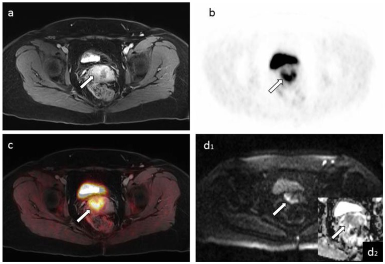
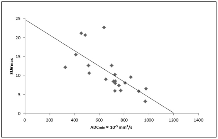
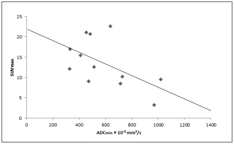
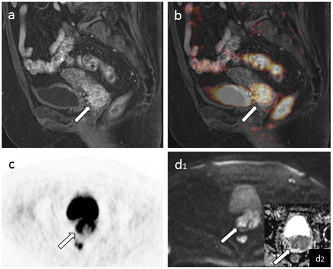
Results: In 15 out of 19 patients cervical cancer lesions (n = 12) or lymph node metastases (n = 42) were detected. Mean SUVmax (12.5 ± 6.5) and ADCmin (644.5 ± 179.7 × 10(-5) mm2/s) values for all assessed tumor lesions showed a significant but weak inverse correlation (R = -0.342, p < 0.05). When subdivided in primary and recurrent tumors, primary tumors and associated primary lymph node metastases revealed a significant and strong inverse correlation between SUVmax and ADCmin (R = -0.692, p < 0.001), whereas recurrent cancer lesions did not show a significant correlation.
Conclusions: These initial results of this emerging hybrid imaging technique demonstrate the high diagnostic potential of simultaneous PET/MR imaging for the assessment of functional biomarkers, revealing a significant and strong correlation of tumor metabolism and higher cellularity in cervical cancer lesions.
Conflict of interest statement
Figures
Similar articles
-
Parameters of simultaneous 18F-FDG-PET/MRI predict tumor stage and several histopathological features in uterine cervical cancer.Oncotarget. 2017 Apr 25;8(17):28285-28296. doi: 10.18632/oncotarget.16043. Oncotarget. 2017. PMID: 28423698 Free PMC article.
-
Simultaneous [18F]FDG-PET/MRI: Correlation of Apparent Diffusion Coefficient (ADC) and Standardized Uptake Value (SUV) in Primary and Recurrent Cervical Cancer.PLoS One. 2015 Nov 9;10(11):e0141684. doi: 10.1371/journal.pone.0141684. eCollection 2015. PLoS One. 2015. PMID: 26551527 Free PMC article.
-
Correlation between standardized uptake value and apparent diffusion coefficient of neoplastic lesions evaluated with whole-body simultaneous hybrid PET/MRI.AJR Am J Roentgenol. 2013 Nov;201(5):1115-9. doi: 10.2214/AJR.13.11304. AJR Am J Roentgenol. 2013. PMID: 24147485
-
Standardized uptake value and apparent diffusion coefficient of endometrial cancer evaluated with integrated whole-body PET/MR: Correlation with pathological prognostic factors.J Magn Reson Imaging. 2015 Dec;42(6):1723-32. doi: 10.1002/jmri.24932. Epub 2015 Apr 27. J Magn Reson Imaging. 2015. PMID: 25919115 Clinical Trial.
-
Correlation of the apparent diffusion coefficient (ADC) with the standardized uptake value (SUV) in hybrid 18F-FDG PET/MRI in non-small cell lung cancer (NSCLC) lesions: initial results.Rofo. 2013 Nov;185(11):1056-62. doi: 10.1055/s-0033-1350110. Epub 2013 Jul 16. Rofo. 2013. PMID: 23860802 Clinical Trial.
Cited by
-
Parameters of simultaneous 18F-FDG-PET/MRI predict tumor stage and several histopathological features in uterine cervical cancer.Oncotarget. 2017 Apr 25;8(17):28285-28296. doi: 10.18632/oncotarget.16043. Oncotarget. 2017. PMID: 28423698 Free PMC article.
-
Simultaneous [18F]FDG-PET/MRI: Correlation of Apparent Diffusion Coefficient (ADC) and Standardized Uptake Value (SUV) in Primary and Recurrent Cervical Cancer.PLoS One. 2015 Nov 9;10(11):e0141684. doi: 10.1371/journal.pone.0141684. eCollection 2015. PLoS One. 2015. PMID: 26551527 Free PMC article.
-
Diagnostic Value of FDG PET/MRI in Females With Pelvic Malignancy-A Systematic Review of the Literature.Front Oncol. 2020 Sep 29;10:519440. doi: 10.3389/fonc.2020.519440. eCollection 2020. Front Oncol. 2020. PMID: 33123460 Free PMC article.
-
Correlations between DW-MRI and 18 F-FDG PET/CT parameters in head and neck squamous cell carcinoma following definitive chemo-radiotherapy.Cancer Rep (Hoboken). 2021 Aug;4(4):e1360. doi: 10.1002/cnr2.1360. Epub 2021 May 7. Cancer Rep (Hoboken). 2021. PMID: 33960739 Free PMC article.
-
The use of PET/MRI in radiotherapy.Insights Imaging. 2024 Feb 27;15(1):63. doi: 10.1186/s13244-024-01627-6. Insights Imaging. 2024. PMID: 38411742 Free PMC article. Review.
References
-
- Jemal A, Bray F (2011) Center MM, Ferlay J, Ward E, et al (2011) Global cancer statistics. CA Cancer J Clin 61: 69–90. - PubMed
-
- Jemal A, Siegel R, Ward E, Hao Y, Xu J, et al. (2008) Cancer statistics, 2008. CA Cancer J Clin 58: 71–96. - PubMed
-
- Bipat S, Glas AS, van der Velden J, Zwinderman AH, Bossuyt PM, et al. (2003) Computed tomography and magnetic resonance imaging in staging of uterine cervical carcinoma: a systematic review. Gynecol Oncol 91: 59–66. - PubMed
-
- Naganawa S, Sato C, Kumada H, Ishigaki T, Miura S, et al. (2005) Apparent diffusion coefficient in cervical cancer of the uterus: comparison with the normal uterine cervix. Eur Radiol 15: 71–78. - PubMed
-
- Thoeny HC, De Keyzer F, King AD (2012) Diffusion-weighted MR imaging in the head and neck. Radiology 263: 19–32. - PubMed
MeSH terms
Substances
LinkOut - more resources
Full Text Sources
Other Literature Sources
Medical
