Calorie restriction decreases murine and human pancreatic tumor cell growth, nuclear factor-κB activation, and inflammation-related gene expression in an insulin-like growth factor-1-dependent manner
- PMID: 24804677
- PMCID: PMC4013119
- DOI: 10.1371/journal.pone.0094151
Calorie restriction decreases murine and human pancreatic tumor cell growth, nuclear factor-κB activation, and inflammation-related gene expression in an insulin-like growth factor-1-dependent manner
Abstract
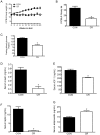
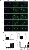
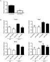
Calorie restriction (CR) prevents obesity and has potent anticancer effects that may be mediated through its ability to reduce serum growth and inflammatory factors, particularly insulin-like growth factor (IGF)-1 and protumorigenic cytokines. IGF-1 is a nutrient-responsive growth factor that activates the inflammatory regulator nuclear factor (NF)-κB, which is linked to many types of cancers, including pancreatic cancer. We hypothesized that CR would inhibit pancreatic tumor growth through modulation of IGF-1-stimulated NF-κB activation and protumorigenic gene expression. To test this, 30 male C57BL/6 mice were randomized to either a control diet consumed ad libitum or a 30% CR diet administered in daily aliquots for 21 weeks, then were subcutaneously injected with syngeneic mouse pancreatic cancer cells (Panc02) and tumor growth was monitored for 5 weeks. Relative to controls, CR mice weighed less and had decreased serum IGF-1 levels and smaller tumors. Also, CR tumors demonstrated a 70% decrease in the expression of genes encoding the pro-inflammatory factors S100a9 and F4/80, and a 56% decrease in the macrophage chemoattractant, Ccl2. Similar CR effects on tumor growth and NF-κB-related gene expression were observed in a separate study of transplanted MiaPaCa-2 human pancreatic tumor cell growth in nude mice. In vitro analyses in Panc02 cells showed that IGF-1 treatment promoted NF-κB nuclear localization, increased DNA-binding of p65 and transcriptional activation, and increased expression of NF-κB downstream genes. Finally, the IGF-1-induced increase in expression of genes downstream of NF-κB (Ccdn1, Vegf, Birc5, and Ptgs2) was decreased significantly in the context of silenced p65. These findings suggest that the inhibitory effects of CR on Panc02 pancreatic tumor growth are associated with reduced IGF-1-dependent NF-κB activation.
Conflict of interest statement
Figures


Similar articles
-
Decreased systemic IGF-1 in response to calorie restriction modulates murine tumor cell growth, nuclear factor-κB activation, and inflammation-related gene expression.Mol Carcinog. 2013 Dec;52(12):997-1006. doi: 10.1002/mc.21940. Epub 2012 Jul 6. Mol Carcinog. 2013. PMID: 22778026
-
Dose-dependent effects of calorie restriction on gene expression, metabolism, and tumor progression are partially mediated by insulin-like growth factor-1.Cancer Med. 2012 Oct;1(2):275-88. doi: 10.1002/cam4.23. Epub 2012 Aug 6. Cancer Med. 2012. PMID: 23342276 Free PMC article.
-
Genetic reduction of insulin-like growth factor-1 mimics the anticancer effects of calorie restriction on cyclooxygenase-2-driven pancreatic neoplasia.Cancer Prev Res (Phila). 2011 Jul;4(7):1030-40. doi: 10.1158/1940-6207.CAPR-11-0027. Epub 2011 May 18. Cancer Prev Res (Phila). 2011. PMID: 21593196 Free PMC article.
-
Diet-gene interactions in p53-deficient mice: insulin-like growth factor-1 as a mechanistic target.J Nutr. 2004 Sep;134(9):2482S-2486S. doi: 10.1093/jn/134.9.2482S. J Nutr. 2004. PMID: 15333746 Review.
-
NF-κB and Pancreatic Cancer; Chapter and Verse.Cancers (Basel). 2021 Sep 7;13(18):4510. doi: 10.3390/cancers13184510. Cancers (Basel). 2021. PMID: 34572737 Free PMC article. Review.
Cited by
-
Sirtuin 6 deficiency transcriptionally up-regulates TGF-β signaling and induces fibrosis in mice.J Biol Chem. 2020 Jan 10;295(2):415-434. doi: 10.1074/jbc.RA118.007212. Epub 2019 Nov 19. J Biol Chem. 2020. PMID: 31744885 Free PMC article.
-
Six-month Calorie Restriction in Overweight Individuals Elicits Transcriptomic Response in Subcutaneous Adipose Tissue That is Distinct From Effects of Energy Deficit.J Gerontol A Biol Sci Med Sci. 2016 Oct;71(10):1258-65. doi: 10.1093/gerona/glv194. Epub 2015 Oct 20. J Gerontol A Biol Sci Med Sci. 2016. PMID: 26486851 Free PMC article. Clinical Trial.
-
When less may be more: calorie restriction and response to cancer therapy.BMC Med. 2017 May 24;15(1):106. doi: 10.1186/s12916-017-0873-x. BMC Med. 2017. PMID: 28539118 Free PMC article. Review.
-
Obesity and Pancreatic Diseases: From Inflammation to Oncogenesis and the Impact of Weight Loss Interventions.Nutrients. 2025 Jul 14;17(14):2310. doi: 10.3390/nu17142310. Nutrients. 2025. PMID: 40732935 Free PMC article. Review.
-
Fasting and cancer: molecular mechanisms and clinical application.Nat Rev Cancer. 2018 Nov;18(11):707-719. doi: 10.1038/s41568-018-0061-0. Nat Rev Cancer. 2018. PMID: 30327499 Free PMC article. Review.
References
-
- Siegel R, Naishadham D, Jemal A (2012) Cancer statistics, 2012. CA: a cancer journal for clinicians 62: 10–29. - PubMed
-
- Shaib YH, Davila JA, El-Serag HB (2006) The epidemiology of pancreatic cancer in the United States: changes below the surface. Aliment Pharmacol Ther 24: 87–94. - PubMed
-
- Calle EE, Rodriguez C, Walker-Thurmond K, Thun MJ (2003) Overweight, obesity, and mortality from cancer in a prospectively studied cohort of U.S. adults. N Engl J Med 348: 1625–1638. - PubMed
-
- Giovannucci E, Michaud D (2007) The role of obesity and related metabolic disturbances in cancers of the colon, prostate, and pancreas. Gastroenterology 132: 2208–2225. - PubMed
Publication types
MeSH terms
Substances
Grants and funding
LinkOut - more resources
Full Text Sources
Other Literature Sources
Medical
Research Materials
Miscellaneous

