. 2014 May 7;9(5):e96559.
doi: 10.1371/journal.pone.0096559. eCollection 2014.

Plicidentine in the Early Permian parareptile Colobomycter pholeter, and its phylogenetic and functional significance among coeval members of the clade

Affiliations

Plicidentine in the Early Permian parareptile Colobomycter pholeter, and its phylogenetic and functional significance among coeval members of the clade

Mark J MacDougall et al. PLoS One. .

Abstract

Once thought to be an exclusively anamniote characteristic, plicidentine, a pattern of infolding of dentine, is now known to be found in various amniote clades, including Parareptilia. In the absence of detailed analyses of parareptilian dentition, most parareptiles were assumed to lack plicidentine due to the absence of external indicators, such as plications on the tooth base. The clear presence of this dentinal feature in the largest premaxillary and maxillary teeth of Colobomycter pholeter, led us to the present detailed study within the dentition of this unusual parareptile, and those of coeval members of this clade. Our study reveals that there is large variability in the degree of dentine infolding within C. pholeter dentition, as well as within those of closely related parareptiles. This variability ranges from a lack of plications, to very complex anamniote-like plicidentine. Utilizing computed tomography scans in conjunction with histological sections we also demonstrate the utility of computed tomography scans in conducting non-destructive sampling in the identification of plicidentine. Given the variability of plicidentine in this sample of parareptiles, we hypothesize that one function of parareptilian plicidentine is to increase the surface area for attachment tissues, and we suggest that the use of plicidentine as a character in phylogenetic analyses of parareptiles may be misleading.


Conflict of interest statement

Competing Interests: The authors have declared that no competing interests exist.

Figures

Figure 1
Figure 1. Cladogram showing the relationships of sampled parareptile taxa.
Bolded taxa represent those sampled in this study. Taxa marked with an asterisk represent taxa that are known to exhibit plicidentine. The cladogram was modified from unpublished data of a forthcoming analysis, that itself was a modification of the MacDougall and Reisz and Reisz et al. analyses.
Figure 2
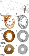
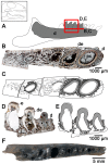
Figure 2. Cross-sectional views of the enlarged premaxillary tooth of Colobomycter pholeter (UWBM 95405).
A: diagram indicating from what parts of the premaxilla the histological sections were taken. B: cross-section taken towards the base of the enlarged premaxillary tooth showing the complex infolding of the dentine and the presence of lamellae. C: interpretation of the cross-section in B. Light grey areas indicate dentine, while darker grey areas indicate alveolar bone. The thick black lines represent the axes of the folds. D: cross-section taken midway up the enlarged premaxillary tooth showing the presence of dark dentine. E: interpretation of the cross-section in D. Light grey areas indicate dentine, while the thick black lines represent the axes of the folds. F: cross-section taken towards the tip of the enlarged maxillary tooth. G: interpretation of the cross-section of F. Light grey areas indicate dentine, while the thick black lines represent the axes of the folds. dd, dark dentine; dl, dentine lamellae pc, pulp cavity; pm, premaxilla.
Figure 3
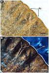
Figure 3. Microstructure in the crown of the enlarged premaxillary tooth of Colobomycter pholeter (UWBM 95405).
A: Crown of the enlarged premaxillary tooth under polarized light. Note the presence of the globular zone of dentine, which tightly infolds to form lamellae. Also visible are dark dentine and the slight grooves formed in the dentine by the folding. B: same image as in A, but under cross-polarized light. Note the presence the thin enamel layer on the exterior of the tooth. dd, dark dentine; dg, dentine groove; dl, dentine lamellae; en, enamel; gzd, globular zone of the dentine.
Figure 4
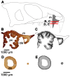
Figure 4. Cross-sectional views of the maxillary teeth of Colobomycter pholeter (ROM 67374).
A: diagram indicating from what parts of the maxilla the histological sections were taken. B: cross-section taken towards the base of the maxillary teeth showing the convoluted dentine infolding of one of the enlarged maxillary teeth, and the simpler infolding of the smaller maxillary teeth. C: interpretation of the cross-section in B. Light grey areas indicate dentine, while darker grey areas indicate alveolar bone. The thick black lines represent the axes of the folds. D: cross-section taken near the midpoint of the enlarged maxillary tooth and towards the tip of the smaller maxillary tooth showing a distinct lack of dentine infolding in both of the teeth. E: interpretation of the cross-section in D. Light grey areas indicate dentine, while the thick black lines represent the axes of the folds. de, dentine; m, maxilla; pc, pulp cavity.
Figure 5
Figure 5. Cross-sectional and longitudinal views of the maxillary and premaxillary dentition of Colobomycter pholeter (OMNH 73535) obtained via X-ray computed tomography (CT) scans.
A: diagram indicating from what parts of maxilla and premaxilla the virtual sections were obtained. B: virtual long-section of the enlarged premaxillary tooth showing its shallow implantation within the premaxilla. C: virtual cross-section of the base of the enlarged premaxillary tooth, which clearly shows the tight infolding of the dentine and the lamellae. D: virtual long-section of the maxillary dentition, shows their shallow implantation within the maxilla regardless of size. E: virtual cross-section of the bases of the maxillary dentition showing the complex dentine infolding of the enlarged maxillary teeth. The looser dentine infolding of the smaller maxillary teeth cannot be seen. de, dentine; dl, dentine lamellae; df, dentine fold; m, maxilla; pc, pulp cavity; pm, premaxilla.
Figure 6
Figure 6. Cross-sectional and longitudinal views of the teeth of a new species of Delorhynchus.
A: diagram indicating from what parts of the maxilla and dentary the histological sections were taken. B: long-section of the maxilla (ROM 67372). C: close up view of one of the posterior maxillary teeth from B. Shows the presence of radial canals surrounded by dentine at the base of the tooth. D: close up view of one of the anterior maxillary teeth from B. Shows how far the dentine folds project into the pulp cavity E: cross-section of the dentary (ROM 67373). F: close up view of some of the dentary teeth from E. Shows the loose dentine folding that is found at the base of the teeth. G: close up view of some of the dentary teeth from E. Shows that as you move towards the tip of the teeth the dentine loses its infolding. c, canal; d, dentary; de, dentine; m, maxilla; pc, pulp cavity.
Figure 7
Figure 7. Cross-sectional views of the maxillary teeth of Microleter mckinzieorum (ROM 67375).
A: diagram indicating from what parts of the maxilla the histological sections were taken. B: cross-section of the maxilla. C: close up view of one of the maxillary teeth from B. Shows the presence of radial canals in the dentine. D: close up view of one of the maxillary teeth from B. Shows the loose dentine infolding that is found at the base of the teeth. c, canal; de, dentine; m, maxilla; pc, pulp cavity.
Figure 8
Figure 8. Cross-sectional views of the maxillary and dentary dentition of Feeserpeton oklahomensis (OMNH 73541) obtained via X-ray computed tomography (CT) scans.
A: skull of F. oklahomensis in right lateral view showing from where the virtual sections of the maxilla were taken. B: skull of F. oklahomensis in ventral view showing from where the virtual sections of the dentary were taken. C: virtual cross-section of the base of one of the enlarged maxillary teeth showing the presence of tight dentine infolding. D: virtual cross-section of the bases of the dentary teeth showing the presence of dentine infolding. d, dentary; de, dentine; df, dentine fold; m, maxilla; pc, pulp cavity.
Figure 9
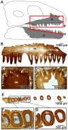
Figure 9. Cross-sectional and longitudinal views of the dentary teeth of Bolosaurus.
A: diagram indicating from what parts of the dentary the histological sections were taken. B: cross-section of a Bolosaurus striatus dentary fragment (StlPB-R 636), showing a complete lack of any dentine infolding. C: interpretation of the cross-section in B. Light grey areas indicate dentine, while darker grey areas indicate alveolar bone. D: long-section of a B. striatus dentary fragment (StlPB-R 637), showing the deep implantation of the associated teeth. E: interpretation of the long-section in D. Light grey areas indicate dentine, while darker grey areas indicate alveolar bone. The white area on the crowns of some of the teeth indicates enamel. F: Occlusal view of the dentary of Bolosaurus grandis showing the deep sockets of the teeth. d, dentary; de, dentine; e, enamel; pc, pulp cavity.

Similar articles

Cited by

References

    1. Tsuji LA, Müller J, Reisz RR (2010) Microleter mckinzieorum gen. et sp. nov. from the Lower Permian of Oklahoma: the basalmost parareptile from Laurasia. J Syst Palaeontol 8: 245–255.
    1. MacDougall MJ, Reisz R (2012) A new parareptile (Parareptilia, Lanthanosuchoidea) from the Early Permian of Oklahoma. J Vertebr Paleontol 32: 1018–1026.
    1. Modesto S, Sues H-D, Damiani R (2001) A new Triassic procolophonoid reptile and its implications for procolophonoid survivorship during the Permo-Triassic extinction event. Proc R Soc Lond B Biol Sci 268: 2047–2052. - PMC - PubMed
    1. Modesto SP, Damiani RJ, Neveling J, Yates AM (2003) A new Triassic owenettid parareptile and the mother of mass extinctions. J Vertebr Paleontol 23: 715–719.
    1. Tsuji LA, Müller J (2009) Assembling the history of the Parareptilia: phylogeny, diversification, and a new definition of the clade. Foss Rec 12: 71–81.

Publication types

LinkOut - more resources