. 2014 May 7;9(5):e97032.
doi: 10.1371/journal.pone.0097032. eCollection 2014.

Progranulin gene delivery protects dopaminergic neurons in a mouse model of Parkinson's disease

Affiliations

Progranulin gene delivery protects dopaminergic neurons in a mouse model of Parkinson's disease

Jackalina M Van Kampen et al. PLoS One. .

Abstract

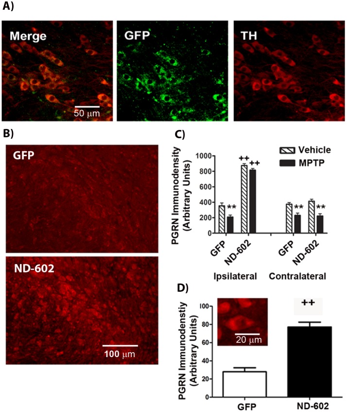
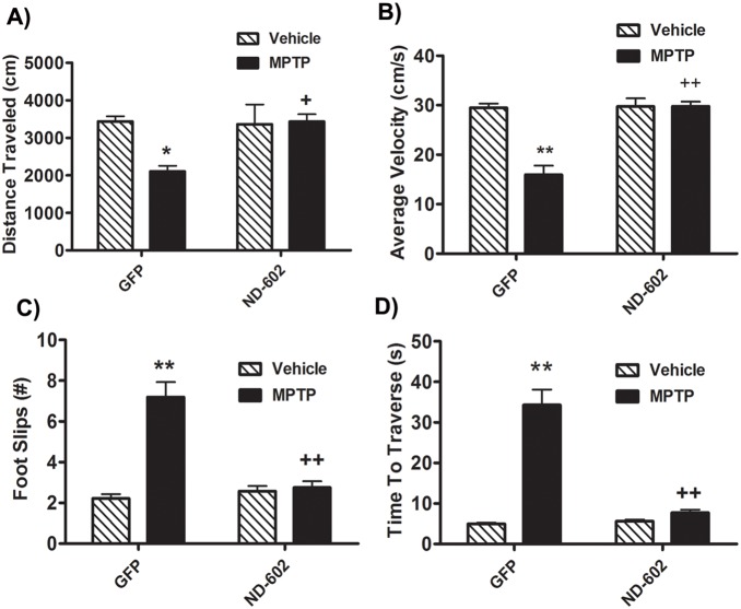
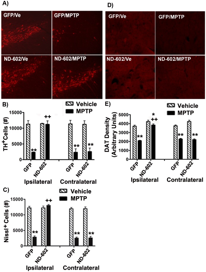
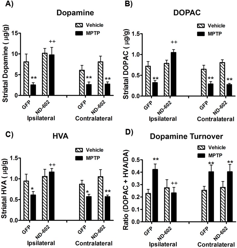
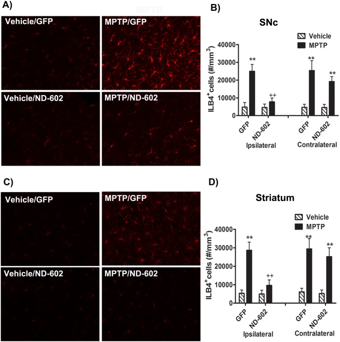
Parkinson's disease (PD) is a progressive neurodegenerative disorder characterized by tremor, rigidity and akinesia/bradykinesia resulting from the progressive loss of nigrostriatal dopaminergic neurons. To date, only symptomatic treatment is available for PD patients, with no effective means of slowing or stopping the progression of the disease. Progranulin (PGRN) is a 593 amino acid multifunction protein that is widely distributed throughout the CNS, localized primarily in neurons and microglia. PGRN has been demonstrated to be a potent regulator of neuroinflammation and also acts as an autocrine neurotrophic factor, important for long-term neuronal survival. Thus, enhancing PGRN expression may strengthen the cells resistance to disease. In the present study, we have used the 1-methyl-4-phenyl-1,2,3,6-tetrahydropyridine (MPTP) model of PD to investigate the possible use of PGRN gene delivery as a therapy for the prevention or treatment of PD. Viral vector delivery of the PGRN gene was an effective means of elevating PGRN expression in nigrostriatal neurons. When PGRN expression was elevated in the SNC, nigrostriatal neurons were protected from MPTP toxicity in mice, along with a preservation of striatal dopamine content and turnover. Further, protection of nigrostriatal neurons by PGRN gene therapy was accompanied by reductions in markers of MPTP-induced inflammation and apoptosis as well as a complete preservation of locomotor function. We conclude that PGRN gene therapy may have beneficial effects in the treatment of PD.


Conflict of interest statement

Competing Interests: The authors are/were employees of Neurodyn Inc. which holds a patent on ND-602 as a potential therapy for eurodegenerative diseases. Patent name: Treating neurodegenerative diseases with progranulin and number: US 20100324127 A1. Patent: Method for increasing neprilysin expression and activity, number WO 2012/065248 A1. There are no further patents, products in development or marketed products to declare. This does not alter the authors’ adherence to all the PLOS ONE policies on sharing data and materials.

Figures

Figure 1
Figure 1. Immunohistochemical analysis of enhanced PGRN expression in the SNC following ND-602.
A) Fluorescent photomicrographs depicting GFP expression in TH+ neurons of the ipsilateral SNC 4 weeks following unilateral intranigral ND-602 infusion. B) Fluorescent photomicrographs depicting PGRN immunolabeling in the ipsilateral SNC 4 weeks following unilateral intranigral infusion of either ND-602 or GFP control. C) In GFP controls, MPTP produced a significant reduction in PGRN immunolabeling within the substantia nigra of both hemispheres. Unilateral intranigral delivery of ND-602 significantly elevated PGRN immunolabeling in the ipsilateral SNC of both vehicle- and MPTP-treated animals. No effect of ND-602 was seen in the contralateral nigra. Each bar represents the mean (±S.E.M., n = 5–9) optical density (arbitrary units) averaged across 4 coronal sections through the SNC. D) The density of PGRN immunolabeling within TH+ neurons of the ipsilateral SNC was significantly elevated in those animals treated with ND-602. Inset fluorescent photomicrograph depicts PGRN immunolabeling in dopaminergic neurons of the ipsilateral SNC following ND-602 treatment. Each bar represents the mean (±S.E.M., n = 5–9) optical density (arbitrary units) measured in 10 cells in each of 4 coronal sections through the ipsilateral SNC. **sig. diff. from vehicle control, p<0.001. ++sig. diff. from GFP control, p<0.001.
Figure 2
Figure 2. Locomotor deficits in MPTP-intoxicated mice treated with ND-602.
A) Following MPTP intoxication, animals displayed significantly reduced locomotor activity, as assessed by total distance traveled in one hour. However, MPTP failed to reduce locomotor activity in those animals treated with ND-602. Each bar represents the mean (±S.E.M., n = 5–6) distance traveled (cm) in one hour. B) The average velocity of locomotion was significantly reduced following MPTP intoxication. However, in those animals treated with viral ND-602, no such reduction was observed. Velocity in those animals was significantly higher than in their GFP-treated counterparts. Each bar represents the mean (±S.E.M., n = 5–6) velocity (cm/s) observed over a one hour period. C) When traversing the horizontal ladder, MPTP-treated animals displayed reduced locomotor coordination as evidenced by significantly more foot slips. However, in those animals treated with ND-602, no such elevation in foot slips was observed. Locomotor coordination in those animals was significantly better than in their GFP-treated counterparts. Each bar represents the mean (±S.E.M., n = 5–6) number of foot slips recorded. D) Following MPTP intoxication, animals took significantly longer to traverse a horizontal ladder. However, those animals treated with ND-602 showed no increase in travel time and were significantly faster than their GFP-treated counterparts. Each bar represents the mean (±S.E.M., n = 5–6) time(s) to traverse a horizontal ladder. **sig. diff. from vehicle control, p<0.001; *p<0.05. ++sig. diff. from GFP control, p<0.001; +p<0.05.
Figure 3
Figure 3. Nigrostriatal integrity of MPTP-intoxicated mice treated with ND-602.
A) Representative fluorescent photomicrographs depicting TH immunolabeling in the ipsilateral SNC following MPTP intoxication in ND-602- and GFP-treated animals. Unbiased stereological counts of B) TH+ and C) Nissl+ cells in the SNC were significantly reduced following MPTP intoxication. However, no significant cell loss was observed ipsilateral to viral vector delivery of PGRN by ND-602. Significantly more TH+ and Nissl+ cells were noted ipsilateral to ND-602 infusion relative to GFP controls. Each bar represents the mean (±S.E.M., n = 5–9) number of TH or Nissl immunopositive cells counted in the SNC. D) Representative fluorescent photomicrographs depicting DAT immunolabeling in the ipsilateral striatum following MPTP intoxication in ND-602- and GFP-treated animals. E) MPTP intoxication resulted in a significant loss of DAT immunolabeling in the striatum. In those animals treated with ND-602, the loss of striatal DAT was significantly attenuated in the ipsilateral hemisphere. Each bar represents the mean (±S.E.M., n = 5–9) optical density (arbitrary units) measured in three striatal regions across 4 coronal sections through the striatum. **sig. diff. from vehicle control, p<0.001; *p<0.05. ++sig. diff. from GFP control, p<0.001.
Figure 4
Figure 4. Striatal dopamine and turnover in MPTP-intoxicated mice following ND-602.
A) Levels of striatal dopamine were significantly reduced following MPTP intoxication. However, in those animals treated with viral vector delivery of PGRN by ND-602, no decline in dopamine was observed ipsilateral to vector delivery. Levels were reduced, however, in the contralateral, untreated, hemisphere. The dopamine metabolites, B) DOPAC and C) HVA, were similarly reduced by MPTP, with no reductions observed ipsilateral to ND-602 delivery. D) Striatal dopamine turnover was significantly elevated by MPTP intoxication. However, no change in turnover was observed ipsilateral to ND-602 treatment. Each bar represents the mean (±S.E.M., n = 5–8) µg of dopamine, DOPAC or HVA detected per gram of wet tissue weight. For dopamine turnover, the calculation, DOPAC + HVA/dopamine was applied. **sig. diff. from vehicle control, p<0.001. ++sig. diff. from GFP control, p<0.001.
Figure 5
Figure 5. Nigrostriatal inflammation in MPTP-intoxicated mice treated with ND-602.
Representative fluorescent photomicrographs of isolectin (ILB4) labeling in the A) SN and C) striatum two weeks following the last MPTP injection. MPTP intoxication resulted in a significant elevation in the number of activated microglia in both the B) SNC and D) striatum. In both regions, this activation was significantly reduced in those animals treated with ND-602. Each bar represents the mean (±S.E.M., n = 5–6) number of ILB4-positive cells estimated per mm3. **sig. diff. from vehicle control, p<0.001; *p<0.05. ++sig. diff. from GFP control, p<0.001.

Similar articles

Cited by

References

    1. Eriksen JL, MacKenzie IR (2008) Progranulin: normal function and role in neurodegeneration. Journal of Neurochemistry 104: 287–297. - PubMed
    1. Jin FY, Nathan C, Radzioch D, Ding A (1997) Secretory leukocyte protease inhibitor: a macrophage product induced by and antagonistic to bacterial lipopolysaccharide. Cell 88: 417–426. - PubMed
    1. Baker M, MacKenzie IR, Pickering-Brown SM, Gass J, Rademakers R, et al. (2006) Mutations in progranulin cause tau-negative frontotemporal dementia linked to chromosome 17. Nature 442: 916–919. - PubMed
    1. Cruts M, Kumar-Singh S, Van Broeckhoven C (2006) Progranulin mutations in ubiquitin-positive frontotemporal dementia linked to chromosome 17q21. Curr Alzheimer Res 3: 485–491. - PubMed
    1. Cruts M, Gijselinck I, van der Zee J, Engelborghs S, Wils H, et al. (2006) Null mutations in progranulin cause ubiquitin-positive frontotemporal dementia linked to chromosome 17q21. Nature 442: 920–924. - PubMed

Publication types

MeSH terms