. 2014 Jul 17;28(11):1555-66.
doi: 10.1097/QAD.0000000000000289.

Reactivation of HIV latency by a newly modified Ingenol derivative via protein kinase Cδ-NF-κB signaling

Affiliations

Reactivation of HIV latency by a newly modified Ingenol derivative via protein kinase Cδ-NF-κB signaling

Guochun Jiang et al. AIDS. .

Abstract

Objective: Although HAART effectively suppresses viral replication, it fails to eradicate latent viral reservoirs. The 'shock and kill' strategy involves the activation of HIV from latent reservoirs and targeting them for eradication. Our goal was to develop new approaches for activating HIV from latent reservoirs.

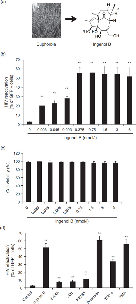
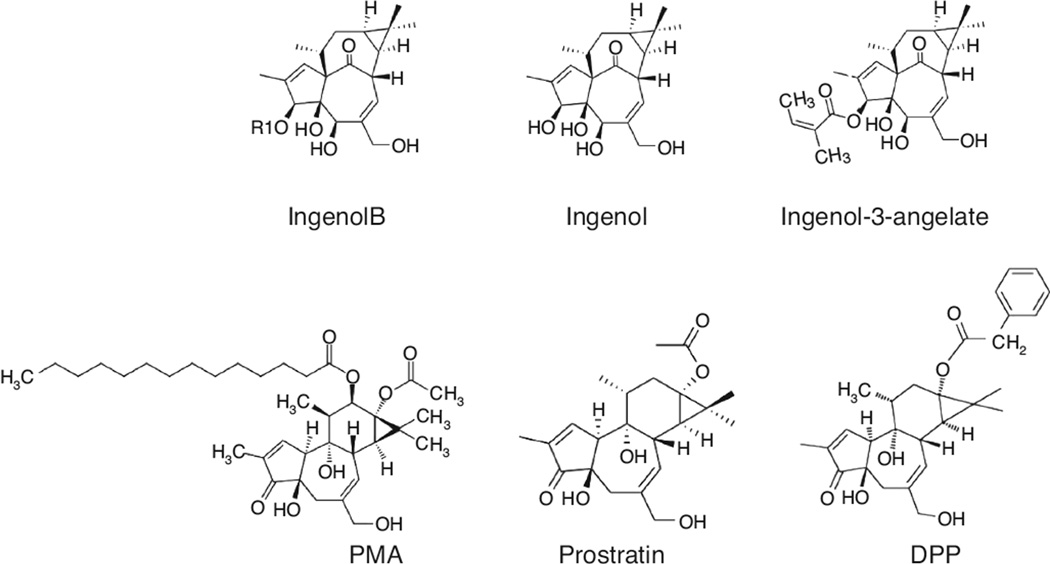
Design: We investigated capacity of Ingenol B (IngB), a newly modified derivative of Ingenol ester that was originally isolated from a Brazilian plant in Amazon, for its capacity and mechanisms of HIV reactivation.

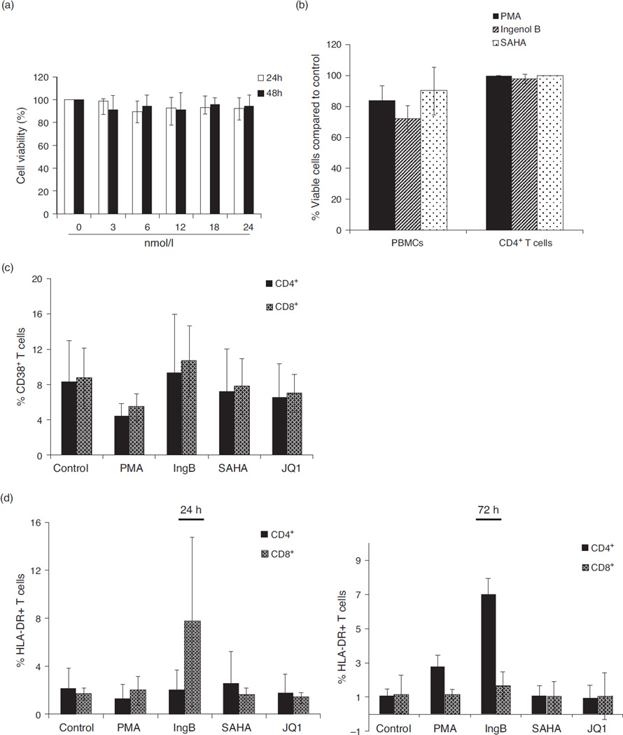
Methods: Reactivation of HIV-1 by IngB was evaluated in J-Lat A1 cell culture model of HIV latency as well as in purified primary CD4 T cells from long-term HAART-treated virologically-suppressed HIV-infected individuals. The underlining molecular mechanisms of viral reactivation were investigated using flow cytometry, RT-qPCR and chromatin immunoprecipitation.

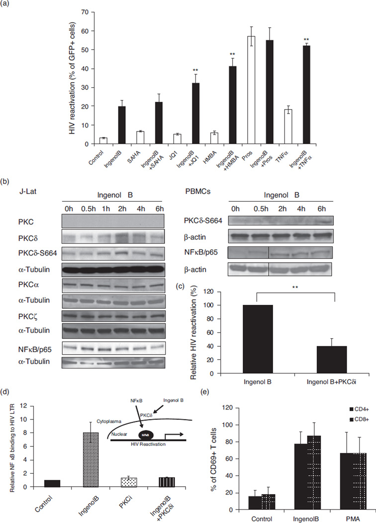
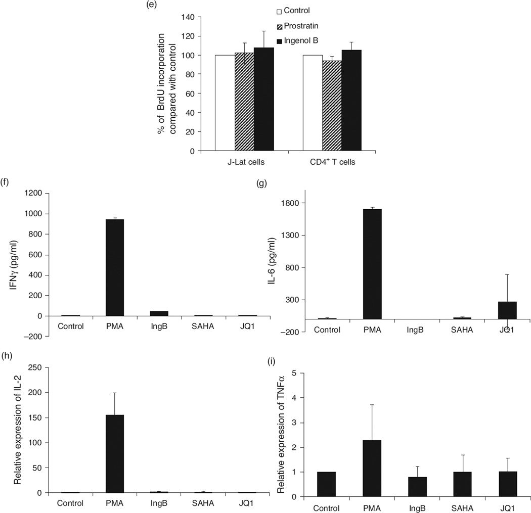
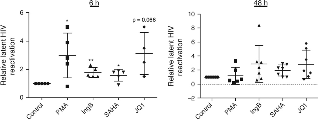
Results: IngB is highly effective in reactivating HIV in J-Lat A1 cells with relatively low cellular toxicity. It is also able to reactivate latent HIV in purified CD4 T cells from HAART-treated HIV-positive individuals ex vivo. Our data show that IngB may reactivate HIV expression by both activating protein kinase C (PKC)δ-nuclear factor kappa-light-chain-enhancer of activated B cells (NF-κB) pathway and directly inducing NF-κB protein expression. Importantly, IngB has a synergistic effect with JQ1, a BET bromodomain inhibitor, in latent HIV reactivation.

Conclusions: IngB is a new promising compound to activate latent HIV reservoirs. Our data suggest that formulating novel derivatives from Ingenol esters may be an innovative approach to develop new lead compounds to reactivate latent HIV.


Conflict of interest statement

Conflicts of interest

There are no conflicts of interest to declare for all authors.

Figures

Fig. 1
Fig. 1. Ingenol B is a potent activator of HIV LTR in J-Lat A1 cell line model for HIV latency
(a) Ingenol B is a modified compound isolated from a Brazilian plant, Euphorbia tirucalli. (b) Ingenol B reactivated latent HIV-1 in J-Lat A1 cells. Increased doses of Ingenol B, from 23 pmol/l to 6 nmol/l, were added in J-Lat A1 cells for 24 h. Latent HIV-1 reactivation was measured by detecting GFP-expressing cells by flow cytometry. (**) P < 0.01. (c) Cell viability during Ingenol B treatment. Increased doses of Ingenol B, from 23 pmol/l to 6 nmol/l, were added to J-Lat A1 cells for 24 h and cell viability was measured using live/dead dye by flow cytometry. (d) Ingenol B has higher potential of HIV LTR reactivation in J-Lat cells compared to SAHA or JQ1. The HIV LTR driven GFP expression in J-Lat A1 cells was determined by flow cytometry at 24 h of treatment with Ingenol B and other compounds. The dosage for the treatment was: Ingenol B 375 pmol/l, PMA 5 ng/ml, TNF-α 25 TNF ng/ml, HMBA 5 mmol/l, JQ1 2 µmol/l, prostratin 10 µmol/l, SAHA 500 nmol/l. HIV LTR reactivation was analyzed in comparison to untreated J-Lat cell controls. TNF, tumor necrosis factor. (*) P < 0.05, (**) P < 0.01.
Fig. 2
Fig. 2. Ingenol B reactivates latent HIV LTR-regulated expression through protein kinase Cd–S664–NF-kB signaling
(a) A single compound or a combination of compounds was added to J-Lat A1 cells for 24 h. HIV-1 LTR-driven GFP expression in the cells was measured by flow cytometry as shown in Fig. 1. Combined treatments included Ingenol B at 46 pmol/l. (**) P < 0.01, compared to single compound. (b) Activation of PKCδ and expression of PKC isoforms in J-Lat cells or human PBMCs from healthy donors. J-Lat A1 cells or human PBMCs were treated with 6 nmol/l Ingenol B for 6 h. The cells were collected and lyzed in RIPA buffer. Western blot analysis was performed to detect expression of isoforms of PKC, expression of NF-κB/p65, or Ser664 phosphorylation at PKCδ with indicated antibodies. (c) PKCδ inhibitor suppresses Ingenol B-induced HIV reactivation. J-Lat A1 cells were treated with 3 nmol/l Ingenol B in the presence of 5 µmol/l PKCδ inhibitor and examined for GFP expression by flow cytometry. (**) P < 0.01. (d) Ingenol B promotes NF-κB/p65 binding to HIV LTR through activation of PKCδ. J-Lat A1 cells were treated with 6 nmol/l Ingenol B with or without 5 µmol/l PKCδ inhibitor Millipore). The cells were collected and ChIP-qPCR assay was performed to detect NF-κB/p65 binding to HIV LTR. Relative NF-κB/p65 binding to HIV LTR was expressed after normalized to untreated controls. (e) Activation of CD69 expression in PBMC following Ingenol B treatment. The CD69 expression was analyzed in CD4+ and CD8+ T-cell subsets in PBMC from HIV-negative controls that were treated with PMA or Ingenol B for 24 h. PMA-treated cells served as the positive control. PKC, protein kinase C.
Fig. 3
Fig. 3. Ingenol B causes minimal T-cell activation and cell toxicity
(a) Lack of cellular toxicity in J-Lat cells during Ingenol B treatment. J-Lat A1 cells were treated with Ingenol B from 3 to 24 nmol/l for 24 or 48 h, and cell proliferation and viability were measured by MTT. (b) Ingenol B has minimal cell toxicity in PBMCs or isolated CD4+ T cells. Human PBMCs or CD4+ T cells were isolated from peripheral blood of healthy HIV-negative donors and treated with 5 ng/ml PMA, 500 nmol/l SAHA, or 3 nmol/l Ingenol B for 20 h, and evaluated for cell proliferation/toxicity by MTT. (c) PBMCs were treated with specified agents for 24 h and examined for CD38 expression on CD4+ and CD8+ T cells by flow cytometry. (d) PBMCs were treated with indicated agents for 24 or 72 h and examined for HLA-DR expression on CD4+ or CD8+ T cells by flow cytometry. (e) J-Lat A1 cells and CD4+ T cells were treated with BrdU with or without Ingenol B or prostratin for 24 h, and BrdU ELISAs were performed with anti-BrdU antibody for quantification of BrdU incorporation. (f–i) Expression of IFNγ, IL-6, IL-2, or TNF-α in PBMCs from healthy donors (n = 3) following treatment with 3 nmol/l Ingenol B for 24 h. The expression of IFNγ and IL-6 proteins was detected by ELISA, and RNA expression of IL-2 and TNF-α was detected by RT-qPCR after normalizing the values with glyceraldehyde 3-phosphate dehydrogenase housekeeping gene and untreated controls. IFN, interferon; IL, interleukin; TNF, tumor necrosis factor.
Fig. 4
Fig. 4. Ingenol B reactivates latent HIV in primary CD4+ T cells from long-term HAART-treated HIV-positive individuals
Primary CD4+ T cells were purified from PBMCs isolated from cohorts of HAART-treated virologically suppressed HIV-infected individuals, and incubated with 200 ng/ml PMA and 2 µmol/l inomycin, 3 nmol/l Ingenol B, 500 nmol/l SAHA or 2 µmol/l JQ1 for 6 or 48 h. Cells were collected and RNAs were extracted by Qiagen RNeasy kit. RT-qPCR was performed for HIV reactivation analysis after normalized with expression of internal control (18S rRNA). The value was expressed relative to control treatment. (*) P < 0.05, (**) P < 0.01.
Fig. 5
Fig. 5. Core chemical structure of Ingenol B is analogous to phorbol esters
Ingenols share core structure analogous to phorbol esters.

Similar articles

Cited by

References

    1. Macal M, Sankaran S, Chun TW, Reay E, Flamm J, Prindiville TJ, et al. Effective CD4+ T-cell restoration in gut-associated lymphoid tissue of HIV-infected patients is associated with enhanced Th17 cells and polyfunctional HIV-specific T-cell responses. Mucosal Immunol. 2008;1:475–488. - PubMed
    1. Lerner P, Guadalupe M, Donovan R, Hung J, Flamm J, Prindiville T, et al. The gut mucosal viral reservoir in HIV-infected patients is not the major source of rebound plasma viremia following interruption of highly active antiretroviral therapy. J Virol. 2011;85:4772–4782. - PMC - PubMed
    1. Finzi D, Hermankova M, Pierson T, Carruth LM, Buck C, Chaisson RE, et al. Identification of a reservoir for HIV-1 in patients on highly active antiretroviral therapy. Science. 1997;278:1295–1300. - PubMed
    1. Chun TW, Carruth L, Finzi D, Shen X, DiGiuseppe JA, Taylor H, et al. Quantification of latent tissue reservoirs and total body viral load in HIV-1 infection. Nature. 1997;387:183–188. - PubMed
    1. Chomont N, El-Far M, Ancuta P, Trautmann L, Procopio FA, Yassine-Diab B, et al. HIV reservoir size and persistence are driven by T cell survival and homeostatic proliferation. Nat Med. 2009;15:893–900. - PMC - PubMed

Publication types