. 2014 Jul;79(1):139-49.
doi: 10.1111/tpj.12547. Epub 2014 Jun 17.

Glycosylinositol phosphorylceramides from Rosa cell cultures are boron-bridged in the plasma membrane and form complexes with rhamnogalacturonan II

Affiliations

Glycosylinositol phosphorylceramides from Rosa cell cultures are boron-bridged in the plasma membrane and form complexes with rhamnogalacturonan II

Aline Voxeur et al. Plant J. 2014 Jul.

Abstract

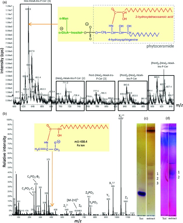
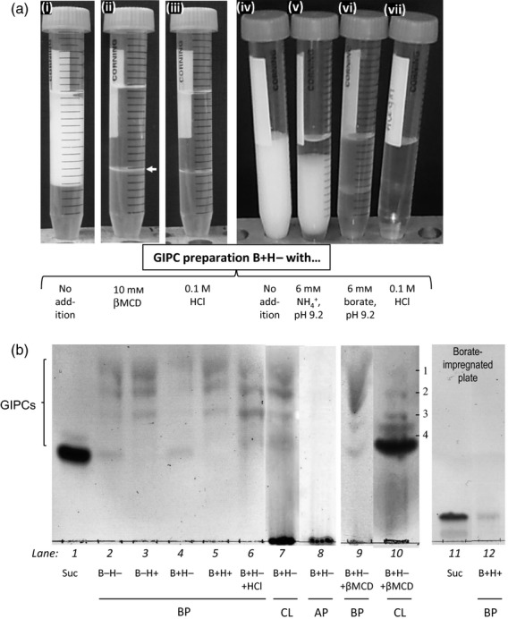
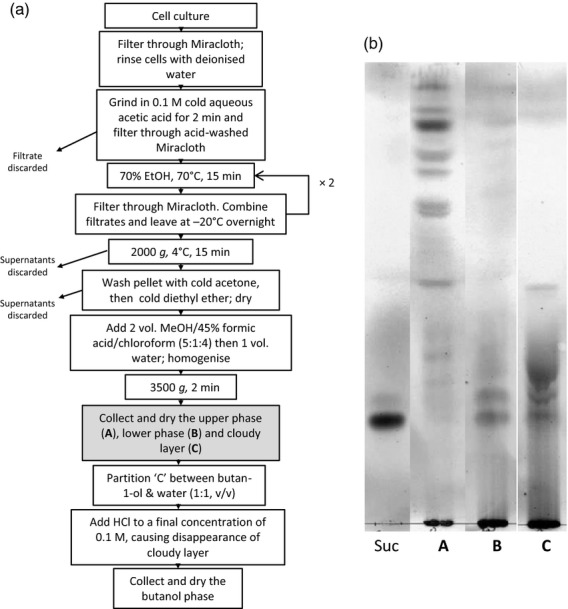
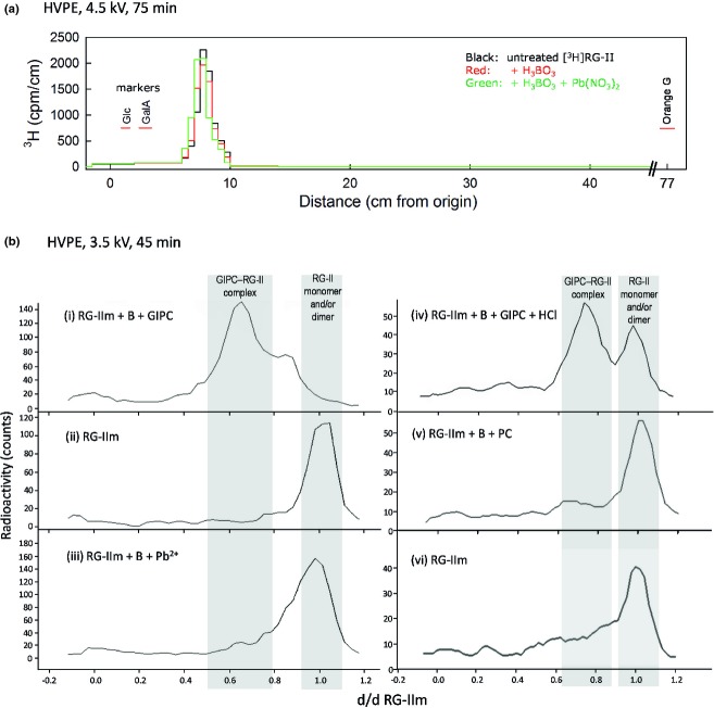
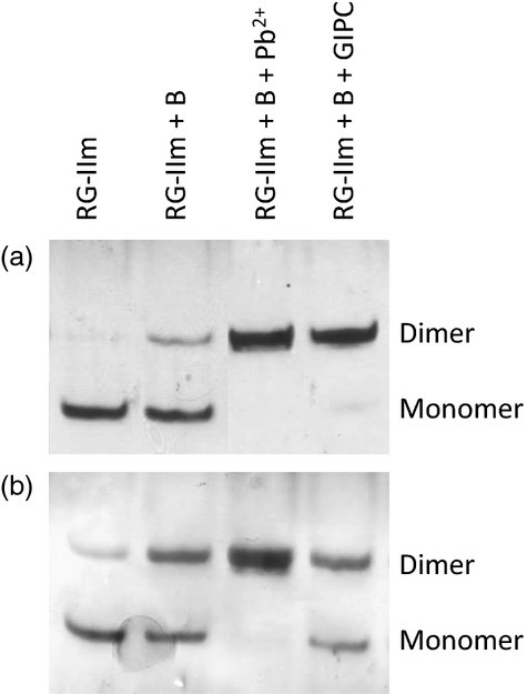
Boron (B) is essential for plant cell-wall structure and membrane functions. Compared with its role in cross-linking the pectic domain rhamnogalacturonan II (RG-II), little information is known about the biological role of B in membranes. Here, we investigated the involvement of glycosylinositol phosphorylceramides (GIPCs), major components of lipid rafts, in the membrane requirement for B. Using thin-layer chromatography and mass spectrometry, we first characterized GIPCs from Rosa cell culture. The major GIPC has one hexose residue, one hexuronic acid residue, inositol phosphate, and a ceramide moiety with a C18 trihydroxylated mono-unsaturated long-chain base and a C24 monohydroxylated saturated fatty acid. Disrupting B bridging (by B starvation in vivo or by treatment with cold dilute HCl or with excess borate in vitro) enhanced the GIPCs' extractability. As RG-II is the main B-binding site in plants, we investigated whether it could form a B-centred complex with GIPCs. Using high-voltage paper electrophoresis, we showed that addition of GIPCs decreased the electrophoretic mobility of radiolabelled RG-II, suggesting formation of a GIPC-B-RG-II complex. Last, using polyacrylamide gel electrophoresis, we showed that added GIPCs facilitate RG-II dimerization in vitro. We conclude that B plays a structural role in the plasma membrane. The disruption of membrane components by high borate may account for the phytotoxicity of excess B. Moreover, the in-vitro formation of a GIPC-B-RG-II complex gives the first molecular explanation of the wall-membrane attachment sites observed in vivo. Finally, our results suggest a role for GIPCs in the RG-II dimerization process.

Keywords: Rosa sp; boron; cell wall; cross-linking; glycosylinositol phosphoceramides; glycosylinositol phosphorylceramides; lipid rafts; plasma membrane; rhamnogalacturonan II.


Figures

Figure 1
Figure 1
Mass spectrometric and thin-layer chromatographic analysis of Rosa glycosylinositol phosphorylceramides (GIPCs). (a) ESI-MS analysis of GIPC extract from Rosa cell culture. The spectrum was acquired in the negative ion mode. Abbreviations: Hex, hexose residue (probably α-mannose); HexA, hexuronic acid residue (probably α-glucuronic acid); Pent, pentose residue; Ins, myo-inositol; P, phosphate; Cer, phytoceramide. Inset: proposed structure of the predominant GIPC species; phytoceramide moiety in grey box. (b) ESI-MS/MS (collision-induced dissociation spectrum) analysis of the predominant Hex-HexA-Ins-P-Cer peak seen in (a) as the [M-2H]2− ion at m/z 630. Nitrogen was used as collision gas in a Q-TRAP instrument, with the collision energy set to −40 eV. The standard nomenclature for glycolipid fragmentation has been applied (Costello and Vath, 1990; Levery et al., 2001). Inset: proposed identity of the ion at m/= 438.4, indicating an h24:0 ceramide moiety. (c, d) Thin-layer chromatography (TLC) of GIPC extract. Lipids were chromatographed in CHCl3/CH3OH/4 m NH4OH (9:7:2, by vol.) with 0.2 m ammonium acetate (Kaul and Lester, 1978) and located by orcinol reagent (c) or periodic acid–Schiff staining (d). Lipid bands are labelled: 1, Hex-HexA-Ins-P-Cer; 2, (Hex)2-HexA-Ins-P-Cer; 3, Pent-(Hex)2-HexA-Ins-P-Cer.
Figure 2
Figure 2
Influence of boron on glycosylinositol phosphorylceramide (GIPC) extraction. (a) A cloudy layer was observed during butanol/water phase-partitioning of a GIPC-enriched lipid sample extracted with neutral ethanol from Rosa cell cultures that had been grown in the usual B concentration (i, iv). This cloudy layer disappeared in the presence of 0.1 m HCl (iii, vii), 10 mm βMCD (ii), or 6 mm borate buffer, pH 9.2 (vi). The horizontal arrow indicates the slight cloudy layer left in the presence of βMCD (butanol above). In contrast, 6 mm ammonium buffer, pH 9.2 (v), only led to a partial disappearance. (b) TLC of the different phases after butanol/water phase-partitioning of a GIPC-rich lipid extract from Rosa cell cultures grown in media with (B+) or without boron (B−). The lipids had been extracted in 70% ethanol that contained 0.1 m HCl (H+) or lacking acid (H−). BP, butanol phase; CL, cloudy layer; AP, aqueous phase; Suc, sucrose (marker). In lanes 9 and 10, 10 mm βMCD was present during the partitioning step. Lipids labelled on lane 10: bands 1–3, as in Figure1; band 4, (Pent)2-(Hex)2-HexA-Ins-P-Cer.
Figure 3
Figure 3
Purification of glycosylinositol phosphorylceramide (GIPC) from Rosa cell culture. (a) GIPC purification scheme adapted from Buré et al. (2011). (b) Thin-layer chromatography (TLC) of the phase A, B and C obtained during the step shaded grey in (a). Suc, sucrose marker.
Figure 4
Figure 4
High-voltage paper electrophoresis of radiolabelled RG-II and the effect of boric acid, Pb2+ and glycosylinositol phosphorylceramides (GIPCs). (a) [3H]RG-II was loaded on to the paper after 9 h of pre-treatment of monomeric [3H]RG-II in 250 mm pyridine buffer, pH 4.7, with or without 1 mm H3BO3 or 1 mm H3BO3 plus 0.5 mm Pb(NO3)2. After electrophoresis, the paper was cut into strips, which were assayed for tritium by scintillation-counting. The positions of non-radioactive markers (glucose, galacturonic acid and Orange G) are also shown. (b) Samples were loaded on the paper after 4 h of pre-treatment of monomeric [3H]RG-II (RGIIm) in 50 mm ammonium acetate, pH 4.8, with or without various combinations of 1.2 mm boric acid (B), 0.5 mm Pb(NO3)2 (Pb2+), GIPCs (purified as described in Figure3), commercial phytoceramide (PC), and 0.1 m hydrochloric acid (HCl). After electrophoresis, papers were read on a LabLogic AR2000 radio-TLC Imaging Scanner. The distance migrated (d) is given relative to dRGIIm (the distance migrated by monomeric RG-II, that is to say approximately 6 cm). Grey bands indicate the positions of monomeric RG-II (right) and the GIPC–RG-II complex (left).
Figure 5
Figure 5
Glycosylinositol phosphorylceramide (GIPC)-induced dimerization of monomeric RG-II. After 4 h of pre-treatment of monomeric RG-II without or with 1.2 mm H3BO3 ± GIPC or with 1.2 mm H3BO3 and 0.5 mm Pb(NO3)2, samples were analysed by polyacrylamide gel electrophoresis and silver stained. Abbreviations as in Figure4. Parts (a) and (b) show the results of two independent experiments.
Figure 6
Figure 6
Co-expression network between genes involved in RG-II and glycosylinositol phosphorylceramide (GIPC) biosynthesis in various species using an edge-weighted force-directed approach, based on data retrieved from ATTED-II and visualised in Cytoscape 2.8 (http://www.cytoscape.org). Circles: white circle, LOH (encoding a ceramide synthase); pale grey, encoding enzymes involved in synthesis of RG-II side-chains; dark grey, encoding enzymes involved in synthesis of the homogalacturonan backbone; black, C4H (encoding cinnamate-4-hydroxylase, involved in lignin synthesis; included as a control). Lines: green lines, Arabidopsis thaliana; blue, Populus trichocarpa; red, Oryza sativa; black, Glycine max; yellow, Zea mays. Solid line, strong co-expression [mutual rank (MR) < 1000]; dotted line, weak co-expression (5000 > MR > 1000); no line, no transcriptomic data available or no co-expression (MR > 5000).

References

    1. Ahn J, Verma R, Kim M, Lee J, Kim Y, Bang J, Reiter W, Pai H. Depletion of UDP-d-apiose/UDP-d-xylose synthases results in rhamnogalacturonan-II deficiency, cell-wall thickening, and cell death in higher plants. J. Biol. Chem. 2006;281:13708–13716. - PubMed
    1. Al-Mustafa WA, Falatah AM, El-Shall AA. Effect of excess boron fertilization on status and availability of boron in calcareous soils. Fertilizer Res. 1993;36:71–78.
    1. Aquea F, Federici F, Moscoso C, Vega A, Jullian P, Haseloff JIM, Arce-Johnson P. A molecular framework for the inhibition of Arabidopsis root growth in response to boron toxicity. Plant, Cell Environ. 2012;35:719–734. - PubMed
    1. Bassil E, Hu H, Brown PH. Use of phenylboronic acids to investigate boron function in plants. possible role of boron in transvacuolar cytoplasmic strands and cell-to-wall adhesion. Plant Physiol. 2004;136:3383–3395. - PMC - PubMed
    1. Blevins DG, Lukaszewski KM. Boron in plant structure and function. Annu. Rev. Plant Physiol. Plant Mol. Biol. 1998;49:481–500. - PubMed

Publication types

MeSH terms