. 2014 May 7;9(5):e96790.
doi: 10.1371/journal.pone.0096790. eCollection 2014.

Co-expression of foreign proteins tethered to HIV-1 envelope glycoprotein on the cell surface by introducing an intervening second membrane-spanning domain

Affiliations

Co-expression of foreign proteins tethered to HIV-1 envelope glycoprotein on the cell surface by introducing an intervening second membrane-spanning domain

Hongyun Wang et al. PLoS One. .

Abstract

The envelope glycoprotein (Env) of human immunodeficiency virus type I (HIV-1) mediates membrane fusion. To analyze the mechanism of HIV-1 Env-mediated membrane fusion, it is desirable to determine the expression level of Env on the cell surface. However, the quantification of Env by immunological staining is often hampered by the diversity of HIV-1 Env and limited availability of universal antibodies that recognize different Envs with equal efficiency. To overcome this problem, here we linked a tag protein called HaloTag at the C-terminus of HIV-1 Env. To relocate HaloTag to the cell surface, we introduced a second membrane-spanning domain (MSD) between Env and HaloTag. The MSD of transmembrane protease serine 11D, a type II transmembrane protein, successfully relocated HaloTag to the cell surface. The surface level of Env can be estimated indirectly by staining HaloTag with a specific membrane-impermeable fluorescent ligand. This tagging did not compromise the fusogenicity of Env drastically. Furthermore, fusogenicity of Env was preserved even after the labeling with the ligands. We have also found that an additional foreign peptide or protein such as C34 or neutralizing single-chain variable fragment (scFv) can be linked to the C-terminus of the HaloTag protein. Using these constructs, we were able to determine the required length of C34 and critical residues of neutralizing scFv for blocking membrane fusion, respectively.


Conflict of interest statement

Competing Interests: The authors have declared that no competing interests exist.

Figures

Figure 1
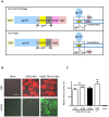
Figure 1. Connection of HaloTag to Env with the addition of the intervening second MSD.
(A) Upper panel: Design of the Env-TM11D-Halo construct. A CMV promoter drives the expression of the tethered construct in all expression vectors. The 21 aa MSD of TM11D was added to the C terminal of gp41 as an MSD2 to help flipping out the HaloTag. Lower panel: Design of the Env-Halo construct, the control tethered construct without the MSD2 of TM11D. The expected membrane topology of the expressed protein with each construct is depicted schematically on the right. ED: ectodomain of gp41, MSD: membrane-spanning domain of gp41, CT: cytoplasmic tail of gp41. The sequences for Env corresponding to HXB2 or JRFL were used. (B) Confocal microscope analysis of tethered HaloTag in transfected 293FT cells stained with membrane-permeable (TMR, red color) or impermeable (AF488, green color) ligands. The transfected DNA is indicated: Mock, control DNA transfection; HXB2-Halo, Halo directly connects with gp41 (without the MSD of TM11D); HXB2-TM11D-Halo, a construct with the MSD of TM11D added between Env and Halo. Scale bar = 20 µm. (C) Effect of Halo ligands on the fusogenicity of HaloTag attached Env by DSP assay. Halo TMR and AF488 ligands were used to label the HXB2-TM11D-Halo fusion protein. The effect on the fusion activity was measured by DSP assay, which measures pore formation during cell-cell fusion by split Renilla luciferase (RL) reporter proteins. DSP activities for the ligand-labeled Env were compared with the HXB2-TM11D-Halo and non-tethered HXB2 Env protein without labeling (only add DMEM culture medium, and the value of HXB2-TM11D-Halo was set at 100%). Error bars represent standard deviations of the results of triplicate experiments. Student’s t-test was used to determine the statistical significance of the measured variables for each differently labeled (open column) and the non-labeled (solid column). ns = nonsignificant.
Figure 2
Figure 2. Flow cytometry analysis of Env-expressing cells labeled with Halo ligands or anti-Env monoclonal antibody.
(A–C) Flow cytometry analysis of Env expression level using different staining strategies. 293FT cells were transfected with expression vectors for tethered Env (HXB2-TM11D-Halo, red line; JRFL-TM11D-Halo, blue line), untethered Env (HXB2-WT, red dashed line; JRFL-WT, blue dashed line), or Mock DNA (grey shade). Cells were stained with membrane-impermeable HaloTag AF488 ligand (A), membrane-permeable HaloTag Oregon Green ligand (C) and anti-Env V3 antibody V3-G2-25 (B). Histograms are representative results from three independent experiments. (D) Positive staining rate of HaloTag labeling and anti-Env antibody immunolabeling of cells transfected with HXB2-TM11D-Halo (solid red bar), HXB2-WT (red shade bar), JRFL-TM11D-Halo (solid blue bar), JRFL-WT (blue shade bar) or Mock (solid gray bar). Error bars represent standard deviations of the results from three independent experiments.
Figure 3
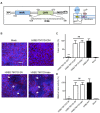
Figure 3. Measurement of fusion inhibition of tethered C34 evaluated by syncytia formation and DSP assay.
(A) Left panel: Schematic view of HIV-1 gp41. FP, fusion peptide; NHR, N-terminal heptad repeat region; CHR, C-terminal heptad repeat region; MSD, membrane-spanning domain; CT, cytoplasmic tail. The residues are numbered according to their position in HXB2 gp160. The amino acid sequence of C34 is shown. Right panel: The expected membrane topology of HXB2-TM11D-C34 is depicted schematically. (B) Syncytia formation assay in transfected 293CD4 cells. 293CD4 cells were transfected with the indicated constructs (HXB2-TM11D-Halo, HXB2-TM11D-2N, and HXB2-TM11D-C34). After 16 h, the nuclei of cells were stained with Hoechst, and the membrane was stained with CellMask Deep Red plasma membrane stain. White arrows indicate typical syncytia formed in transfected 293CD4 cells. Scale bar = 50 µm. (C) Relative fusion activity was quantified using a fusion index (see Materials and Methods). Fusion activities for each plasmid are shown after normalization to that of the non-tethered Env expression construct (HXB2-WT). The activity of HXB2-WT was set at 100%. Error bars represent standard deviations of the results of five fields. Student’s t-test was used to determine the statistical significance of the measured variables for each construct (open column) and control (solid column). Statistical significance was indicated when p<0.001 (***). ns = nonsignificant. (D) Fusion activity measured by DSP assay. Relative fusion activity was measured by DSP assay. DSP activities for each construct were compared with that of the non-tethered Env expression construct (HXB2-WT was set at 100%). Error bars represent standard deviations of the results of triplicate experiments. Student’s t-test was used to determine the statistical significance of the measured variables for each construct (open column) and control (solid column). Statistical significance was indicated when p<0.01 (**). ns = nonsignificant.
Figure 4
Figure 4. Measurement of fusion inhibition of nested serial C-terminal deletion mutants of C34 by syncytia formation and DSP assay.
(A) Syncytia formation assay in transfected 293CD4 cells. 293CD4 cells were transfected with the indicated constructs (HXB2-TM11D-C24, C26, C28, C30, C32, C34, and HXB2-TM11D-Halo). Relative fusion activity was quantified using a fusion index. Fusion activities for each plasmid are shown after normalization to that of the HXB2-TM11D-Halo construct (set at 100%). Error bars represent standard deviations of the results of five fields. Student’s t-test was used to determine the statistical significance of the measured variables for each construct (open column) and control (solid column). Statistical significance was indicated when p<0.001 (***). (B) Fusion activity measured by DSP assay. The relative fusion activity was measured by DSP assay. DSP activities for each construct were compared with that of the HXB2-TM11D-Halo construct (set at 100%). Error bars represent standard deviations of the results of triplicate experiments. Student’s t-test was used to determine the statistical significance of the measured variables for each construct (open column) and control (solid column). Statistical significance was indicated when p<0.01 (**) or p<0.001 (***). (C) Amino acid sequence of all C-terminal deletion mutants of C34 are listed.
Figure 5
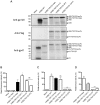
Figure 5. Effect of tethered neutralizing antibodies evaluated by syncytia formation and DSP assay.
(A) Immunoblotting analysis of tethered fusion protein expression in 293FT cells with anti-gp120 (upper panel), anti-Flag (middle panel) or Chessie 8 anti-gp41 antibodies (lower panel). The expression vector used is indicated above the lane and the position of different fusion proteins is shown to the right. The anti-gp120 antibody detected the precursor form of tethered proteins and processed gp120 band; the anti-Flag antibody detected the tethered precursor and processed gp41-TM11D-scFv band; and the Chessie 8 anti-gp41antibody detected the processed gp41 (including gp41-TM11D-Halo and gp41-TM11D-scFv) bands. (B) The MFI of different constructs determined by flow cytometry. HaloTag Alexa Fluor 488 ligand was used to stain proteins expressed on the cell surface of 293FT cells transfected with different tethered constructs. The Tac-Halo vector (Halo is expressed in the cytoplasm) was used as a negative control for surface staining. Data was acquired with a BD FACSCalibur system and at least 12,000 events were collected and analyzed using FlowJo software. The MFI of HXB2-TM11D-Halo was set at 100%. Error bars represent standard deviations of the results of triplicate experiments. Student’s t test was used for the statistical analysis of the measured variables between individual construct (open column) and control (solid column). Significance was reported with p<0.05(*). ns = nonsignificant. (C) Fusion activity measured by DSP assay. The relative fusion activity was measured by DSP assay. DSP activities for each construct were compared with that of Env tethered with TM11D-MSD and HaloTag (HXB2-TM11D-Halo was set at 100%). Error bars represent standard deviations of the results of triplicate experiments. Student’s t-test was used to determine the statistical significance of the measured variables for each construct (open column) and control (solid column). Statistical significance was indicated when p<0.05(*), p<0.001 (***). ns = nonsignificant. (D) Normalization of DSP activity with surface expression level of Env. The DSP activities of each tethered construct shown in panel C were normalized by respective surface expression level defined by MFI measured by flow cytometry shown in panel B. Student’s t-test was used to determine the statistical significance of the calculated variables for each construct (open column) and HXB2-TM11D-13H11 (solid grey column). Statistical significance was indicated when p<0.001 (***).
Figure 6
Figure 6. Mutations in the CDR3 region of tethered neutralizing scFvs recover fusion activity.
(A) B12 and 2F5 CDR H3 loop mutants are listed. Left panel, mutations introduced into the tip of b12 CDR H3 (mutated amino acids are shown in red). The CDR H3 of b12 contains a number of residues important for gp120 binding. These residues are designated D100A, D100B. M100J (Kabat numbering). Right panel, mutations introduced in the tip of the 2F5 CDR H3 region (mutated amino acids are shown in blue). The CDR H3 of 2F5 contains a patch of hydrophobic residues affecting its neutralizing activity, including residues L100A, F100B, V100D, and I100F (Kabat numbering). (B) DSP assay with tethered scFv mutants. DSP activities for each mutant scFv construct are shown after normalization to that of the HXB2-TM11D-13H11construct (the activity of 13H11 was set at 100%). Error bars represent standard deviations of the results of triplicate experiments. Student’s t-test was used for statistical analysis between each construct (open column) and control (solid column). Statistical significance was indicated when p<0.01 (**). ns = nonsignificant. (C) Syncytia formation assay was performed at the indicated time after the transfection of scFv mutants. The relative fusion activity of mutant constructs was quantified using a fusion index. Fusion activities for each mutant plasmid are shown after normalization to that of the tethered HXB2-TM11D-13H11 expression construct (fusion activity of tethered 13H11 24 h after transfection was set at 100%). Error bars represent standard deviations of the results of observing five fields. Student’s t-test was used for statistical analysis between each construct (open column) and control (solid column). Statistical significance was indicated when p<0.05 (*), p<0.01 (**) or p<0.001 (***). ns = nonsignificant.

Similar articles

Cited by

References

    1. Dettenhofer M, Yu XF (2001) Characterization of the biosynthesis of human immunodeficiency virus type 1 Env from infected T-cells and the effects of glucose trimming of Env on virion infectivity. J Biol Chem 276: 5985–5991. - PubMed
    1. Otteken A, Earl PL, Moss B (1996) Folding, assembly, and intracellular trafficking of the human immunodeficiency virus type 1 envelope glycoprotein analyzed with monoclonal antibodies recognizing maturational intermediates. J Virol 70: 3407–3415. - PMC - PubMed
    1. Dash B, McIntosh A, Barrett W, Daniels R (1994) Deletion of a single N-linked glycosylation site from the transmembrane envelope protein of human immunodeficiency virus type 1 stops cleavage and transport of gp160 preventing env-mediated fusion. J Gen Virol 75 (Pt 6): 1389–1397. - PubMed
    1. Wyatt R, Sodroski J (1998) The HIV-1 envelope glycoproteins: fusogens, antigens, and immunogens. Science 280: 1884–1888. - PubMed
    1. Shang L, Yue L, Hunter E (2008) Role of the membrane-spanning domain of human immunodeficiency virus type 1 envelope glycoprotein in cell-cell fusion and virus infection. J Virol 82: 5417–5428. - PMC - PubMed

Publication types

MeSH terms

Substances