. 2014 Oct 1;23(19):2364-76.
doi: 10.1089/scd.2013.0617. Epub 2014 Jun 26.

Induction of mixed chimerism using combinatory cell-based immune modulation with mesenchymal stem cells and regulatory T cells for solid-organ transplant tolerance

Affiliations

Induction of mixed chimerism using combinatory cell-based immune modulation with mesenchymal stem cells and regulatory T cells for solid-organ transplant tolerance

Keon-Il Im et al. Stem Cells Dev. .

Abstract

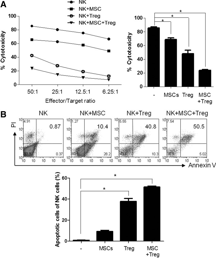
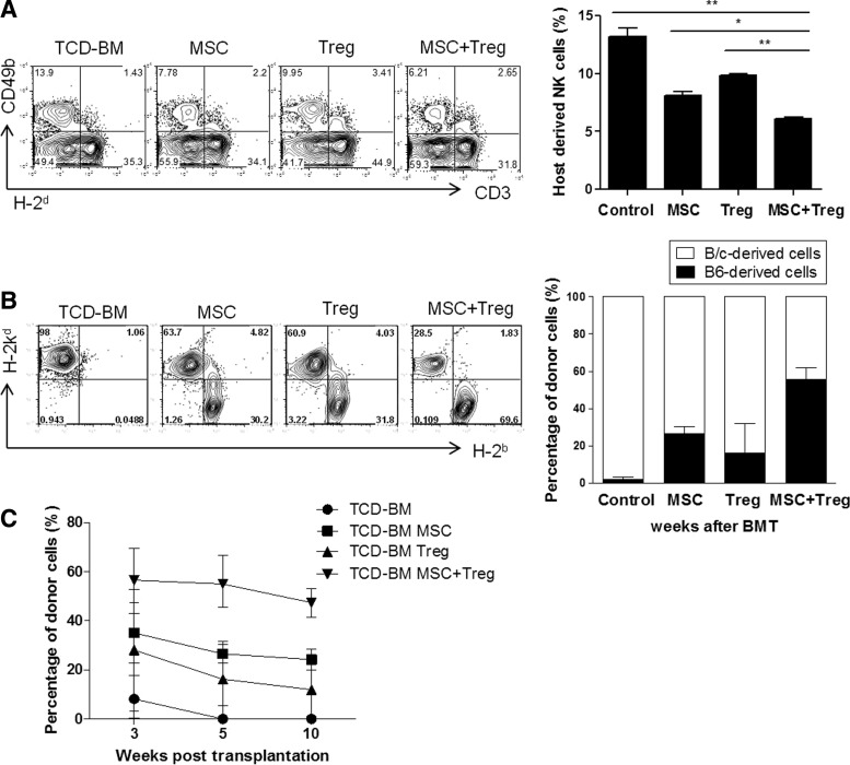
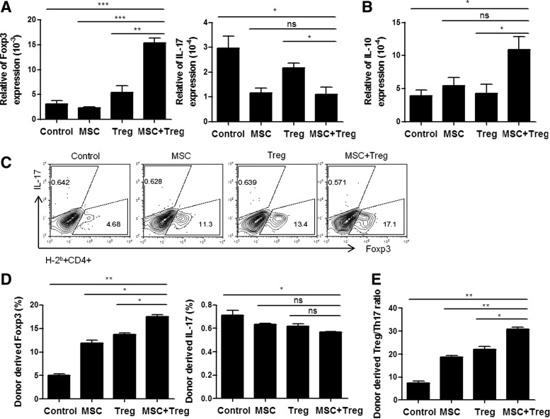
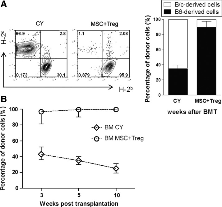
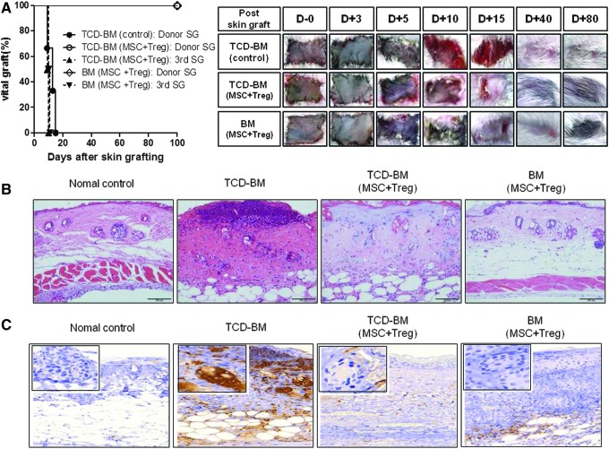
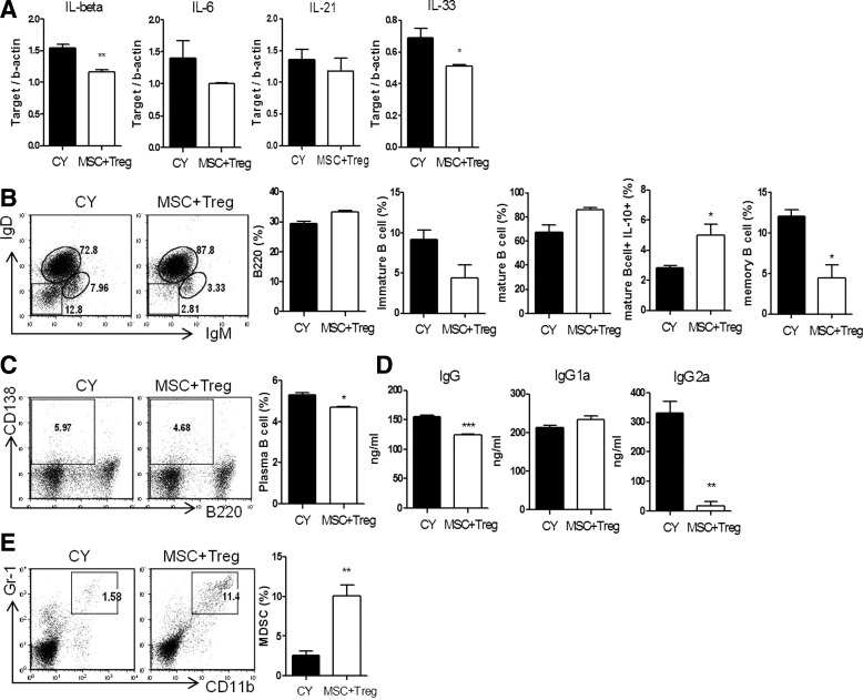
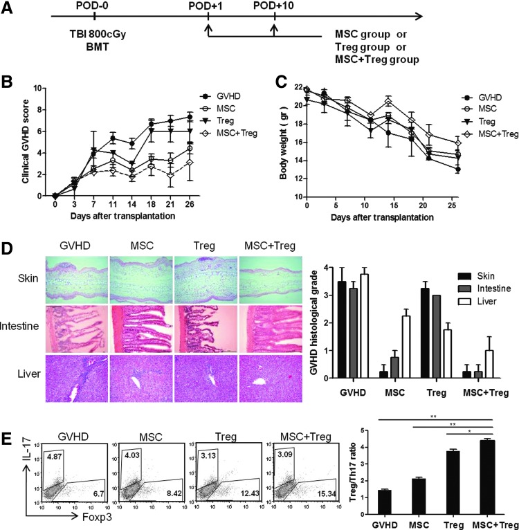
Establishment of mixed chimerism is an ideal approach to induce donor-specific tolerance while expanding its potential in various clinical settings. Despite the developments in partial conditioning regimens, improvements are still needed in reducing toxicity and bone marrow transplantation-related complications. Recently, cell-based therapies, including mesenchymal stem cells (MSCs), have been incorporated in establishing noncytoreductive mixed chimerism protocols; however, its efficacy is only partial and shows reversed immunosuppressive properties. This study demonstrates a novel approach to induce mixed chimerism and tolerance through combinatory cell-based immune modulation (CCIM) of MSCs and regulatory T cells (Tregs). We hypothesize that the interaction between these cells may lead to greater inhibition of host immune responses. Compared with single cell therapy, CCIM induced a higher engraftment rate and robust donor-specific tolerance to skin allografts across full major histocompatibility complex barriers. These regulatory effects were associated with inhibition of natural killer cell cytotoxic activity, CD4(+)IL-17(+) cells, memory B cells, plasma cells, and immunoglobulin production levels along with increased frequencies of CD4(+)Foxp3(+) cells, IL-10-producing mature B cells, and myeloid-derived suppressor cells. Furthermore, CCIM was able to regulate mortality in a graft-versus-host disease model through reciprocal regulation of Treg/Th17. Taken together, we suggest CCIM as a clinically applicable strategy for facilitating the induction of mixed chimerism and permanent tolerance.


Figures

<b>FIG. 1.</b>
FIG. 1.
Natural killer (NK) activity regulation by combinatory cell-based immune modulation (CCIM) with mesenchymal stem cells (MSCs) and regulatory T cells (Tregs). (A) Cytolytic activity of NK cells was evaluated by chromium release assay of labeled target Yac-1 cells in the presence of different ratios of effector cells (NK cells). NK cells isolated from BALB/c mice were cultured with irradiated MSCs or Tregs or MSCs plus Tregs before incubation with 51Cr-labeled Yac-1 tumor targets. Results of the chromium release assay are represented as the means±SEM of triplicate wells from one representative of three experiments. (B) Incubation of NK cells with MSCs or Tregs or MSCs plus Tregs for 48 h. After staining of cells with annexin V-propidium iodide (PI), apoptotic cells (annexin V+/PI and annexin V+/PI+ cells) were analyzed as a dot plot using a flow cytometer. The numbers in the quadrants of each plot indicate the percentage of annexin-positive (apoptotic) NK cells. The figures are representative of three replicates. *P<0.05.
<b>FIG. 2.</b>
FIG. 2.
CCIM with MSCs and Tregs in the early post-transplant period induces stable hematopoietic chimerism without graft-versus-host disease (GVHD) in full major histocompatibility complex (MHC)-mismatched murine models. On day 0, recipients received 3×10⁷ T-cell-depleted (TCD) bone marrow (BM) cells from MHC-mismatched C57BL/6 (H-2b) donors. On days+1 and +3 after bone marrow transplantation (BMT), recipients received 2×10⁶ MSCs, 2×10⁶ Treg cells, or 2×10⁶ MSCs plus 2×10⁶ Treg cells. (A) The splenocytes was collected at 7 days after BM transplantation to detect host-derived NK cells by staining with anti-H-2d, anti-CD3, and anti-CD49b. (B) To obtain peripheral blood mononuclear cells (PBMCs), leukocytes isolated from recipients of these all groups were stained with MHC class I (H-2b and H-2d) at 5 weeks post-transplantation. The bars show the ratios of C57BL6-originated cells (■) and BALB/c-originated cells (□). (C) Percentages of donor cells (H-2d) in recipients in each group were assessed by flow cytometry. Data are shown as the mean±SEM; results are representative of four independent experiments. *P<0.05; **P<0.01.
<b>FIG. 3.</b>
FIG. 3.
Immunological effects of CCIM after nonmyeloablative conditioning of BMT. The CCIM with MSCs and Tregs results in a significant reduction in Th17 cells, but enhancement in Treg cells. Expression level of transcription factor mRNA is measured in the spleen cells by real-time PCR at 1 week post-transplantation. (A) Expression of IL-17 was decreased and Foxp3 and (B) IL-10 were increased in CCIM treatment groups than in single cell therapy groups. (C, D) The following endogenous T-cell differentiation subsets in the all groups were identified by FACS analysis with antibodies: endogenous Treg cells (H-2b+CD4+Foxp3+) and endogenous Th17 cells (H-2b+CD4+IL-17+). (E) Data are presented as the ratio of Treg/Th17 among CD4+ T cells, which was calculated as the ratio of IL-17+ CD4+ T cells divided by the percentage of Foxp3+ CD4+ T cells. The purity of all cell subsets was >95% as determined by fluorescence-activated cell sorting (FACS) analysis. Data are shown as the mean±SEM; results are representative of four independent experiments. *P<0.05; **P<0.01; ***P<0.001.
<b>FIG. 4.</b>
FIG. 4.
CCIM with MSCs and Tregs in the early post-transplant period induced immune tolerance without post-transplantation cyclophosphamide (CY) conditioning. On day 0, they received 2.5×10⁷ BM cells from MHC-mismatched C57BL/6 (H-2b) donors. On day+1 after BMT, recipients received CY or 2×10⁶ MSCs plus 2×10⁶ Treg cells. (A) To obtain PBMCs, leukocytes isolated from recipients of these all groups were stained with MHC class I (H-2b and H-2d) at 5 weeks post-transplantation. The bars show the ratios of C57BL6-originated cells (■) and BALB/c-originated cells (□). (B) Percentages of donor cells (H-2d) in recipients in each group were assessed by flow cytometry. Data are shown as the mean±SEM; results are representative of three independent experiments.
<b>FIG. 5.</b>
FIG. 5.
Allograft tolerance in recipients co-infused with MSCs and Tregs. (A) After T-cell depletion BMT, the groups treated with or without MSCs plus Tregs (BALB/c) were transplanted with skin grafts (SG) from C57BL/6 (H-2b) mice and C3H (H-2k). The untreated MSCs plus Tregs control group (●) rejected the donor graft. These grafts were significantly contracted for wound remodeling at 12 days after skin grafting. The mixed chimerism group co-infused with MSCs and Tregs accepted donor skin from C57BL/6 mice (○) but rejected third-party skin from C3H mice (▲).In addition, the full chimerism group that exhibited no GVHD at all survived, and their hair grew during the early phase (◊). The third-party skin grafts from C3H mice were rejected at average 10 days post-transplantation (▼). (B) Skin allografts were removed from recipients at 7 days post-transplantation for histological analysis through hematoxylin and eosin staining. Original magnification ×200. (C) Immunohistochemistry staining for neutrophils in the SG (original magnification ×200). Rejection of grafts was correlated with the degree of the inflammatory response, as evidenced by markedly increased neutrophil numbers. Data represent the pool of two independent experiments.
<b>FIG. 6.</b>
FIG. 6.
Differences in differentiation and functions of T, B cells and myeloid-derived suppressor cells (MDSCs) in hematopoietic chimeras between the CY and MSC plus Treg-treated groups. Splenocytes were assessed at 6 weeks after allogeneic transplantation. (A) Total mRNA was extracted from spleen cells and subjected to quantitative real-time PCR analysis of the indicated genes. Data represent the relative amount of target mRNA normalized to β-actin. (B) The following B cell differentiation subsets in the CY and MSCs plus Tregs groups were identified by FACS analysis with antibodies: B cells (B220), immature B cells (IgM+IgDB220+), mature B cells (IgM+IgD+B220+), IL-10-expressing B cells (mature IL-10+B220+), and memory B cells (IgMIgDB220+). (C) The proportions of plasma cells (CD138+B220+) and (E) MDSCs (CD11b+Gr-1+) were determined by FACS as indicated. (D) Serum levels of total IgG, IgG1, and IgG2a were measured by ELISA. Data are shown as the mean±SEM; results are representative of three independent experiments. *P<0.05; **P<0.01; ***P<0.001.
<b>FIG. 7.</b>
FIG. 7.
Reduction of GVHD severity by post-transplantation infusion of MSCs and Tregs. (A) Lethally irradiated (800 cGy) BALB/c (H-2d) mice received 5×10⁶ whole BM cells and 5×10⁶ spleen cells from C57BL/6 (H-2b) donors on day 0. On days+1 and +10, mice in the untreated group (●, 1×10⁶, n=6), the MSC group (○, 1×10⁶, n=6), the Treg group (▼, 1×10⁶, n=6), and the group co-infused with MSCs and Tregs (◊, 1×10⁶, n=6) received transplants. (B) All animals were monitored for clinical signs and (C) mean serial weight measurements. (D) Histological GVHD scores at 2 weeks after transplantation were evaluated in skin, intestine, and liver tissues. (E) The splenocytes were stained with anti-CD4 antibody followed by intracellular Foxp3 and IL-17 antibodies and examined by flow cytometry. Treg/Th17 ratio in spleen. Data are shown as the mean±SEM; results are representative of three independent experiments. *P<0.05; **P<0.01.

Similar articles

Cited by

References

    1. Copelan EA. (2006). Hematopoietic stem-cell transplantation. N Engl J Med 354:1813–1826 - PubMed
    1. Welniak LA, Blazar BR. and Murphy WJ. (2007). Immunobiology of allogeneic hematopoietic stem cell transplantation. Annu Rev Immunol 25:139–170 - PubMed
    1. Pilat N. and Wekerle T. (2010). Mechanistic and therapeutic role of regulatory T cells in tolerance through mixed chimerism. Curr Opin Organ Transplant 15:725–730 - PubMed
    1. Asari S, Itakura S, Rawson J, Ito T, Todorov I, Nair I, Shintaku J, Liu CP, Kandeel F. and Mullen YS. (2011). Mesenchymal stem cells facilitate mixed hematopoietic chimerism induction and prevent onset of diabetes in nonobese diabetic mice. Pancreas 40:846–854 - PMC - PubMed
    1. Pilat N, Baranyi U, Klaus C, Jaeckel E, Mpofu N, Wrba F, Golshayan D, Muehlbacher F. and Wekerle T. (2010). Treg-therapy allows mixed chimerism and transplantation tolerance without cytoreductive conditioning. Am J Transplant 10:751–762 - PMC - PubMed

Publication types

MeSH terms