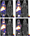
The imaging of insulinomas using a radionuclide-labelled molecule of the GLP-1 analogue liraglutide: a new application of liraglutide
- PMID: 24805918
- PMCID: PMC4013070
- DOI: 10.1371/journal.pone.0096833
The imaging of insulinomas using a radionuclide-labelled molecule of the GLP-1 analogue liraglutide: a new application of liraglutide
Abstract
Objective: This study explores a new, non-invasive imaging method for the specific diagnosis of insulinoma by providing an initial investigation of the use of 125I-labelled molecules of the glucagon-like peptide-1 (GLP-1) analogue liraglutide for in vivo and in vitro small-animal SPECT/CT (single-photon emission computed tomography/computed tomography) imaging of insulinomas.
Methods: Liraglutide was labelled with 125I by the Iodogen method. The labelled 125I-liraglutide compound and insulinoma cells from the INS-1 cell line were then used for in vitro saturation and competitive binding experiments. In addition, in a nude mouse model, the use of 125I-liraglutide for the in vivo small-animal SPECT/CT imaging of insulinomas and the resulting distribution of radioactivity across various organs were examined.
Results: The labelling of liraglutide with 125I was successful, yielding a labelling rate of approximately 95% and a radiochemical purity of greater than 95%. For the binding between 125I-liraglutide and the GLP-1 receptor on the surface of INS-1 cells, the equilibrium dissociation constant (Kd) was 128.8 ± 30.4 nmol/L(N = 3), and the half-inhibition concentration (IC50) was 542.4 ± 187.5 nmol/L(N = 3). Small-animal SPECT/CT imaging with 125I-liraglutide indicated that the tumour imaging was clearest at 90 min after the 125I-liraglutide treatment. An examination of the in vivo distribution of radioactivity revealed that at 90 min after the 125I-liraglutide treatment, the target/non-target (T/NT) ratio for tumour and muscle tissue was 4.83 ± 1.30(N = 3). Our study suggested that 125I-liraglutide was predominantly metabolised and cleared by the liver and kidneys.
Conclusion: The radionuclide 125I-liraglutide can be utilised for the specific imaging of insulinomas, representing a new non-invasive approach for the in vivo diagnosis of insulinomas.
Conflict of interest statement
Figures


Similar articles
-
18F-radiolabeled GLP-1 analog exendin-4 for PET/CT imaging of insulinoma in small animals.Nucl Med Commun. 2013 Jul;34(7):701-8. doi: 10.1097/MNM.0b013e3283614187. Nucl Med Commun. 2013. PMID: 23652208
-
PET of insulinoma using ¹⁸F-FBEM-EM3106B, a new GLP-1 analogue.Mol Pharm. 2011 Oct 3;8(5):1775-82. doi: 10.1021/mp200141x. Epub 2011 Aug 19. Mol Pharm. 2011. PMID: 21800885 Free PMC article.
-
[Lys40(Ahx-DTPA-111In)NH2]exendin-4, a very promising ligand for glucagon-like peptide-1 (GLP-1) receptor targeting.J Nucl Med. 2006 Dec;47(12):2025-33. J Nucl Med. 2006. PMID: 17138746
-
Exendin-4 analogs in insulinoma theranostics.J Labelled Comp Radiopharm. 2019 Aug;62(10):656-672. doi: 10.1002/jlcr.3750. J Labelled Comp Radiopharm. 2019. PMID: 31070270 Free PMC article. Review.
-
Radiolabeled glucagon-like peptide-1 analogues: a new pancreatic β-cell imaging agent.Nucl Med Commun. 2012 Mar;33(3):223-7. doi: 10.1097/MNM.0b013e32834e7f47. Nucl Med Commun. 2012. PMID: 22262216 Review.
Cited by
-
Efficacy, Pharmacokinetics, Biodistribution and Excretion of a Novel Acylated Long-Acting Insulin Analogue INS061 in Rats.Drug Des Devel Ther. 2021 Aug 10;15:3487-3498. doi: 10.2147/DDDT.S317327. eCollection 2021. Drug Des Devel Ther. 2021. PMID: 34408401 Free PMC article.
-
Interaction Between Glucagon-like Peptide 1 and Its Analogs with Amyloid-β Peptide Affects Its Fibrillation and Cytotoxicity.Int J Mol Sci. 2025 Apr 25;26(9):4095. doi: 10.3390/ijms26094095. Int J Mol Sci. 2025. PMID: 40362335 Free PMC article.
-
Targets and probes for non-invasive imaging of β-cells.Eur J Nucl Med Mol Imaging. 2017 Apr;44(4):712-727. doi: 10.1007/s00259-016-3592-1. Epub 2016 Dec 26. Eur J Nucl Med Mol Imaging. 2017. PMID: 28025655 Free PMC article. Review.
-
Advances in GLP-1 receptor targeting radiolabeled agent development and prospective of theranostics.Theranostics. 2020 Jan 1;10(1):437-461. doi: 10.7150/thno.38366. eCollection 2020. Theranostics. 2020. PMID: 31903131 Free PMC article. Review.
References
-
- Tucker ON, Crotty PL, Conlon KC (2006) The management of insulinoma. Br J Surg, 93(3): , 264–275. doi: 10.1002/bjs.5280 - PubMed
-
- La Rosa S, Pariani D, Calandra C, Marando A, Sessa F, et al..(2013) Ectopic duodenal insulinoma: a very rare and challenging tumor type. Description of a case and review of the literature. Endocr Pathol, 24(4): , 213–219. doi: 10.1007/s12022-013-9262-y - PubMed
-
- An L, Li W, Yao KC, Liu R, Lv F, et al.. (2011) Assessment of contrast-enhanced ultrasonography in diagnosis and preoperative localization of insulinoma. Eur J Radiol, 80(3): , 675–680. doi: 10.1016/j.ejrad.2010.09.014 - PubMed
-
- Joseph AJ, Kapoor N, Simon EG, Chacko A, Thomas EM, et al.. (2013) Endoscopic ultrasonography—a sensitive tool in the preoperative localization of insulinoma. Endocr Pract, 19(4): , 602–608. doi: 10.4158/EP12122.OR - PubMed
-
- Versari A, Camellini L, Carlinfante G, Frasoldati A, Nicoli F, et al.. (2010) Ga-68 DOTATOC PET, endoscopic ultrasonography, and multidetector CT in the diagnosis of duodenopancreatic neuroendocrine tumors: a single-centre retrospective study. Clin Nucl Med, 35(5): , 321–328. doi: 10.1097/RLU.0b013e3181d6677c - PubMed
Publication types
MeSH terms
Substances
LinkOut - more resources
Full Text Sources
Other Literature Sources
Research Materials

