. 2015 May;73(5):1764-74.
doi: 10.1002/mrm.25291. Epub 2014 May 7.

Self-gated fat-suppressed cardiac cine MRI

Affiliations

Self-gated fat-suppressed cardiac cine MRI

R Reeve Ingle et al. Magn Reson Med. 2015 May.

Abstract

Purpose: To develop a self-gated alternating repetition time balanced steady-state free precession (ATR-SSFP) pulse sequence for fat-suppressed cardiac cine imaging.

Methods: Cardiac gating is computed retrospectively using acquired magnetic resonance self-gating data, enabling cine imaging without the need for electrocardiogram (ECG) gating. Modification of the slice-select rephasing gradients of an ATR-SSFP sequence enables the acquisition of a one-dimensional self-gating readout during the unused short repetition time (TR). Self-gating readouts are acquired during every TR of segmented, breath-held cardiac scans. A template-matching algorithm is designed to compute cardiac trigger points from the self-gating signals, and these trigger points are used for retrospective cine reconstruction. The proposed approach is compared with ECG-gated ATR-SSFP and balanced steady-state free precession in 10 volunteers and five patients.

Results: The difference of ECG and self-gating trigger times has a variability of 13 ± 11 ms (mean ± SD). Qualitative reviewer scoring and ranking indicate no statistically significant differences (P > 0.05) between self-gated and ECG-gated ATR-SSFP images. Quantitative blood-myocardial border sharpness is not significantly different among self-gated ATR-SSFP ( 0.61±0.15 mm -1), ECG-gated ATR-SSFP ( 0.61±0.15 mm -1), or conventional ECG-gated balanced steady-state free precession cine MRI ( 0.59±0.15 mm -1).

Conclusion: The proposed self-gated ATR-SSFP sequence enables fat-suppressed cardiac cine imaging at 1.5 T without the need for ECG gating and without decreasing the imaging efficiency of ATR-SSFP.

Keywords: alternating repetition time; cardiac imaging; cine magnetic resonance imaging; self gating; steady-state free precession.


Figures

Figure 1
Figure 1
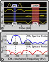
Self-gated ATR-SSFP pulse sequence. (a) Bridging the slice-select rephasing lobes during TR2 enables acquisition of 1D self-gating (SG) readouts along kz with no time penalty compared to standard ATR-SSFP. (b) The ATR-SSFP spectral profile yields fat suppression during TR1 while maintaining high fat and water signal during TR2.
Figure 2
Figure 2
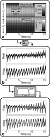
Representative self-gating signal from a single anterior coil located near the heart. (a) Raw self-gating k-space data shows periodic uctuation with the cardiac cycle. (b) Summation along kz yields the self-gating signal from the center of the imaged slice. (c) Bandpass filtering (BPF) with a 0.6 – 10 Hz passband removes baseline drift and high-frequency noise from eddy currents and other unwanted sources.
Figure 3
Figure 3
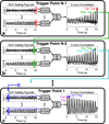
Template-matching algorithm for self-gating trigger detection. (a) Working backward from the end of the self-gating signals, template regions (red rectangles) spanning half of the mean RR interval are extracted directly from the filtered self-gating signals (real part shown for coils 1 and 8). The cross correlation is computed between the template region and the rest of the corresponding self-gating signal. Templates are normalized to yield unity cross correlation for a relative shift of 0. The results from all coils are summed to yield a cross-correlation waveform, and peak detection is used to find the previous trigger point (green circle). (b) This trigger point is used to update the template regions (green rectangles), and the process is repeated to compute the previous trigger point (cyan circle). (c) This procedure is repeated to detect all trigger points, ending with the first template region (blue rectangle) and trigger point (purple circle).
Figure 4
Figure 4
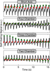
Comparison of ECG R-wave trigger points and self-gating trigger points. Filtered self-gating signals (real part) from an anterior coil element of a representative volunteer study are shown for four different scan planes. ECG trigger points (red diamonds) and self-gating trigger points (green circles) are overlaid on each waveform. The SAX acquisition (top) has a highly periodic self-gating signal and the smallest trigger variability (3 ms) among the four acquisitions. The long-axis acquisitions (bottom three rows) have self-gating signals with a slowly varying cyclic pattern from beginning to end of the acquisition. The trigger variabilities (11, 19, and 12 ms for 4CH, 3CH, 2CH, respectively) are larger than that of the SAX acquisition.
Figure 5
Figure 5
(a) Short-axis, (b) four-chamber, (c) three-chamber, and (d) two-chamber ATR-SSFP acquisitions from a single volunteer study were reconstructed with ECG gating and self gating (SG). Systolic (Sys) and diastolic (Dia) images are shown for each of the four scan planes. The difference of ECG and self-gating reconstructions is shown for each case, scaled by a factor of 10. For each scan plane, the same window and level settings were used to display all images.
Figure 6
Figure 6
Short-axis (left) and four-chamber (right) ATR-SSFP images from two different patient studies (a–b) performed after contrast injection. (a) Systolic (Sys) and diastolic (Dia) images of a patient with an apical septal infarct were reconstructed with ECG gating and self gating (SG). The difference of ECG and SG reconstructions is shown for each case, scaled by a factor of 10. Interestingly, a region of hyperintensity in the interventricular septal wall (black arrows) and apex corresponded to regions of scarring found in clinical delayed enhancement scans. (b) Systolic and diastolic images of a patient with aortic valve replacement were reconstructed with ECG gating and self gating. The difference of ECG and SG reconstructions is shown for each case, scaled by a factor of 10. Signal dropouts (arrowheads) are due to the aortic valve replacement.
Figure 7
Figure 7
Self-gated ATR-SSFP images (top) and bSSFP images (bottom) from the same cardiac phase are shown for three different volunteers (a–c). (a) ATR-SSFP suppresses the signal from epicardial fat surrounding the left and right ventricles, which appears bright in the bSSFP image (black arrows, magnified in inset images). (b) ATR-SSFP suppresses the signal from epicardial fat located at the apex of the heart, which appears bright in the bSSFP image (white arrows, magnified in inset images). Ghosting artifacts near vessels with high flow are slightly worse for ATR-SSFP than bSSFP (bracketed column). (c) A portion of the right coronary artery can be seen in the ATR-SSFP image due to suppression of the signal from surrounding fat (white arrowhead, magnified in inset images). In the bSSFP image, the artery is hypointense due to signal cancellation from the surrounding epicardial fat.

References

    1. Manning WJ, Pennell DJ. Cardiovascular magnetic resonance. 2nd ed. Philadelphia, PA: Saunders/Elsevier; 2010.
    1. Polson MJ, Barker AT, Gardiner S. The effect of rapid rise-time magnetic fields on the ECG of the rat. Clin Phys Physiol Meas. 1982;3:231–234. - PubMed
    1. Shetty AN. Suppression of radiofrequency interference in cardiac gated MRI: a simple design. Magn Reson Med. 1988;8:84–88. - PubMed
    1. Becker M, Frauenrath T, Hezel F, Krombach GA, Kremer U, Koppers B, Butenweg C, Goemmel A, Utting JF, Schulz-Menger J, Niendorf T. Comparison of left ventricular function assessment using phonocardiogram- and electrocardiogram-triggered 2D SSFP CINE MR imaging at 1.5 T and 3.0 T. Eur Radiol. 2010;20:1344–1355. - PubMed
    1. Spraggins TA. Wireless retrospective gating: application to cine cardiac imaging. Magn Reson Imaging. 1990;8:675–681. - PubMed

Publication types