. 2014 May 7;9(5):e96947.
doi: 10.1371/journal.pone.0096947. eCollection 2014.

Protein kinase D1 (PKD1) phosphorylation promotes dopaminergic neuronal survival during 6-OHDA-induced oxidative stress

Affiliations

Protein kinase D1 (PKD1) phosphorylation promotes dopaminergic neuronal survival during 6-OHDA-induced oxidative stress

Arunkumar Asaithambi et al. PLoS One. .

Abstract

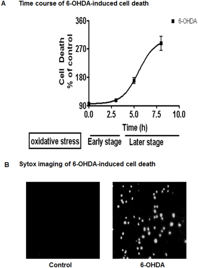
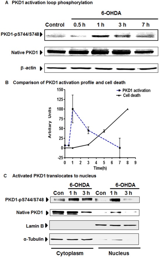
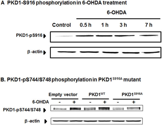
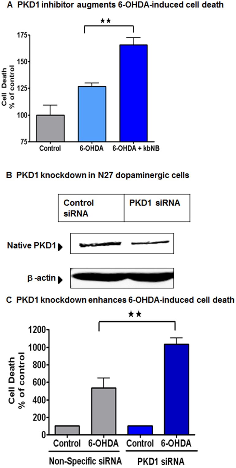
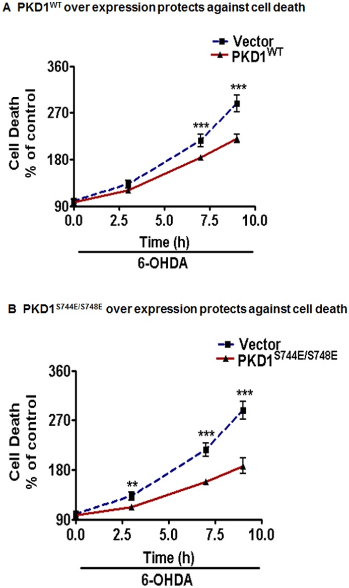
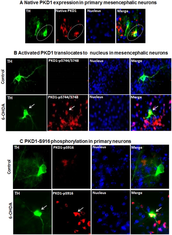
Oxidative stress is a major pathophysiological mediator of degenerative processes in many neurodegenerative diseases including Parkinson's disease (PD). Aberrant cell signaling governed by protein phosphorylation has been linked to oxidative damage of dopaminergic neurons in PD. Although several studies have associated activation of certain protein kinases with apoptotic cell death in PD, very little is known about protein kinase regulation of cell survival and protection against oxidative damage and degeneration in dopaminergic neurons. Here, we characterized the PKD1-mediated protective pathway against oxidative damage in cell culture models of PD. Dopaminergic neurotoxicant 6-hydroxy dopamine (6-OHDA) was used to induce oxidative stress in the N27 dopaminergic cell model and in primary mesencephalic neurons. Our results indicated that 6-OHDA induced the PKD1 activation loop (PKD1S744/S748) phosphorylation during early stages of oxidative stress and that PKD1 activation preceded cell death. We also found that 6-OHDA rapidly increased phosphorylation of the C-terminal S916 in PKD1, which is required for PKD1 activation loop (PKD1S744/748) phosphorylation. Interestingly, negative modulation of PKD1 activation by RNAi knockdown or by the pharmacological inhibition of PKD1 by kbNB-14270 augmented 6-OHDA-induced apoptosis, while positive modulation of PKD1 by the overexpression of full length PKD1 (PKD1WT) or constitutively active PKD1 (PKD1S744E/S748E) attenuated 6-OHDA-induced apoptosis, suggesting an anti-apoptotic role for PKD1 during oxidative neuronal injury. Collectively, our results demonstrate that PKD1 signaling plays a cell survival role during early stages of oxidative stress in dopaminergic neurons and therefore, positive modulation of the PKD1-mediated signal transduction pathway can provide a novel neuroprotective strategy against PD.


Conflict of interest statement

Competing Interests: The authors have declared that no competing interests exist.

Figures

Figure 1
Figure 1. 6-OHDA-induced oxidative stress causes cell death in N27 dopaminergic neuronal cell models.
N27 dopaminergic cells were treated with 6-OHDA (100 µM) for 0–8 h and assayed for cytotoxicity using Sytox green fluorescence assay. The fluorescence was measured by using a fluorescence plate reader. Non-linear regression was performed from two or more independent experiments (A), and visualized by fluorescence microscopy (B).
Figure 2
Figure 2. PKD1 is activated in N27 cells during 6-OHDA-induced oxidative stress.
N27 dopaminergic neuronal cells were treated with 6-OHDA (100 µM) for 1–7 h and probed for PKD1 activation loop phosphorylation pS744/pS748 and native PKD1 expression (A). A comparative time course graph depicting the reciprocal relationship between PKD1 activation and cytotoxicity during 6-OHDA exposure (B). Cytoplasmic and nuclear extracts from control and 6-OHDA-treated N27 cells were prepared and probed for PKD1 activation loop phosphorylation pS744/pS748 and native PKD1 expression (C). Lamin B (nuclear fraction) or α-Tubulin (cytoplasmic fraction) was used as loading control.
Figure 3
Figure 3. PKCδ-dependent S916-phosphorylation of PKD1 precedes PKD1 S744/S748 active loop phosphorylation in N27 dopaminergic cells.
N27 dopaminergic cells were treated with 100 µM 6-OHDA for 1–7 h and monitored for PKD1 S916 phosphorylation (A). N27 cells transiently expressing PKD1S916A mutant prevent PKD1 activation during oxidative stress, as seen by Western blotting for PKD1 S744/S748 phosphorylation (B).
Figure 4
Figure 4. PKD1 activation is essential for cell survival during 6-OHDA-induced oxidative stress in N27 dopaminergic neurons.
N27 cells were co-treated with the PKD1 inhibitor kb-NB 14270 and monitored for cell death during 6-OHDA treatment for 4 h (A). N27 dopaminergic cells were transfected with 1 µM PKD1 siRNA and non-specific siRNA (B) and treated with 100 µM 6-OHDA for 9 h and monitored for cytotoxicity using Sytox green assay. Fluorescence measurements for the incorporation of Sytox green dye were performed using a fluorescence plate reader. **, p<0.01 denotes a significant difference between non-specific siRNA-6-OHDA and PKD1 siRNA-6-OHDA treated groups (C).
Figure 5
Figure 5. Modulation of PKD1 offers neuroprotection in N27 dopaminergic neuronal cells.
N27 dopaminergic cells transiently transfected with 5 µg of full-length PKD1 plasmid (PKD1WT) and 5 µg of vector plasmid were treated with or without 100 µM 6-OHDA and monitored for cytotoxicity at various time points using Sytox green assay. ***, p<0.001 denotes a significant difference between treatment groups from n≥6 (A). N27 dopaminergic cells transiently transfected with 5 µg of constitutively active PKD1S744E/S748E mutant and 5 µg of vector plasmid were treated with or without 100 µM 6-OHDA and monitored for cytotoxicity at various time points using Sytox green assay. ***, p<0.001 denotes a significant difference between treatment groups from n≥6 (B).
Figure 6
Figure 6. PKD1 is highly expressed in primary dopaminergic neurons and activated during 6-OHDA-induced oxidative stress.
(A) Primary dopaminergic neurons stained for tyrosine hydroxylase (TH) show co-localization of native PKD1 with TH in the cytosol. TH-Green, PKD1 native-Red, Nucleus-Blue, Merge-Yellow. Nuclei were stained with Hoechst dye. (B) Immunofluorescence analysis of primary dopaminergic neurons stained for TH shows translocation of activated PKD1 to the nucleus during 6-OHDA exposure. TH-Green, PKD1pS744/S748-Red, Nucleus-Blue, Merge-Pink. Nuclei were stained with Hoechst dye. White arrows indicate the TH+ neurons, in which activated PKD1 translocates into nucleus after 6-OHDA treatment. (C) Primary dopaminergic neurons staining for TH show the presence of PKD1pS916 in both the cytosol and nucleus during 6-OHDA exposure. TH-Green, PKD1pS916-Red, red/yellow-Merge.
Figure 7
Figure 7. A proposed model for PKD1 neuroprotection in dopaminergic neurons during early stages of oxidative insult.
Oxidative insult rapidly induces PKD1 activation loop phosphorylation at pS744/pS748, which then translocates from the cytoplasm to the nucleus. The PKD1 activation and nuclear translocation lead to counteracting oxidative injury and subsequent initiation of cell survival processes.

References

    1. Dawson TM, Dawson VL (2003) Molecular pathways of neurodegeneration in Parkinson’s disease. Science 302: 819–822. - PubMed
    1. Przedborski S (2005) Pathogenesis of nigral cell death in Parkinson’s disease. Parkinsonism Relat Disord 11 Suppl 1S3–7. - PubMed
    1. Dauer W, Przedborski S (2003) Parkinson’s disease: mechanisms and models. Neuron 39: 889–909. - PubMed
    1. Malkus KA, Tsika E, Ischiropoulos H (2009) Oxidative modifications, mitochondrial dysfunction, and impaired protein degradation in Parkinson’s disease: how neurons are lost in the Bermuda triangle. Mol Neurodegener 4: 24. - PMC - PubMed
    1. Jin H, Kanthasamy A, Ghosh A, Anantharam V, Kalyanaraman B, et al. (2013) Mitochondria-targeted antioxidants for treatment of Parkinson’s disease: Preclinical and clinical outcomes. Biochim Biophys Acta. http://dx.doi.org/10.1016/j.bbadis.2013.09.007. - DOI - PMC - PubMed

Publication types