

c-kit+ Cardiac stem cells alleviate post-myocardial infarction left ventricular dysfunction despite poor engraftment and negligible retention in the recipient heart
- PMID: 24806457
- PMCID: PMC4013035
- DOI: 10.1371/journal.pone.0096725
c-kit+ Cardiac stem cells alleviate post-myocardial infarction left ventricular dysfunction despite poor engraftment and negligible retention in the recipient heart
Abstract
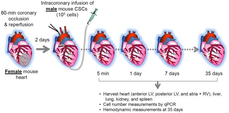
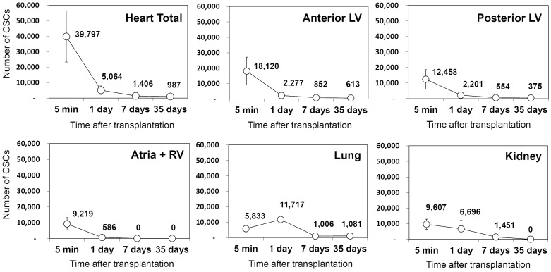
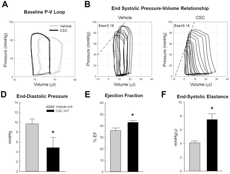
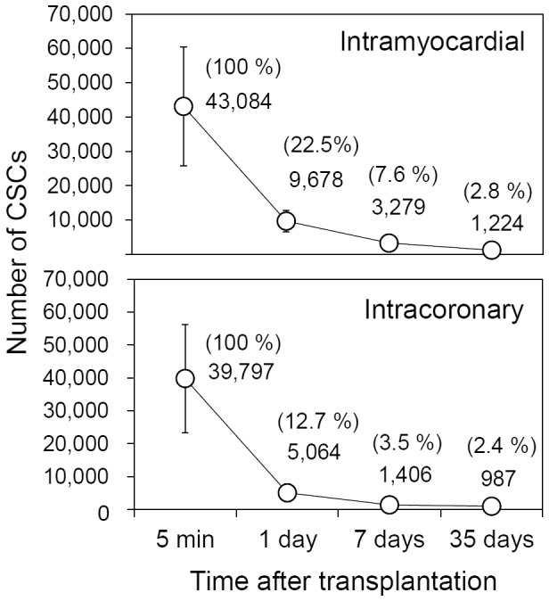
Although transplantation of c-kit+ cardiac stem cells (CSCs) has been shown to alleviate left ventricular (LV) dysfunction induced by myocardial infarction (MI), the number of exogenous CSCs remaining in the recipient heart following transplantation and their mechanism of action remain unclear. We have previously developed a highly sensitive and accurate method to quantify the absolute number of male murine CSCs in female recipient organs after transplantation. In the present study, we used this method to monitor the number of donor CSCs in the recipient heart after intracoronary infusion. Female mice underwent a 60-min coronary occlusion followed by reperfusion; 2 days later, 100,000 c-kit+/lin- syngeneic male mouse CSCs were infused intracoronarily. Only 12.7% of the male CSCs present in the heart immediately (5 min) after infusion were still present in the heart at 24 h, and their number declined rapidly thereafter. By 35 days after infusion, only ∼ 1,000 male CSCs were found in the heart. Significant numbers of male CSCs were found in the lungs and kidneys, but only in the first 24 h. The number of CSCs in the lungs increased between 5 min and 24 h after infusion, indicating recirculation of CSCs initially retained in other organs. Despite the low retention and rapid disappearance of CSCs from the recipient heart, intracoronary delivery of CSCs significantly improved LV function at 35 days (Millar catheter). These results suggest that direct differentiation of CSCs alone cannot account for the beneficial effects of CSCs on LV function; therefore, paracrine effects must be the major mechanism. The demonstration that functional improvement is dissociated from survival of transplanted cells has major implications for our understanding of cell therapy. In addition, this new quantitative method of stem cell measurement will be useful in testing approaches of enhancing CSC engraftment and survival after transplantation.
Conflict of interest statement
Figures
References
-
- Beltrami AP, Barlucchi L, Torella D, Baker M, Limana F, et al. (2003) Adult cardiac stem cells are multipotent and support myocardial regeneration. Cell 114: 763–776. - PubMed
-
- Ellison GM, Vicinanza C, Smith AJ, Aquila I, Leone A, et al. (2013) Adult c-kit(pos) Cardiac Stem Cells Are Necessary and Sufficient for Functional Cardiac Regeneration and Repair. Cell 154: 827–842. - PubMed
Publication types
MeSH terms
Substances
Grants and funding
LinkOut - more resources
Full Text Sources
Other Literature Sources
Medical
