. 2014 May 7;34(19):6470-9.
doi: 10.1523/JNEUROSCI.4305-13.2014.

β-Catenin is required for hair-cell differentiation in the cochlea

Affiliations

β-Catenin is required for hair-cell differentiation in the cochlea

Fuxin Shi et al. J Neurosci. .

Abstract

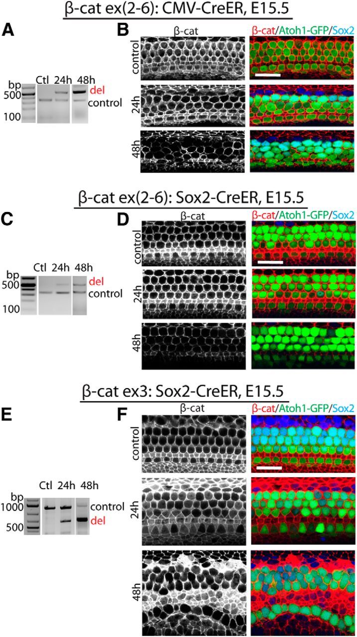
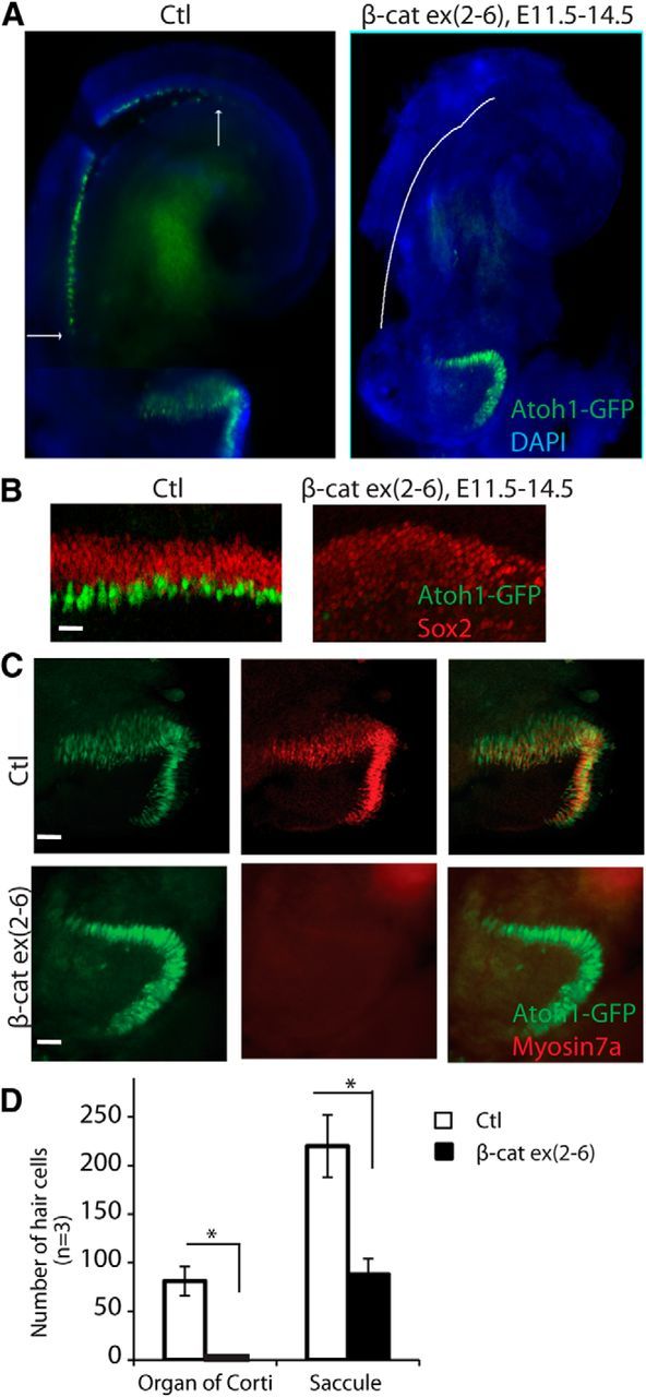
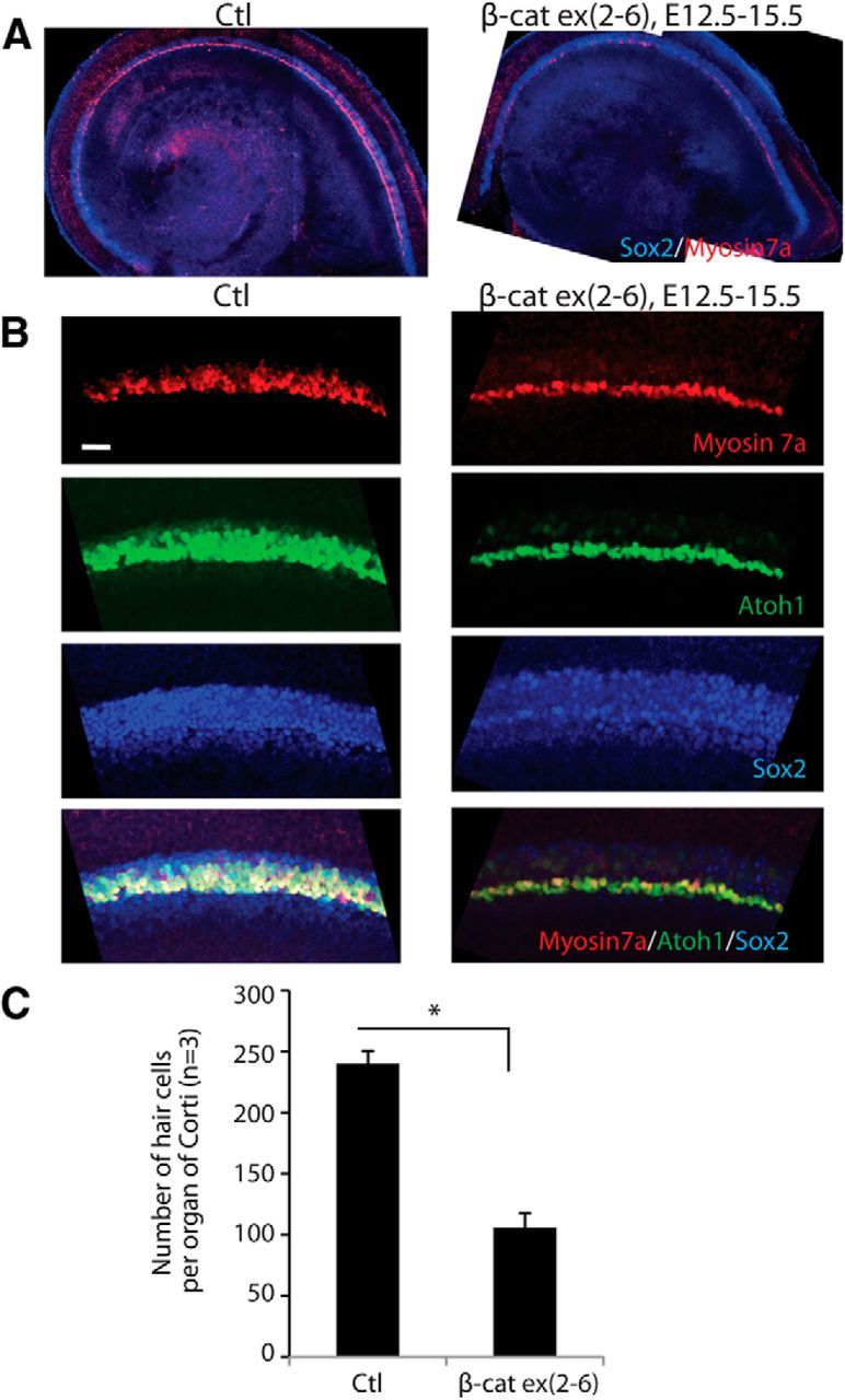
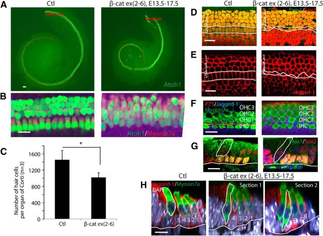
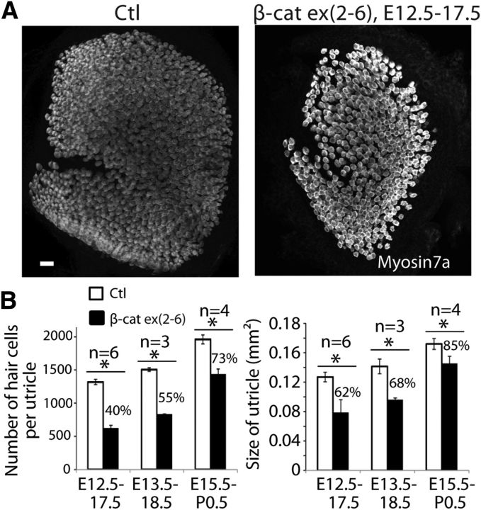
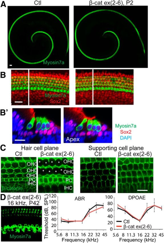
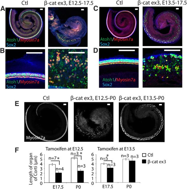
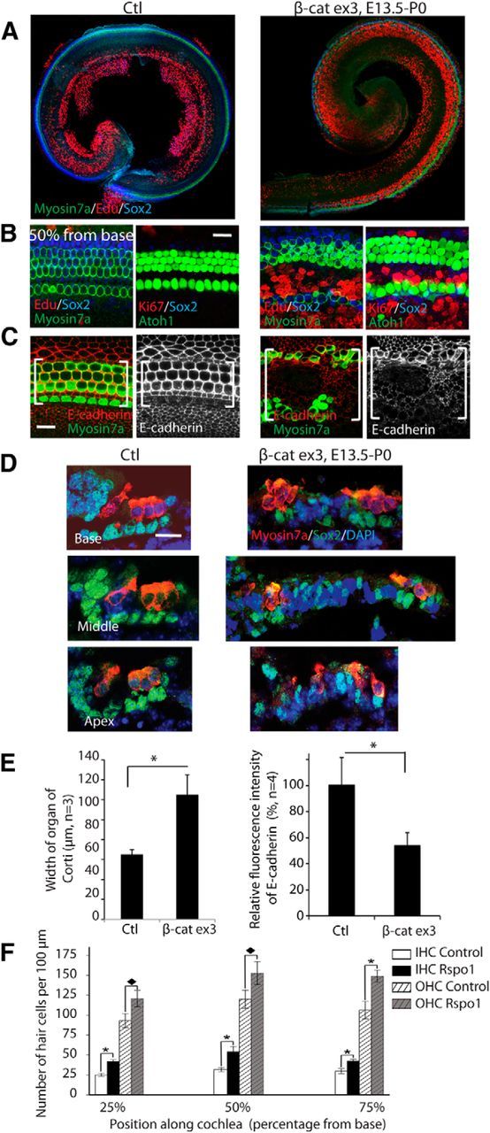
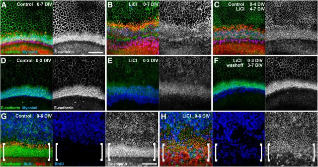
The development of hair cells in the auditory system can be separated into steps; first, the establishment of progenitors for the sensory epithelium, and second, the differentiation of hair cells. Although the differentiation of hair cells is known to require the expression of basic helix-loop-helix transcription factor, Atoh1, the control of cell proliferation in the region of the developing cochlea that will ultimately become the sensory epithelium and the cues that initiate Atoh1 expression remain obscure. We assessed the role of Wnt/β-catenin in both steps in gain- and loss-of-function models in mice. The canonical Wnt pathway mediator, β-catenin, controls the expression of Atoh1. Knock-out of β-catenin inhibited hair-cell, as well as pillar-cell, differentiation from sensory progenitors but was not required to maintain a hair-cell fate once specified. Constitutive activation of β-catenin expanded sensory progenitors by inducing additional cell division and resulted in the differentiation of extra hair cells. Our data demonstrate that β-catenin plays a role in cell division and differentiation in the cochlear sensory epithelium.


Figures

Figure 1.
Figure 1.
Manipulation of β-catenin expression in developing sensory epithelium. A, In CMV-CreER;β-cateninflox(exon2–6 mutants, exons 2–6 of β-catenin were deleted in sensory epithelium 24 and 48 h after the first dose of tamoxifen at E15.5. B, The decrease of β-catenin expression in sensory epithelium was at its highest point 48 h after the initial dose of tamoxifen. Hair cells were positive for GFP (compound mutants also expressed GFP under the control of Atoh1); β-catenin (red) was expressed in the entire sensory epithelium; Sox2 (blue) was expressed in supporting cells. C, In Sox2-CreER;β-cateninflox(exon2–6) mutants, exons 2–6 of β-catenin were deleted in sensory epithelium 24 and 48 h after the first dose of tamoxifen. D, β-catenin expression in sensory epithelium decreased 48 h after the first dose of tamoxifen. E, Following the initial tamoxifen injection at E15.5 in Sox2-CreER;β-cateninflox(exon3) mutants, deletion of exon 3 of β-catenin was seen at 24 and 48 h in sensory epithelium. F, A greater increase in β-catenin was apparent in sensory epithelium 48 h after the first dose of tamoxifen. The controls are littermates without Cre expression at 48 h after tamoxifen. Scale bar, 20 μm.
Figure 2.
Figure 2.
Disruption of hair cell development in β-catenin knock-out mice. A, No hair cells were evident in a cochlea analyzed at E14.5 after deletion of β-catenin at E11.5 (before hair-cell differentiation) by administration of tamoxifen to a CMV-CreER;β-cateninflox(exon2–6) embryo that also expressed nGFP under control of Atoh1. B, Hair cells were detected at E14.5 in the Cre-negative β-cateninflox(exon2–6) organ of Corti (Ctl) at the midbasal region. Cells in the prosensory domain of β-catenin knock-out ears were positive for Sox2 but did not contain Atoh1-positive cells. C, Hair cells in the saccule were Atoh1-nGFP-positive. Some were also myosin VIIa-positive in the control. The β-catenin knock-out ears had fewer Atoh1-positive cells and no myosin VIIa-positive cells. D, The number of hair cells was significantly decreased in the β-catenin mutants (p < 0.01). Scale bar, 20 μm.
Figure 3.
Figure 3.
Knock-out of β-catenin in sensory progenitors inhibits hair cell development. A, The number of hair cells was decreased in the sensory region after deletion of β-catenin was induced at E12.5 in sensory progenitors (Sox2-positive) of Sox2-CreER;β-cateninflox(exon2–6) mice. Low-magnification views are shown at E15.5. B, Two to three rows of Atoh1-positive outer hair cells were observed lateral to the myosin VIIa-positive inner hair cells in the midbasal region; some outer hair cells expressed myosin VIIa in a littermate lacking Cre. A single shortened row of hair cells had developed in β-cateninΔΔexon2–6 mutants. No change in Sox2 expression was seen. C, The difference in hair cell number was significant (p < 0.01).
Figure 4.
Figure 4.
β-Catenin knock-out delays hair cell development and patterning. A, Fewer hair cells differentiated and the organ of Corti was shorter in Sox2-CreER; β-cateninflox(exon2–6); mice at E17.5 when β-catenin deletion was induced at E13.5. B, The hair cells were less organized and inner and outer hair cells had not separated in β-catenin knock-out mice. C, The organ of Corti in the mutants was 29% shorter and had 33% less hair cells (p < 0.05). D, The number of pillar cells decreased and a continuous row of inner pillar cells was not apparent (white lines) when supporting cells were labeled with Sox2 and Prox1. E, An antibody to jagged-1 revealed irregular patterning in the inner pillar cell area (white lines). F, No staining (P75) was observed in the pillar cell area. G, Staining for Prox1, a pillar cell marker, was absent and the inner hair cells contacted the basilar membrane (white line). H, Inner hair cells (outlined; Section 1) or the first outer hair cells (outlined; Section 2) contacted the basilar membrane (white line) as shown by myosin VIIa staining. Scale bar, 20 μm; H1, H2, H3 are Dieters' cells and H4 and H5 are pillar cells. IHC, Inner hair cells; OHC, outer hair cells.
Figure 5.
Figure 5.
Knock-out of β-catenin in sensory progenitors inhibits utricular hair cell development. A, Examination of the ear at E17.5 after deletion of β-catenin at E12.5 in Sox2-CreER;β-cateninflox(exon2–6) mice revealed a smaller utricle with less hair cells. B, The decrease in utricular hair cell number and utricular size were significant after β-catenin knock-out from E12.5 to E17.5, E13.5 to E18.5, and E15.5 to P0.5 (p < 0.01). Scale bar, 20 μm.
Figure 6.
Figure 6.
Morphology is normal after β-catenin knock-out in hair cells. A, No gross change of hair-cell morphology was seen at P2 in a Gfi-Cre;β-cateninflox(exon2–6) ear (knock-out of β-catenin in hair cells). B, Hair cells and supporting cells were stained, respectively, with myosin VIIa and Sox2. B′, An XZ scan of B at the white line shows a cross-section of the organ of Corti. C, β-catenin was expressed in both hair cells and supporting cells of the Cre-negative animal. β-catenin was not detected in hair cells (hair cell plane) but was retained in supporting cells in the hair cell-specific β-catenin knock-out. D, At P42, the cochlea in the Gfi-Cre;β-cateninflox(exon2–6) mouse had regular hair cell rows, and ABR and DPOAE thresholds did not differ from control mice.
Figure 7.
Figure 7.
Overexpression of β-catenin expands and inhibits elongation of the sensory epithelium. A, When β-catenin was overexpressed in sensory progenitors at E12.5 in Sox2-CreER;β-cateninflox(exon3) mice, the sensory epithelium expanded with extra hair cells in the midbasal region, but failed to extend to the apex by E17.5, when hair cells had nearly completely developed in a Cre-negative littermate. B, At high-magnification, in the midbasal region, all Atoh1-positive cells expressed myosin VIIa. In β-catenin expression mutants, the sensory epithelium was wider, but only some of the Atoh1-positive cells expressed myosin VIIa. C, When β-catenin overexpression was initiated at E13.5, the sensory epithelium also expanded and extended toward the apex by E17.5. D, Higher-magnification of B (overexpression of β-catenin) showed an expanded area between inner and outer hair cells. E, At P0 the sensory epithelium extended nearly to the apex when β-catenin overexpression was initiated at E13.5 but not at E12.5. F, When overexpression of β-catenin was initiated at E12.5, the difference in the length of the sensory epithelium was significant at E17.5 and P0 (p < 0.01), whereas it was shorter at E17.5 but similar to the Cre-negative littermate by P0 when initiated at E13.5 (p < 0.05 at E17.5). Scale bar, 100 μm.
Figure 8.
Figure 8.
Overexpression of β-catenin expands sensory epithelium and generates more hair cells. A, The organ of Corti in β-catenin overexpression mutants was expanded when β-catenin was overexpressed from E13.5 to P0 in Sox2-CreER;β-cateninflox(exon3) mice. B, EdU was incorporated, and Ki67-positive cells were seen in the inner pillar cell region in β-catenin overexpression mutants. C, β-catenin overexpression reduced E-cadherin expression in the expanded organ of Corti (bracket). D, The cells in the pillar cell region were expanded dramatically in the β-catenin overexpressing ear. The expanded cells had decreased or abolished Sox2 expression. E, β-catenin overexpression significantly expanded the organ of Corti, and decreased the expression of E-cadherin, with p < 0.01. F, The number of hair cells in organ of Corti explant culture was increased by Rspo1 (p < 0.01).
Figure 9.
Figure 9.
In vitro activation of Wnt/β-catenin signaling reduces the level of membranous E-cadherin expression in the developing organ of Corti. A–C, Organ of Corti explants established at E13 and maintained for 7 DIV were immunostained for E-cadherin (green), Sox2 (red), and myosin 6 (blue); merged colors are shown at left and E-cadherin is shown alone at right. Compared with explants maintained for 7 DIV in control media (A), explants maintained in 10 mm LiCl for 7 DIV (B) showed a reduction in membranous E-cadherin expression. Similarly, delayed addition of LiCl until the last 3 DIV (C) also resulted in reduced membranous E-cadherin expression within the organ of Corti domain. D–F, High levels of membranous E-cadherin could be detected after 3 DIV (D), but were significantly reduced if explants were maintained in LiCl for the first 3 DIV (E). If the LiCl was washed from the media and explants were maintained under control conditions for an additional 4 DIV, membranous E-cadherin expression was restored (F). G, H, In E13 explants maintained for 6 DIV, BrdU incorporation (blue) was only detected in cells outside of the organ of Corti domain which has the highest levels of membranous E-cadherin expression (G, brackets), whereas numerous BrdU-positive cells were found within the organ of Corti domain in LiCl-treated explants (H, brackets) where the levels of membranous E-cadherin were significantly reduced. Scale bars, 100 μm.

References

    1. Arnold K, Sarkar A, Yram MA, Polo JM, Bronson R, Sengupta S, Seandel M, Geijsen N, Hochedlinger K. Sox2(+) adult stem and progenitor cells are important for tissue regeneration and survival of mice. Cell Stem Cell. 2011;9:317–329. doi: 10.1016/j.stem.2011.09.001. - DOI - PMC - PubMed
    1. Barker N, van Es JH, Kuipers J, Kujala P, van den Born M, Cozijnsen M, Haegebarth A, Korving J, Begthel H, Peters PJ, Clevers H. Identification of stem cells in small intestine and colon by marker gene Lgr5. Nature. 2007;449:1003–1007. doi: 10.1038/nature06196. - DOI - PubMed
    1. Bermingham NA, Hassan BA, Price SD, Vollrath MA, Ben-Arie N, Eatock RA, Bellen HJ, Lysakowski A, Zoghbi HY. Math1: an essential gene for the generation of inner ear hair cells. Science. 1999;284:1837–1841. doi: 10.1126/science.284.5421.1837. - DOI - PubMed
    1. Brault V, Moore R, Kutsch S, Ishibashi M, Rowitch DH, McMahon AP, Sommer L, Boussadia O, Kemler R. Inactivation of the beta-catenin gene by Wnt1-Cre-mediated deletion results in dramatic brain malformation and failure of craniofacial development. Development. 2001;128:1253–1264. - PubMed
    1. Cai T, Seymour ML, Zhang H, Pereira FA, Groves AK. Conditional deletion of Atoh1 reveals distinct critical periods for survival and function of hair cells in the organ of Corti. J Neurosci. 2013;33:10110–10122. doi: 10.1523/JNEUROSCI.5606-12.2013. - DOI - PMC - PubMed

Publication types

MeSH terms

LinkOut - more resources