. 2014 May 7;11(96):20140277.
doi: 10.1098/rsif.2014.0277. Print 2014 Jul 6.

Synchrotron imaging reveals bone healing and remodelling strategies in extinct and extant vertebrates

Affiliations

Synchrotron imaging reveals bone healing and remodelling strategies in extinct and extant vertebrates

Jennifer Anné et al. J R Soc Interface. .

Abstract

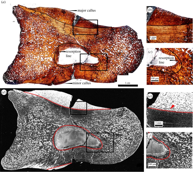
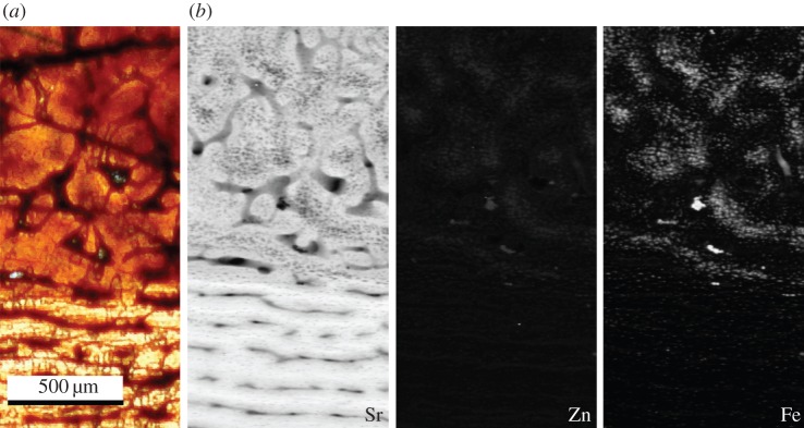
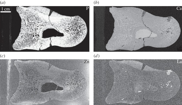
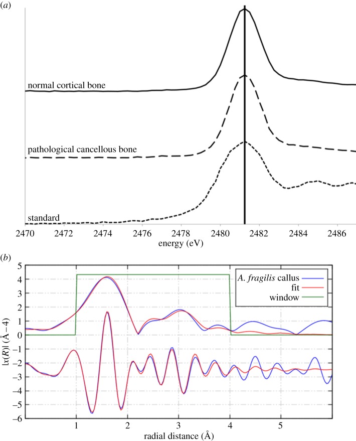
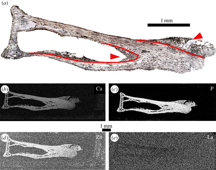
Current understanding of bone healing and remodelling strategies in vertebrates has traditionally relied on morphological observations through the histological analysis of thin sections. However, chemical analysis may also be used in such interpretations, as different elements are known to be absorbed and used by bone for different physiological purposes such as growth and healing. These chemical signatures are beyond the detection limit of most laboratory-based analytical techniques (e.g. scanning electron microscopy). However, synchrotron rapid scanning-X-ray fluorescence (SRS-XRF) is an elemental mapping technique that uniquely combines high sensitivity (ppm), excellent sample resolution (20-100 µm) and the ability to scan large specimens (decimetre scale) approximately 3000 times faster than other mapping techniques. Here, we use SRS-XRF combined with microfocus elemental mapping (2-20 µm) to determine the distribution and concentration of trace elements within pathological and normal bone of both extant and extinct archosaurs (Cathartes aura and Allosaurus fragilis). Results reveal discrete chemical inventories within different bone tissue types and preservation modes. Chemical inventories also revealed detail of histological features not observable in thin section, including fine structures within the interface between pathological and normal bone as well as woven texture within pathological tissue.

Keywords: SRS–XRF; archosaur; bone; fracture healing; histology.


Figures

Figure 1.
Figure 1.
Thin section of Allosaurus fragilis (specimen UMNH 6282) pedal phalanx as seen in optical observation under plain polarized light (a–c) and in an elemental map of iron taken at the SSRL beamline 6-2 (d–f; grey scale relative image with white as high concentration, black as low). Calli are present on the dorsal (major callus) and ventral (minor callus) surfaces in both optical and elemental images. Photomicrographs (b,c,e,f) represent magnified views of areas of interests (boxed areas), including the major callus (b,e) and resorption cavity (c,f). Laminar orientation of bone tissue is observed in the map of iron around the interface of the major callus and normal cortical bone (e) that is not seen in thin section (b). The extent of resorption and woven bone infill of the medullary cavity are also enhanced in iron (f) compared with thin section (c).
Figure 2.
Figure 2.
(a) Histological section as observed under plane polarized light and (b) false colour elemental maps of strontium, zinc and iron at the boundary between normal and pathological bone of the major callus in specimen UMNH 6282. The map of strontium highlights the woven texture of the callus, boundary of the bone tissues and laminar orientation of the normal bone. Zinc and iron are found mainly within the woven bone tissue of the callus.
Figure 3.
Figure 3.
Elemental maps of phosphorus (a), calcium (b), zinc (c) and lanthanum (d) in UMNH 6282 taken at the SSRL beamline 6-2. Phosphorus is concentrated within the original tissue (a), whereas calcium concentrations are uniform throughout the entire specimen excluding the glass mounting slide of UMNH 6282 (b). Lanthanum is enriched in the pathological tissues of the calli (d). Owing to spectral overlap, the map of lanthanum convolves both lanthanum and barium. Lanthanum was distinguished from barium using PyMCA analysis [44].
Figure 4.
Figure 4.
(a) XANES spectra for normal cortical bone taken from the dorsal surface and pathological bone taken from the main callus of UMNH 6282 compared with a sulfate standard. The peak for the sulfur species sulfate is marked [45]. (b) EXAFS spectrum for zinc taken from the main callus of UMNH 6282 revealing a tetrahedral coordination with phosphate.
Figure 5.
Figure 5.
Thin section of DMNH 83356 as seen in optical observation under plane polarized light (a), calcium (b), phosphorus (c), zinc (d) and lanthanum (e) taken at SSRL (beamline 6-2). Woven pathological bone growth is present along both the endosteal surface of the mid-shaft and the distal articulation surface (arrowheads). The interface between pathological growth and the normal bone tissue outline is more distinct in elemental maps compared with optical observation (outlined in red), especially in determining the extent of pathological growth along the endosteal surface (b–d). Lanthanum is below detection limit within the bone tissue, which is expected in modern samples (e).

References

    1. Chinsamy-Turan A. 2005. The microstructure of dinosaur bone: deciphering biology with fine-scale techniques. Baltimore, MD: The John Hopkins University Press.
    1. Currey J. 2002. Bones: structure and biomechanics. Princeton, NJ: Princeton University Press.
    1. de Ricqlés A. 1974. Evolution of endothermy: histological evidence. Evol. Theor. 1, 51–80.
    1. Tumarkin-Deratzian AR, Vann DV, Dodson P. 2006. Bone surface texture as an ontogenetic indicator in long bones of the Canada goose Branta Canadensis (Anseriformes: Anatidae). Zool. J. Linn. Soc. Lond. 148, 133–168. ( 10.1111/j.1096-3642.2006.00232.x) - DOI
    1. Madsen J. 1976. Allosaurus fragilis: a revised osteology. Utah Geol. Surv. Bull. 109, 1–163.

Publication types