Review
. 2014 May;197(1):19-31.
doi: 10.1534/genetics.112.148254.

Genetic and genomic tools for the marine annelid Platynereis dumerilii

Affiliations
Review

Genetic and genomic tools for the marine annelid Platynereis dumerilii

Juliane Zantke et al. Genetics. 2014 May.

Abstract

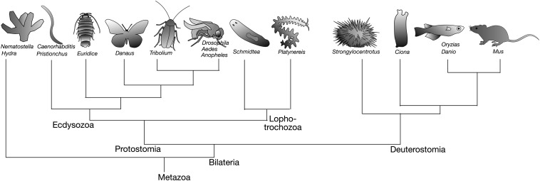
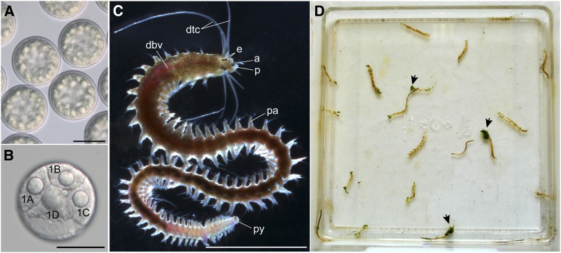
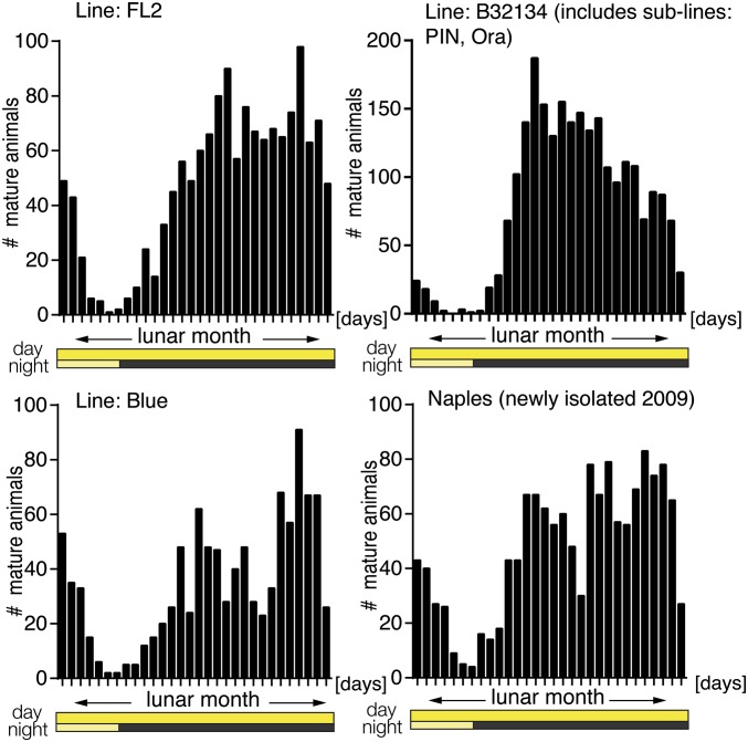
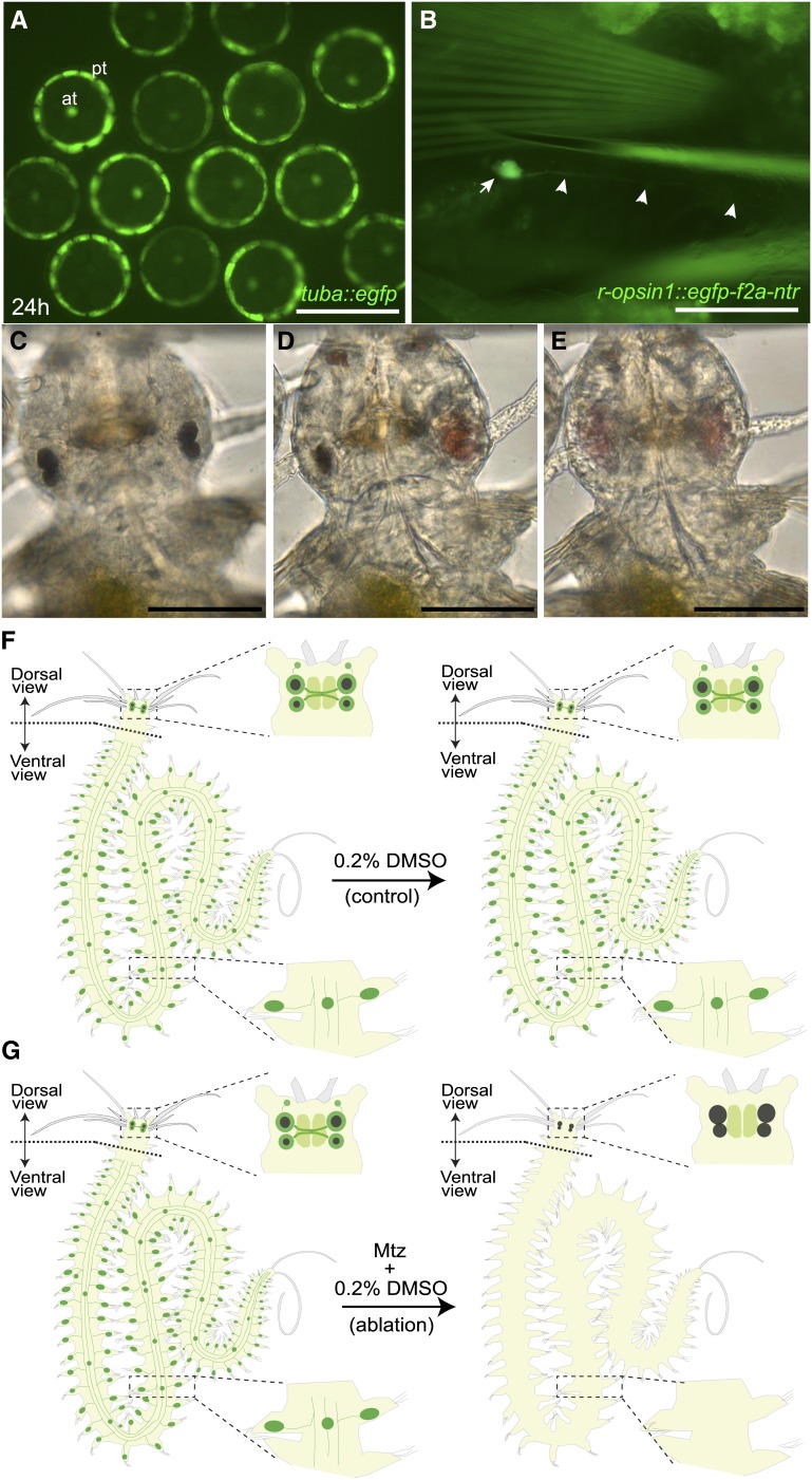
The bristle worm Platynereis dumerilii displays many interesting biological characteristics. These include its reproductive timing, which is synchronized to the moon phase, its regenerative capacity that is hormonally controlled, and a slow rate of evolution, which permits analyses of ancestral genes and cell types. As a marine annelid, Platynereis is also representative of the marine ecosystem, as well as one of the three large animal subphyla, the Lophotrochozoa. Here, we provide an overview of the molecular resources, functional techniques, and behavioral assays that have recently been established for the bristle worm. This combination of tools now places Platynereis in an excellent position to advance research at the frontiers of neurobiology, chronobiology, evo-devo, and marine biology.


Figures

Figure 1
Figure 1
The phylogenetic position of P. dumerilii. A schematized phylogenetic tree of Bilateria with its three main branches: Ecdysozoa, Lophotrochozoa, and Deuterostomia (Aguinaldo et al. 1997; de Rosa et al. 1999). Platynereis is an annelid worm positioned within the Lophotrochozoan group. The species represented in the tree have at least one functional tool established (transgenesis, RNAi, morpholino-based gene knockdown, genome mutagenesis using Zn-fingers, TALENs or Cas9/CRISR).
Figure 2
Figure 2
The annelid P. dumerilii. (A and B) Early developmental stages of P. dumerilii. (A) Unfertilized eggs. (B) Cleaving embryo at stereoblastula stage (6 hpf) at 18° ±1°, 4-cell stage (anterior view, blastomeres 1A, 1B, 1C, 1D). The refractive spherical structures in A and B are lipid droplets. (C) Premature adult worm removed from the tube, dorsal view: a, antennae; dbv, dorsal blood vessel; dtc, dorsal tentacular cirri; e, eyes; p, palps; pa, parapodia; py, pygidium. (D) Adult worms >2 months of age in worm box (25 × 25 cm) living in self-made tubes feeding on spinach (arrows). Scale bars: A and B, 100 µm; C, 0.5 cm.
Figure 3
Figure 3
Circalunar reproductive periodicity of different P. dumerilii strains. Maturation curves under the displayed light regime. The y-axis represents the number of mature spawning animals recorded each day of the lunar month (duration ∼29.5 days). The x-axis shows the days of the lunar month and the illumination conditions for day (top bar) and night (bottom bar). Yellow, daylight; black, nights without moon (new moon, NM); light yellow; nights with dim light simulating full moon (FM). FL2 and Blue strains share a common origin, but were subsequently independently inbred. Vio, PIN, and Ora share a common origin (B3213); Ora and PIN diverged one generation later (B32134). Maturation data are pooled from data collected over the course of several months. For further details on the illumination regime and scoring see Zantke et al. (2013).
Figure 4
Figure 4
Transgenesis and specific cell ablation in P. dumerilii. (A and B) Examples of cells marked by stable transgenesis with EGFP. (A) Trochophore larvae (24 hpf, apical view): larval protroch (pt) and apical tuft (at) cells are labeled by a construct containing the regulatory region of the Platynereis α-tubulin locus (tuba::egfp; Backfisch et al. 2013, 2014). (B) Peripheral neuron (arrow, soma; arrowheads, neurite) in appendage of an immature adult worm highlighted by an r-opsin1::egfp-f2a-ntr construct (Backfisch et al. 2013, 2014; Veedin Rajan et al. 2013). Orientation: ventral view, anterior to the top. (C–E) Laser ablation of Platynereis adult eyes performed on a 2- to 3-week-old juvenile worm. (F and G) Scheme of chemical ablation using metronidazole (mtz) to induce apoptosis in cells expressing eGFP and nitroreductase (Ntr) from an integrated r-opsin1::egfp-f2a-ntr transgene (Veedin Rajan et al. 2013). Cells expressing r-opsin1::egfp-f2a-ntr are depicted in green. (F) DMSO control. (G) Incubation with mtz. Scale bars: A and B, 100 µm; C–E, 50 µm.
Figure 5
Figure 5
Workflow for targeted mutagenesis using TALENs in P. dumerilii. (A) TALENs can be designed to target specific DNA sequences given their modular composition: each repeat in the DNA binding domain contains a repeat variable di-residue (RVD) that recognizes a single nucleotide (blue, HD = C; yellow, NI = A; purple, NG = T; green, NN = G). (i) TALEN pairs are designed to recognize a specified locus using a web-based prediction tool (https://tale-nt.cac.cornell.edu/node/add/talen). (ii) Genotyping PCR of target loci (arrows, primers) to test for SNPs at target site (yellow). (iii) Target sites comprise 15- to 20-bp binding sites for each TALEN separated by a 15- to 16-bp spacer, with unique restriction endonuclease sites in the spacer, to facilitate screening. (B) TALEN expression plasmids are constructed via two-step GoldenGate (Cermak et al. 2011). (C) In vitro cleavage assay to validate that the constructed TALENs are catalytically active against the intended target. TALEN activity is confirmed by the presence of smaller (gray, “cut target”) bands visible by gel electrophoresis (red arrows). (D) Equal amounts of left and right TALEN mRNA are delivered into Platynereis zygotes via micro-injection. (E) Single or small pools of injected larvae are digested with proteinase K to produce a DNA lysate used as template for screening PCR. Genomic DNA from adult worm tail-clip tissue samples are prepared by conventional genomic DNA extraction kits. (F) Mutation screening is performed by PCR amplification of the target locus (yellow) and (i) digestion of the PCR product with the restriction enzyme cutting within the wild-type spacer sequence (jagged line): if mutations are present in the spacer that disrupts the restriction site, some or all of the PCR product will be resistant to digestion, resulting in “uncut” bands. (ii) Larger deletions are detected as smaller PCR bands (red arrow). (G) Subcloning of uncut bands or smaller deletion bands and subsequent sequencing is used to confirm the presence of mutations centered at the target site.

Similar articles

Cited by

References

    1. Ackermann C., 2003. Markierung der Zelllinien im Embryo von Platynereis. Ph.D. Thesis, University of Mainz, Mainz.
    1. Ackermann C., Dorresteijn A., Fischer A., 2005. Clonal domains in postlarval Platynereis dumerilii (Annelida: Polychaeta). J. Morphol. 266: 258–280. - PubMed
    1. Aguinaldo A. M., Turbeville J. M., Linford L. S., Rivera M. C., Garey J. R., et al. , 1997. Evidence for a clade of nematodes, arthropods and other moulting animals. Nature 387: 489–493. - PubMed
    1. Arendt D., Technau U., Wittbrodt J., 2001. Evolution of the bilaterian larval foregut. Nature 409: 81–85. - PubMed
    1. Arendt D., Tessmar K., de Campos-Baptista M. I., Dorresteijn A., Wittbrodt J., 2002. Development of pigment-cup eyes in the polychaete Platynereis dumerilii and evolutionary conservation of larval eyes in Bilateria. Development 129: 1143–1154. - PubMed

Publication types