Review
. 2014 Sep;27(5):699-720.
doi: 10.1111/pcmr.12257. Epub 2014 May 30.

MC1R, the cAMP pathway, and the response to solar UV: extending the horizon beyond pigmentation

Affiliations
Review

MC1R, the cAMP pathway, and the response to solar UV: extending the horizon beyond pigmentation

Jose C García-Borrón et al. Pigment Cell Melanoma Res. 2014 Sep.

Abstract

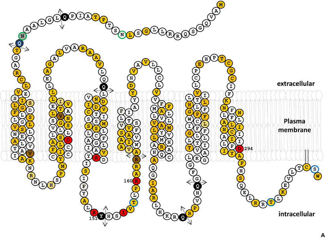
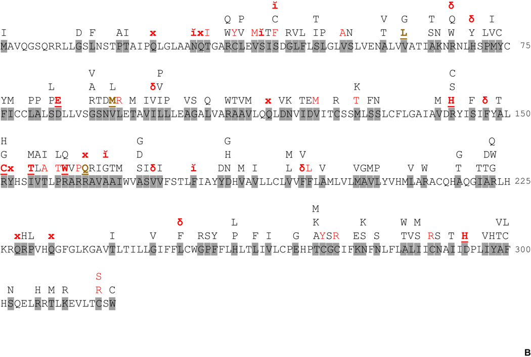
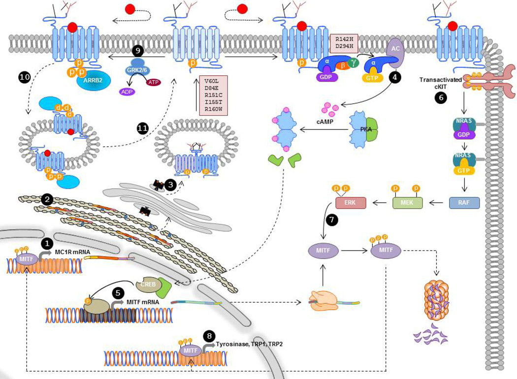
The melanocortin 1 receptor (MC1R) is a G protein-coupled receptor crucial for the regulation of melanocyte proliferation and function. Upon binding melanocortins, MC1R activates several signaling cascades, notably the cAMP pathway leading to synthesis of photoprotective eumelanin. Polymorphisms in the MC1R gene are a major source of normal variation of human hair color and skin pigmentation, response to ultraviolet radiation (UVR), and skin cancer susceptibility. The identification of a surprisingly high number of MC1R natural variants strongly associated with pigmentary phenotypes and increased skin cancer risk has prompted research on the functional properties of the wild-type receptor and frequent mutant alleles. We summarize current knowledge on MC1R structural and functional properties, as well as on its intracellular trafficking and signaling. We also review the current knowledge about the function of MC1R as a skin cancer, particularly melanoma, susceptibility gene and how it modulates the response of melanocytes to UVR.

Keywords: MC1R variants; Melanocortin 1 receptor; cAMP; melanocytes; melanoma; ultraviolet radiation.


Figures

Figure 1
Figure 1. Structure of MC1R and protein sequence changing polymorphisms
A. Primary structure and transmembrane topology. The sequence corresponds to transcript MC1R-001 (ID number ENST00000555147). Polymorphic positions for which no reliable association studies are available are indicated in yellow. Positions of R and r variants are shown in red and brown, respectively. Residues shown in gray correspond to indels, and black circles with white lettering followed by broken arrows to premature stop codons. Positions where both an indel and a point mutation have been found are shown as yellow circles hatched in gray. Ser/Thr residues presumably phosphorylated are highlighted with a blue border. The two Asn residues glycosylated in WT MC1R are indicated with a green border. B. MC1R protein sequence polymorphisms. Variant positions with described sequence alterations are shown on a gray background. x stands for premature stop, ĭ for insertion and δ for deletion. Known R (red letters) and r (brown letters) variants are indicated with bold underlined letters specifying the mutant amino acid. Substitutions presumably behaving as either R or r mutations based on functional studies, but for which no genetic association data are available are shown in red capital letters. Further information is provided in Supplementary Table 1.
Figure 2
Figure 2. Scheme of the main steps in MC1R biosynthesis and functional coupling
Transcription of the MC1R gene leads preferentially to the MC1R-001 transcript (1). Translation occurs in the rough ER, where post-translational modifications including oligomerization and glycosylation are also performed (2). Glycosylated oligomers proceed to the Golgi, then to the plasma membrane, and forward trafficking is dependent on Thr157 phosphorylation (3). For simplicity, the receptor is depicted as a monomer on the cell surface, but its structure is likely dimeric/oligomeric. Upon agonist binding, MC1R activates AC via the Gs protein, thus triggering cAMP synthesis and PKA activation (4). Active PKA catalytic subunits move to the nucleus to activate CREB transcription factors, which in turn increase the rate of transcription of the MITF gene (5). Agonist-activated MC1R also transactivates cKIT or a related RTK to trigger the NRAS-BRAF-MEK-ERK cascade (6). Active ERKs can phosphorylate MITF to increase its transcriptional activity and its proteasome-dependent degradation (7). Active MITF promotes transcription of the genes encoding for melanogenic enzymes (8), and for MC1R. Agonist-bound MC1R can also be desensitized by GRK2 or GRK6-dependent phosphorylation and ARRB2 recruitment (9), followed by sequestration in endocytic vesicles (10) whose likely destination is recycling to the cell surface (11).

References

    1. Abdel-Malek Z, Swope VB, Suzuki I, Akcali C, Harriger MD, Boyce ST, Urabe K, Hearing VJ. Mitogenic and melanogenic stimulation of normal human melanocytes by melanotropic peptides. Proc. Natl. Acad. Sci. U. S. A. 1995;92:1789–1793. - PMC - PubMed
    1. Abdel-Malek ZA, Kadekaro AL, Kavanagh RJ, et al. Melanoma prevention strategy based on using tetrapeptide alpha-MSH analogs that protect human melanocytes from UV-induced DNA damage and cytotoxicity. FASEB J. 2006;20:1561–1563. - PubMed
    1. Abdel-Malek ZA, Ruwe A, Kavanagh-Starner R, Kadekaro AL, Swope V, Haskell-Luevano C, Koikov L, Knittel JJ. alpha-MSH tripeptide analogs activate the melanocortin 1 receptor and reduce UV-induced DNA damage in human melanocytes. Pigment Cell Melanoma Res. 2009;22:635–644. - PubMed
    1. Aberdam E, Bertolotto C, Sviderskaya EV, de TV, Hemesath TJ, Fisher DE, Bennett DC, Ortonne JP, Ballotti R. Involvement of microphthalmia in the inhibition of melanocyte lineage differentiation and of melanogenesis by agouti signal protein. J. Biol. Chem. 1998;273:19560–19565. - PubMed
    1. Abrisqueta M, Herraiz C, Perez Oliva AB, Sanchez-Laorden BL, Olivares C, Jimenez-Cervantes C, Garcia-Borron JC. Differential and competitive regulation of human melanocortin 1 receptor signaling by beta-arrestin isoforms. J. Cell Sci. 2013;126:3724–3737. - PubMed

Publication types

MeSH terms

LinkOut - more resources