. 2014 Jul;88(14):8091-101.
doi: 10.1128/JVI.00944-14. Epub 2014 May 7.

Novel roles of cytoplasmic ICP0: proteasome-independent functions of the RING finger are required to block interferon-stimulated gene production but not to promote viral replication

Affiliations

Novel roles of cytoplasmic ICP0: proteasome-independent functions of the RING finger are required to block interferon-stimulated gene production but not to promote viral replication

Kathryne E Taylor et al. J Virol. 2014 Jul.

Abstract

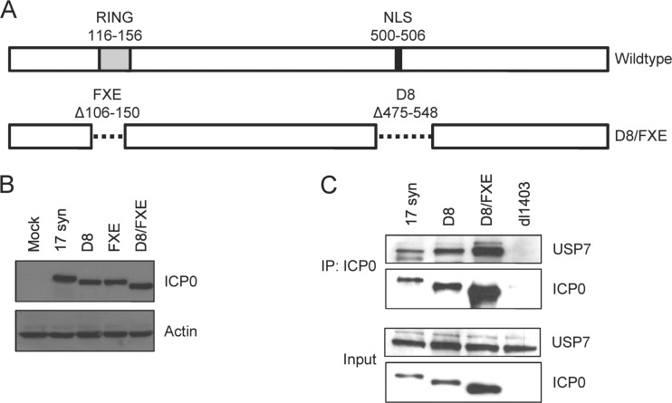
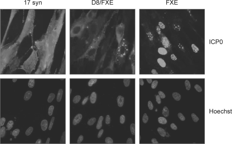
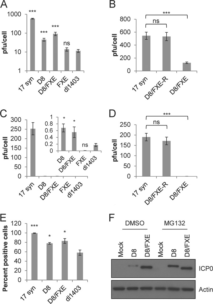
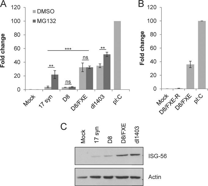
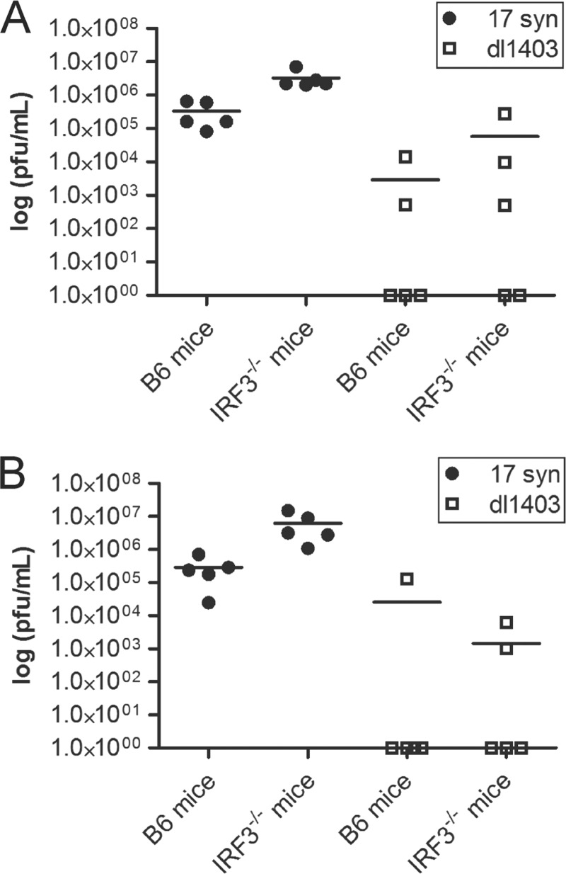
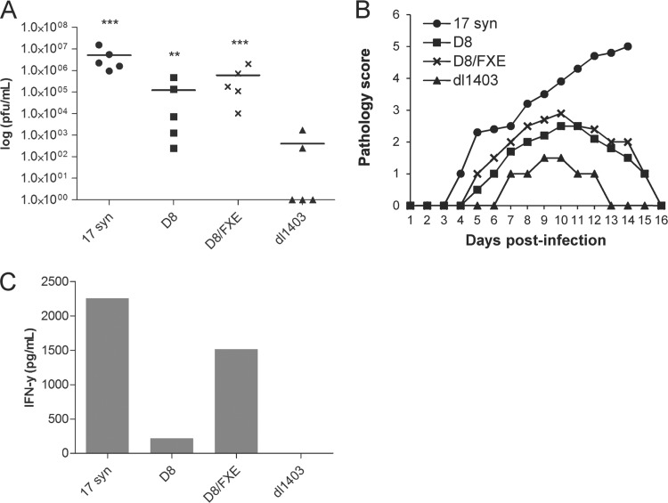
The immediate-early protein ICP0 from herpes simplex virus 1 (HSV-1) plays pleiotropic roles in promoting viral lytic replication and reactivation from latency. Most of the known actions of ICP0 occur in the nucleus and are thought to involve the E3 ubiquitin ligase activity of its RING finger domain, which targets proteins for degradation via the proteasome. Although ICP0 translocates to the cytoplasm as the infection progresses, little is known about its activities in this location. Here, we show that cytoplasmic ICP0 has two distinct functions. In primary cell cultures and in an intravaginal mouse model, cytoplasmic ICP0 promotes viral replication in the absence of an intact RING finger domain. Additionally, ICP0 blocks the activation of interferon regulatory factor 3 (IRF3), a key transcription factor of the innate antiviral response, in a mechanism that requires the RING finger domain but not the proteasome. To our knowledge, this is the first observation of a proteasome-independent function of the RING finger domain of ICP0. Collectively, these results underscore the importance of cytoplasm-localized ICP0 and the diverse nature of its activities. Importance: Despite ICP0 being a well-studied viral protein, the significance of its cytoplasmic localization has been largely overlooked. This is, in part, because common experimental manipulations result in the restriction of ICP0 to the nucleus. By overcoming this constraint, we both further characterize the ability of cytoplasmic ICP0 to inhibit antiviral signaling and show that ICP0 at this site has unexpected activities in promoting viral replication. This demonstrates the importance of considering location when analyzing protein function and adds a new perspective to our understanding of this multifaceted protein.


Figures

FIG 1
FIG 1
Generation of an HSV mutant expressing cytoplasmic ICP0 lacking the RING finger. (A) Schematic of the deletions in the icp0 gene found in the wild-type and D8/FXE viruses. (B and C) HEL cells were infected with the indicated viruses at an MOI of 10 for 8 h. (B) Cells were harvested with RIPA extract and analyzed for ICP0 size and expression relative to actin by Western blotting. (C) Cytoplasmic extracts were obtained, and samples were immunoprecipitated with an αICP0 antibody. The eluant and input extracts were analyzed by Western blotting for ICP0 and USP7.
FIG 2
FIG 2
An ICP0 NLS mutant lacking the RING finger localizes to the cytoplasm. HEL cells were infected with the indicated viruses at an MOI of 10 for 8 h, and immunofluorescence microscopy was performed to determine the subcellular location of ICP0. Nuclei were stained with Hoechst dye. Magnification, ×400.
FIG 3
FIG 3
Cytoplasmic ICP0 promotes viral replication in the absence of the RING finger domain in primary fibroblasts. (A to D) HEL cells were infected with the indicated viruses at a high MOI of 10 (A and B) or a low MOI of 0.1 (C and D) for 24 h. Cells and supernatant were harvested and sent through 3 freeze-thaw cycles, and the titer of U20S cells in the presence of HMBA was determined. (E) HEL cells were infected with the indicated viruses at an MOI of 10 for 12 h, and immunofluorescence microscopy was performed to determine the number of cells expressing ICP4 relative to the total number of cells, as determined by staining nuclei with Hoechst dye. The data are the averages of three independent experiments ± standard errors of the mean (SEM). Statistical analysis was performed using one-way analysis of variance (ANOVA) and Tukey's posttest (B and D) or Dunnett's posttest (A and C) relative to dl1403. *, P < 0.05; ***, P < 0.001; ns, not significant. (F) HEL cells were infected with the indicated viruses at an MOI of 10 for 8 h in the presence of MG132 or the carrier DMSO and then harvested via cytoplasmic extract. Western blot analysis was performed to determine the levels of ICP0 and actin.
FIG 4
FIG 4
Cytoplasmic ICP0 requires the RING finger but not the proteasome to block ISG expression. HEL cells were infected with the indicated viruses at an MOI of 10. (A) Infections were performed in the presence or absence of the proteasome inhibitor MG132, as indicated. (A and B) RNA was harvested after 6 h, and the expression of ISG-56 relative to GAPDH was determined using the TaqMan system of quantitative RT-PCR. Values are reported relative to poly(I·C) treated cells, whose fold change was set to 100. The data are the averages of three independent experiments ± SEM. Statistical analysis was performed using one-way ANOVA and Tukey's posttest. **, P < 0.01; ***, P < 0.001; ns, not significant. (C) Protein was harvested after 8 h and analyzed for ISG-56 and actin expression by Western blotting.
FIG 5
FIG 5
IRF3 deficiency cannot compensate for the loss of ICP0 in a mouse model of genital HSV infection. Wild-type B6 or irf3−/− mice were intravaginally infected with 1 × 10⁵ PFU of the indicated viruses. Vaginal washes were collected after 1 day (A) and 2 days (B), and the titer on U2OS cells was determined in the presence of HMBA. Scatter plot and mean values are shown.
FIG 6
FIG 6
Cytoplasmic ICP0 promotes viral replication in the absence of the RING finger domain in a mouse genital model of HSV infection. The indicated viruses (1 × 10⁵ PFU) were used to intravaginally infect irf3−/− mice. (A) Vaginal washes were collected after 2 days, and the titer on U2OS cells was determined in the presence of HMBA. Statistical analysis was performed using one-way ANOVA and Dunnett's posttest relative to dl1403. **, P < 0.01; ***, P < 0.001. Scatter plot and mean values are shown. (B) Vaginal pathology was monitored daily and scored on a 5-point scale. (C) Vaginal washes collected after 2 days were pooled, and ELISA was used to measure IFN-γ levels.

Similar articles

Cited by

References

    1. Everett RD. 2000. ICP0, a regulator of herpes simplex virus during lytic and latent infection. Bioessays 22:761–770. 10.1002/1521-1878(200008)22:8<761::AID-BIES10>3.0.CO;2-A - DOI - PubMed
    1. Boutell C, Sadis S, Everett RD. 2002. Herpes simplex virus type 1 immediate-early protein ICP0 and is isolated RING finger domain act as ubiquitin E3 ligases in vitro. J. Virol. 76:841–850. 10.1128/JVI.76.2.841-850.2002 - DOI - PMC - PubMed
    1. Everett RD. 2000. ICP0 induces the accumulation of colocalizing conjugated ubiquitin. J. Virol. 74:9994–10005. 10.1128/JVI.74.21.9994-10005.2000 - DOI - PMC - PubMed
    1. Everett RD, Orr A, Preston CM. 1998. A viral activator of gene expression functions via the ubiquitin-proteasome pathway. EMBO J. 17:7161–7169. 10.1093/emboj/17.24.7161 - DOI - PMC - PubMed
    1. Mullen MA, Ciufo DM, Hayward GS. 1994. Mapping of intracellular localization domains and evidence for colocalization interactions between the IE110 and IE175 nuclear transactivator proteins of herpes simplex virus. J. Virol. 68:3250–3266 - PMC - PubMed

Publication types

Substances

LinkOut - more resources