. 2014 Jul;88(14):8153-65.
doi: 10.1128/JVI.00894-14. Epub 2014 May 7.

Analysis of recombinant H7N9 wild-type and mutant viruses in pigs shows that the Q226L mutation in HA is important for transmission

Affiliations

Analysis of recombinant H7N9 wild-type and mutant viruses in pigs shows that the Q226L mutation in HA is important for transmission

Qinfang Liu et al. J Virol. 2014 Jul.

Abstract

The fact that there have been more than 300 human infections with a novel avian H7N9 virus in China indicates that this emerging strain has pandemic potential. Furthermore, many of the H7N9 viruses circulating in animal reservoirs contain putative mammalian signatures in the HA and PB2 genes that are believed to be important in the adaptation of other avian strains to humans. To date, the definitive roles of these mammalian-signature substitutions in transmission and pathogenesis of H7N9 viruses remain unclear. To address this we analyzed the biological characteristics, pathogenicity, and transmissibility of A/Anhui/1/2013 (H7N9) virus and variants in vitro and in vivo using a synthetically created wild-type virus (rAnhui-WT) and two mutants (rAnhui-HA-226Q and rAnhui-PB2-627E). All three viruses replicated in lungs of intratracheally inoculated pigs, yet nasal shedding was limited. The rAnhui-WT and rAnhui-PB2-627E viruses were transmitted to contact animals. In contrast, the rAnhui-HA-226Q virus was not transmitted to sentinel pigs. Deep sequencing of viruses from the lungs of infected pigs identified substitutions arising in the viral population (e.g., PB2-T271A, PB2-D701N, HA-V195I, and PB2-E627K reversion) that may enhance viral replication in pigs. Collectively, the results demonstrate that critical mutations (i.e., HA-Q226L) enable the H7N9 viruses to be transmitted in a mammalian host and suggest that the myriad H7N9 genotypes circulating in avian species in China and closely related strains (e.g., H7N7) have the potential for further adaptation to human or other mammalian hosts (e.g., pigs), leading to strains capable of sustained human-to-human transmission. Importance: The genomes of the zoonotic avian H7N9 viruses emerging in China have mutations in critical genes (PB2-E627K and HA-Q226L) that may be important in their pandemic potential. This study shows that (i) HA-226L of zoonotic H7N9 strains is critical for binding the α-2,6-linked receptor and enables transmission in pigs; (ii) wild-type A/Anhui/1/2013 (H7N9) shows modest replication, virulence, and transmissibility in pigs, suggesting that it is not well adapted to the mammalian host; and (iii) both wild-type and variant H7N9 viruses rapidly develop additional mammalian-signature mutations in pigs, indicating that they represent an important potential intermediate host. This is the first study analyzing the phenotypic effects of specific mutations within the HA and PB2 genes of the novel H7N9 viruses created by reverse genetics in an important mammalian host model. Finally, this study illustrates that loss-of-function mutations can be used to effectively identify residues critical to zoonosis/transmission.


Figures

FIG 1
FIG 1
Synthetic generation of the eight full-length genomic segments of the A/Anhui/1-JCVI.1/2013 virus. The products were assembled from oligonucleotides and error corrected. L, 1-kb Plus DNA ladder from Life Technologies.
FIG 2
FIG 2
Experimental design for the pig infection study. Forty-four 3- to 4-week-old pigs were randomly assigned to 4 groups housed in separate rooms (12 pigs in each inoculated group and 8 pigs in the mock-inoculated control group). Nine pigs in each group were intratracheally inoculated with 106 TCID50 of the appropriate H7N9 virus on day 0. Three contact pigs were commingled with each inoculated group at 3 days postinfection (dpi). Three inoculated pigs from each group were necropsied at 3, 5 and 7 dpi, and the 3 contact pigs were sacrificed at 4 days postcontact (dpc).
FIG 3
FIG 3
Comparison of RNA polymerase activity of the H7N9 virus with contemporary human or avian viruses and analysis of the effect of the PB2-K627E substitution on the rAnhui-WT (H7N9-WT) polymerase activity. RNA polymerase activities from a range of human and avian viruses were compared: H1N1, A/PR/8/1934; H3N2, A/New York/238/2005; pH1N1, A/New York/1682/2009; H5N1, A/Hong Kong/213/2003; H7N9-WT (rAnhui-WT); H7N9-627E (rAnhui-PB2-627E) polymerase substitution mutant. For each cell line at each temperature, the polymerase activity of H7N9-WT was set at 100%, and all other viruses were compared to H7N9-WT. Each virus was significantly different than the H7N9-WT using a one-way ANOVA with Dunnett's multiple-comparison test (P < 0.05), unless noted on the figure as having no significant difference (P > 0.05). Bars represent means ± SD for 3 independent replicates.
FIG 4
FIG 4
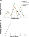
Fever in inoculated and contact pigs. (A) Body temperature changes in pigs that were inoculated with the indicated H7N9 viruses; (B) body temperature changes in contact pigs commingled with pigs that were inoculated with the indicated H7N9 viruses.
FIG 5
FIG 5
Gross lung lesions from pigs inoculated with the indicated H7N9 viruses at 5 days postinfection (dpi). Lung lesions due to influenza were characterized by multifocal to coalescing, red-plum-colored, consolidated areas. No gross lesions were seen in the mock-inoculated control group (A). Pigs inoculated with the rAnhui-WT virus and variants typically had multifocal, red-plum-colored lesions on the right apical and cardiac lung lobes (B, C, and D).
FIG 6
FIG 6
Microscopic lung sections from pigs inoculated with various H7N9 viruses at 5 days postinfection (dpi). (A) The bronchioles are lined by normal cuboidal epithelium (arrow), and the alveoli are clear (asterisk) in the control group; (B) the rAnhui-WT group has damage to bronchioles and mild bronchiolar epithelial hyperplasia (arrow) and moderate numbers of neutrophils in the alveoli (asterisk); (C) the rAnhui-HA-226Q group shows mild degeneration of bronchiolar epithelium (arrow), alveoli infiltrated by moderate numbers of neutrophils (asterisk), and moderate interstitial, peribronchiolar, and perivascular lymphocytic infiltration; (D) the rAnhui-PB2-627E group has minimal to mild bronchiolar epithelial degeneration, as shown by mild attenuation of the bronchiolar wall (arrow), and lesions are alveole-centric and characterized by infiltration of the alveolar lumen with neutrophils and by damage to the alveolar epithelium (asterisk). Bar, 50 μm.
FIG 7
FIG 7
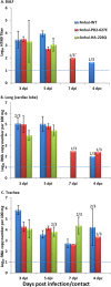
Detection of influenza virus and genomic RNA in inoculated and contact pigs. The bronchoalveolar lavage fluid (A), lungs (B), and tracheas (C) was harvested (3 animals) at 3, 5, or 7 dpi or 4 dpc. Virus titers were determined by TCID50 using MDCK cells (A). Tissue homogenates were processed for RNA extraction and quantitative real-time RT-PCR to detect the influenza virus HA gene (B and C). Dotted lines represent the limit of detection. *, All data represent 3/3 positive animals unless otherwise indicated (e.g., 2/3 means that 2 of 3 animals were positive). Error bars represent standard errors of the means (SEM).
FIG 8
FIG 8
Structure of the H7N9 HA highlighting specific amino acid substitutions. The crystal structure of the hemagglutinin (with Asn-133 glycosylation) from an H7N9 influenza virus isolated from humans (Protein Data Base [PDB] code 4BSA) (45) is shown. The structure was rendered using Pymol, and the surface is shown as transparent. 226L (using H3 numbering; 235L, based on a starting methionine of H7), which was mutated to the glutamine (Q) in rAnhui-HA-226Q, is highlighted in red. The substitutions R149I, A156T, and N167D were found in viruses from the BALF of pigs infected by rAnhui-PB2-627E. F87L and V195I were observed in rAnhui-WT-infected pigs. The single substitution N443K was found in rAnhui-HA-226Q-inoculated pigs. All substitutions found in pig BALF samples are shown in blue. Designations in parentheses indicate the residue based on the starting methionine of the H7 HA.

References

    1. WHO. 2009. Influenza (seasonal) fact sheet 211. http://www.who.int/mediacentre/factsheets/fs211/en/.
    1. Thompson WW, Shay DK, Weintraub E, Brammer L, Cox N, Anderson LJ, Fukuda K. 2003. Mortality associated with influenza and respiratory syncytial virus in the United States. JAMA 289:179–186. 10.1001/jama.289.2.179 - DOI - PubMed
    1. Johnson NP, Mueller J. 2002. Updating the accounts: global mortality of the 1918–1920 “Spanish” influenza pandemic. Bull. Hist. Med. 76:105–115. 10.1353/bhm.2002.0022 - DOI - PubMed
    1. To KK, Chan JF, Chen H, Li L, Yuen KY. 2013. The emergence of influenza A H7N9 in human beings 16 years after influenza A H5N1: a tale of two cities. Lancet Infect. Dis. 13:809–821. 10.1016/S1473-3099(13)70167-1 - DOI - PMC - PubMed
    1. Zhou J, Wang D, Gao R, Zhao B, Song J, Qi X, Zhang Y, Shi Y, Yang L, Zhu W, Bai T, Qin K, Lan Y, Zou S, Guo J, Dong J, Dong L, Zhang Y, Wei H, Li X, Lu J, Liu L, Zhao X, Li X, Huang W, Wen L, Bo H, Xin L, Chen Y, Xu C, Pei Y, Yang Y, Zhang X, Wang S, Feng Z, Han J, Yang W, Gao GF, Wu G, Li D, Wang Y, Shu Y. 2013. Biological features of novel avian influenza A (H7N9) virus. Nature 499:500–503. 10.1038/nature12379 - DOI - PubMed

Publication types

MeSH terms

Substances

Associated data

LinkOut - more resources