. 2014 Jul;88(14):8129-38.
doi: 10.1128/JVI.00732-14. Epub 2014 May 7.

Cytosolic PrP can participate in prion-mediated toxicity

Affiliations

Cytosolic PrP can participate in prion-mediated toxicity

Alana M Thackray et al. J Virol. 2014 Jul.

Abstract

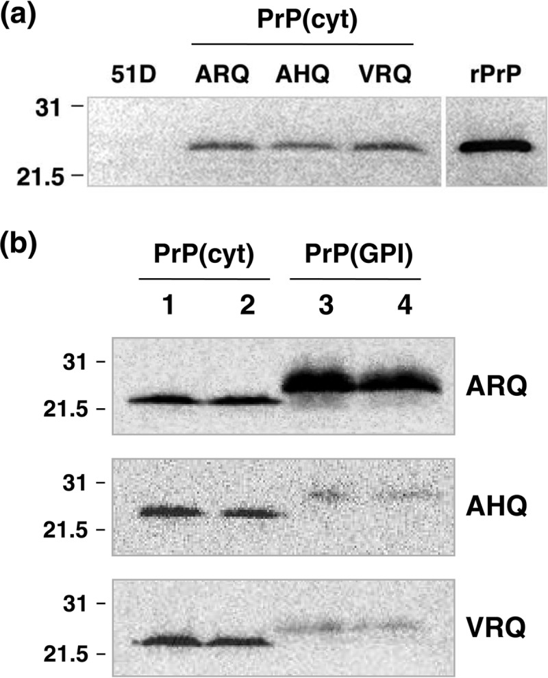
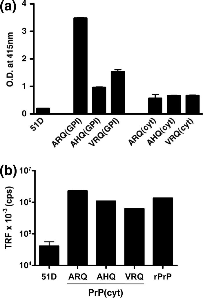
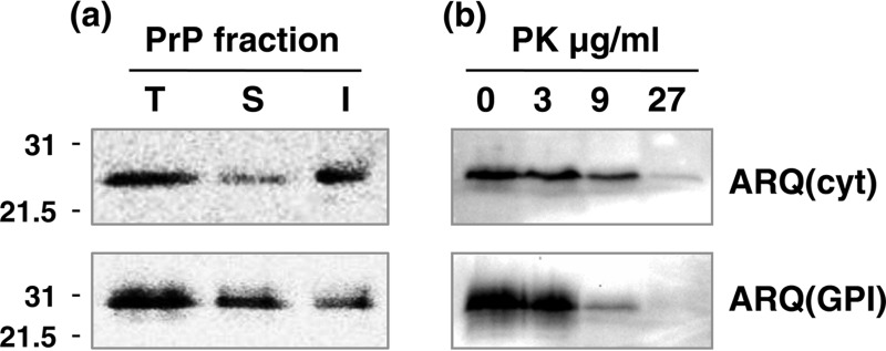
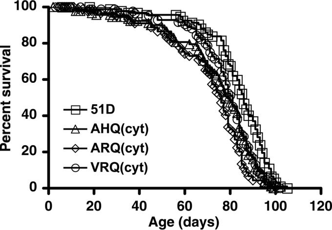
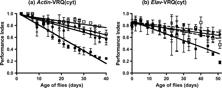
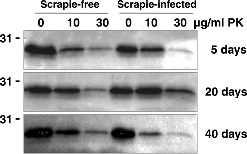
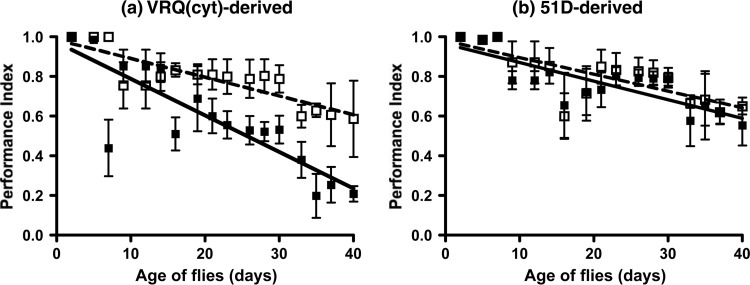
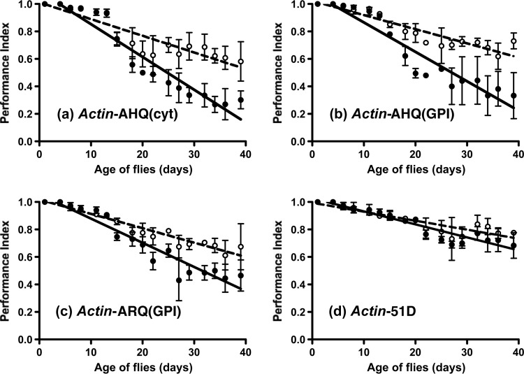
Prion diseases are characterized by a conformational change in the normal host protein PrPC. While the majority of mature PrPC is tethered to the plasma membrane by a glycosylphosphatidylinositol anchor, topological variants of this protein can arise during its biosynthesis. Here we have generated Drosophila transgenic for cytosolic ovine PrP in order to investigate its toxic potential in flies in the absence or presence of exogenous ovine prions. While cytosolic ovine PrP expressed in Drosophila was predominantly detergent insoluble and showed resistance to low concentrations of proteinase K, it was not overtly detrimental to the flies. However, Drosophila transgenic for cytosolic PrP expression exposed to classical or atypical scrapie prion inocula showed a faster decrease in locomotor activity than similar flies exposed to scrapie-free material. The susceptibility to classical scrapie inocula could be assessed in Drosophila transgenic for panneuronal expression of cytosolic PrP, whereas susceptibility to atypical scrapie required ubiquitous PrP expression. Significantly, the toxic phenotype induced by ovine scrapie in cytosolic PrP transgenic Drosophila was transmissible to recipient PrP transgenic flies. These data show that while cytosolic PrP expression does not adversely affect Drosophila, this topological PrP variant can participate in the generation of transmissible scrapie-induced toxicity. These observations also show that PrP transgenic Drosophila are susceptible to classical and atypical scrapie prion strains and highlight the utility of this invertebrate host as a model of mammalian prion disease. Importance: During prion diseases, the host protein PrPC converts into an abnormal conformer, PrPSc, a process coupled to the generation of transmissible prions and neurotoxicity. While PrPC is principally a glycosylphosphatidylinositol-anchored membrane protein, the role of topological variants, such as cytosolic PrP, in prion-mediated toxicity and prion formation is undefined. Here we generated Drosophila transgenic for cytosolic PrP expression in order to investigate its toxic potential in the absence or presence of exogenous prions. Cytosolic ovine PrP expressed in Drosophila was not overtly detrimental to the flies. However, cytosolic PrP transgenic Drosophila exposed to ovine scrapie showed a toxic phenotype absent from similar flies exposed to scrapie-free material. Significantly, the scrapie-induced toxic phenotype in cytosolic transgenic Drosophila was transmissible to recipient PrP transgenic flies. These data show that cytosolic PrP can participate in the generation of transmissible prion-induced toxicity and highlight the utility of Drosophila as a model of mammalian prion disease.


Figures

FIG 1
FIG 1
Western blot detection of cytosolic ovine PrP expression in Drosophila. Head homogenates were prepared from 5-day-old ovine PrP transgenic Drosophila or 51D control flies crossed with the Elav-GAL4 driver fly line. Samples were analyzed by SDS-PAGE and Western blot analysis with anti-PrP monoclonal antibody Sha31. (a) Molecular profile of ARQ(cyt), AHQ(cyt), and VRQ(cyt), all at the equivalent of 10 fly heads per track. Mature length ovine VRQ recombinant PrP (rPrP) was used at 10 ng. Molecular mass marker values (kDa) are shown on the left. (b) Comparison of ovine PrP(cyt) and PrP(GPI) expression in Drosophila. Tracks 1 and 2, PrP(cyt); tracks 3 and 4, PrP(GPI); tracks 1 and 3; male flies; tracks 2 and 4; female flies. The equivalent of five fly heads were run per track. Molecular mass marker values (kDa) are shown on the left. The ovine PrP genotype is indicated on the right.
FIG 2
FIG 2
Capture-detector immunoassay analysis of cytosolic ovine PrP expression in Drosophila. Head homogenates were prepared from 5-day-old ovine PrP transgenic Drosophila or 51D control flies crossed with the Elav-GAL4 driver fly line. Samples were analyzed by ELISA with anti-PrP monoclonal antibody 245 as the capture reagent and biotinylated anti-PrP monoclonal antibody SAF32 as the detector (a). The equivalent of 10 fly heads was measured per well, and the results are shown as the mean optical density (O.D.) at 415 nm ± SD of duplicate wells. (b) CDI. Fly head homogenates were treated with 8 M GdnHCl prior to immunoassay with anti-PrP monoclonal antibody 245 as the capture reagent and biotinylated anti-PrP monoclonal antibody SAF32 as the detector antibody. The equivalent of 20 fly heads was measured per well. Mature-length ovine ARQ recombinant PrP (rPrP) was used at 122 ng/well. The results are shown as average time-resolved fluorescence (TRF) counts per second (cps) ± SD for duplicate wells.
FIG 3
FIG 3
Cytosolic ovine PrP is characterized by reduced solubility and increased PK resistance. Head homogenates were prepared from 5-day-old ovine ARQ(cyt) or ARQ(GPI) PrP transgenic Drosophila crossed with the Elav-GAL4 driver fly line. After various treatments, fly head homogenate samples were analyzed by SDS-PAGE and Western blot analysis with anti-PrP monoclonal antibody Sha31. The equivalent of 10 fly heads was loaded per track. Molecular mass marker values (kDa) are shown on the left of each gel. (a) Total (T), soluble (S), and insoluble (I) fractions of PrP were prepared from fly heads as described in Materials and Methods. (b) Reaction products of fly head homogenates incubated with various concentrations of PK at 37°C for 30 min.
FIG 4
FIG 4
Survival curves of cytosolic ovine PrP transgenic Drosophila. Groups of 100 age-matched Elav-PrP or control Elav-51D flies were selected for survival assays. The number of surviving flies was recorded three times a week as described in Materials and Methods. Survival curves were calculated by using Kaplan-Meier plots, and differences between them were analyzed by the log rank method with Prism (GraphPad Software Inc., San Diego, CA).
FIG 5
FIG 5
Primary transmission of classical ovine scrapie in cytosolic VRQ PrP transgenic Drosophila. VRQ(cyt) PrP transgenic (squares) or 51D control flies (circles) crossed with either the Actin-GAL4 or Elav-GAL4 driver line were assessed for locomotor activity by a negative-geotaxis climbing assay following exposure at the larval stage to VRQ/VRQ scrapie-infected (filled symbols) or scrapie-free (open symbols and dashed lines) sheep brain homogenate. Actin-GAL4-VRQ(cyt) PrP flies did not contain the gene for RFP. The mean PI ± SD is shown for three groups of 15 flies of each genotype per time point (a total of 45 flies of each genotype). Statistical analysis of the linear regression plots was performed by one-way ANOVA and Tukey's honest significant difference test for post hoc analysis.
FIG 6
FIG 6
PK digestion of prion-exposed cytosolic VRQ fly head homogenate. Fly head homogenates were prepared from ovine VRQ(cyt) PrP transgenic Drosophila crossed with the Elav-GAL4 driver fly line following exposure at the larval stage to VRQ/VRQ scrapie-free (tracks 1 to 3) or scrapie-infected (tracks 4 to 6) sheep brain homogenate. Samples were incubated with 0, 10, or 30 μg/ml PK at 37°C for 15 min, and the reaction products were analyzed by SDS-PAGE and Western blot analysis with anti-PrP monoclonal antibody Sha31. The equivalent of 10 fly heads was loaded per track. Molecular mass marker values (kDa) are shown on the left of each gel. Ages of flies (in days) are shown on the right.
FIG 7
FIG 7
Fly-to-fly transmission of the prion-induced toxic phenotype. VRQ(cyt) PrP (without the gene for RFP) transgenic flies crossed with the Actin-GAL4 driver line were assessed for locomotor activity by a negative-geotaxis climbing assay following exposure at the larval stage to a 10% (vol/vol) dilution of head homogenate derived from 30-day-old Drosophila exposed at the larval stage to either scrapie-infected (filled squares) or scrapie-free (open squares and dashed line) sheep brain homogenate. The mean PI ± SD is shown for three groups of 15 flies of each genotype per time point (a total of 45 flies of each genotype). Statistical analysis of the linear regression plots was performed by one-way ANOVA and Tukey's honest significant difference test for post hoc analysis.
FIG 8
FIG 8
Primary transmission of atypical scrapie in Actin-driven PrP transgenic Drosophila. PrP transgenic or 51D control flies crossed with the Actin-GAL4 driver line were assessed for locomotor activity by a negative-geotaxis climbing assay following exposure at the larval stage to AHQ/AHQ scrapie-infected (filled circles) or scrapie-free (open circles and dashed lines) sheep brain homogenate. The mean PI ± SD is shown for three groups of 15 flies of each genotype per time point (a total of 45 flies of each genotype). Statistical analysis of the scrapie-infected and scrapie-free linear regression plots for each fly line was compared by the unpaired-sample t test.
FIG 9
FIG 9
Lack of response by Elav-driven AHQ PrP transgenic Drosophila to atypical scrapie. PrP transgenic or 51D control flies crossed with the Elav-GAL4 driver line were assessed for locomotor activity by a negative-geotaxis climbing assay following exposure at the larval stage to AHQ/AHQ scrapie-infected (closed squares and continuous line) or scrapie-free (closed circles and dashed line) sheep brain homogenate or PBS (closed triangles and dotted line). The mean PI ± SD is shown for three groups of 15 flies of each genotype per time point (a total of 45 flies of each genotype). Statistical analysis of the linear regression plots was performed by one-way ANOVA and Tukey's honest significant difference test for post hoc analysis.

Similar articles

Cited by

References

    1. Prusiner SB. (ed). 2004. Prion biology and diseases, second edition. Cold Spring Harbor Laboratory Press, Cold Spring Harbor, NY
    1. Brandner S, Isenmann S, Raeber A, Fischer M, Sailer A, Kobayashi Y, Marino S, Weissmann C, Aguzzi A. 1996. Normal host prion protein necessary for scrapie-induced neurotoxicity. Nature 379:339–343. 10.1038/379339a0 - DOI - PubMed
    1. Büeler H, Aguzzi A, Sailer A, Greiner RA, Autenried P, Aguet M, Weissmann C. 1993. Mice devoid of PrP are resistant to scrapie. Cell 73:1339–1347. 10.1016/0092-8674(93)90360-3 - DOI - PubMed
    1. Mallucci G, Dickinson A, Linehan J, Klohn PC, Brandner S, Collinge J. 2003. Depleting neuronal PrP in prion infection prevents disease and reverses spongiosis. Science 302:871–874. 10.1126/science.1090187 - DOI - PubMed
    1. Ciccotosto GD, Cappai R, White AR. 2008. Neurotoxicity of prion peptides on cultured cerebellar neurons. Methods Mol. Biol. 459:83–96. 10.1007/978-1-59745-234-2_6 - DOI - PubMed

Publication types

LinkOut - more resources