

Natural history of small index lesions suspicious for prostate cancer on multiparametric MRI: recommendations for interval imaging follow-up
- PMID: 24808435
- PMCID: PMC4463272
- DOI: 10.5152/dir.2014.13319
Natural history of small index lesions suspicious for prostate cancer on multiparametric MRI: recommendations for interval imaging follow-up
Abstract
Purpose: We aimed to determine the natural history of small index lesions identified on multiparametric-magnetic resonance imaging (MP-MRI) of the prostate by evaluating lesion-specific pathology and growth on serial MP-MRI.
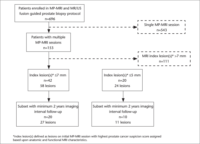
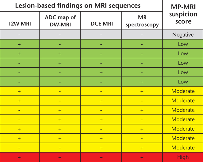
Materials and methods: We performed a retrospective review of 153 patients who underwent a minimum of two MP-MRI sessions, on an institutional review board-approved protocol. Index lesion is defined as the lesion(s) with the highest cancer suspicion score based on initial MP-MRI of a patient, irrespective of size. Two study cohorts were identified: (1) patients with no index lesion or index lesion(s) ≤7 mm and (2) a subset with no index lesion or index lesion(s) ≤5 mm. Pathological analysis of the index lesions was performed following magnetic resonance/ultrasound fusion-guided biopsy. Growth rate of the lesions was calculated based on MP-MRI follow-up.
Results: Patients with small index lesions measuring ≤7 mm (n=42) or a subset with lesions ≤5 mm (n=20) demonstrated either benign findings (86.2% and 87.5%, respectively) or low grade Gleason 6 prostate cancer (13.8% and 12.5%, respectively) on lesion-specific targeted biopsies. These lesions demonstrated no significant change in size (P = 0.93 and P = 0.36) over a mean imaging period of 2.31±1.56 years and 2.40±1.77 years for ≤7 mm and ≤5 mm index lesion thresholds, respectively. These findings held true on subset analyses of patients who had a minimum of two-year interval follow-up with MP-MRI.
Conclusion: Small index lesions of the prostate are pathologically benign lesions or occasionally low-grade cancers. Slow growth rate of these small index lesions on serial MP-MRI suggests a surveillance interval of at least two years without significant change.
Figures
References
-
- Siegel R, Naishadham D, Jemal A. Cancer statistics, 2013. CA Cancer J Clin. 2013;63:11–30. - PubMed
-
- Hernandez J, Thompson IM. Prostate-specific antigen: a review of the validation of the most commonly used cancer biomarker. Cancer. 2004;101:894–904. - PubMed
-
- Stamey TA, Freiha FS, McNeal JE, Redwine EA, Whittemore AS, Schmid HP. Localized prostate cancer. Relationship of tumor volume to clinical significance for treatment of prostate cancer. Cancer. 1993;71:933–938. - PubMed
-
- Epstein JI, Walsh PC, Carmichael M, Brendler CB. Pathologic and clinical findings to predict tumor extent of nonpalpable (stage T1c) prostate cancer. JAMA. 1994;271:368–374. - PubMed
Publication types
MeSH terms
Substances
Grants and funding
LinkOut - more resources
Full Text Sources
Other Literature Sources
Medical
