Review
. 2014 Jul 1;307(1):F1-F11.
doi: 10.1152/ajprenal.00067.2014. Epub 2014 May 7.

pH-responsive, gluconeogenic renal epithelial LLC-PK1-FBPase+cells: a versatile in vitro model to study renal proximal tubule metabolism and function

Affiliations
Review

pH-responsive, gluconeogenic renal epithelial LLC-PK1-FBPase+cells: a versatile in vitro model to study renal proximal tubule metabolism and function

Norman P Curthoys et al. Am J Physiol Renal Physiol. .

Abstract

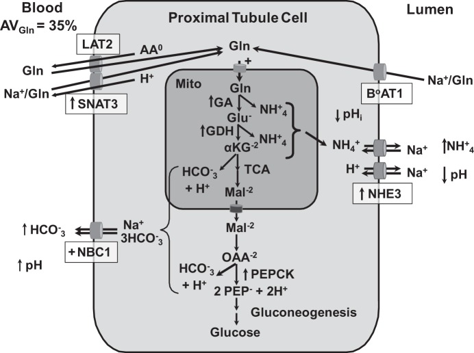
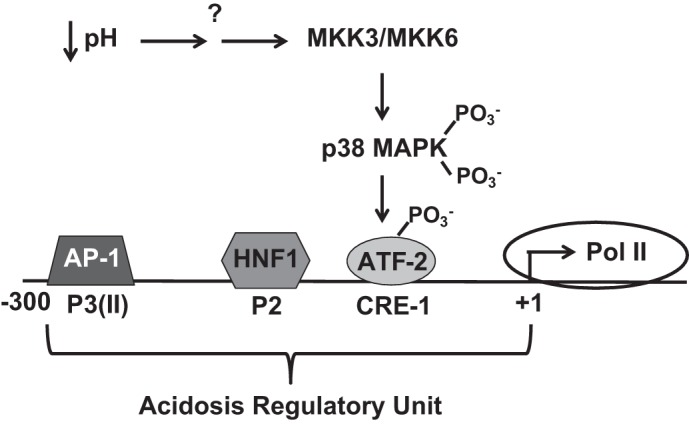
Ammoniagenesis and gluconeogenesis are prominent metabolic features of the renal proximal convoluted tubule that contribute to maintenance of systemic acid-base homeostasis. Molecular analysis of the mechanisms that mediate the coordinate regulation of the two pathways required development of a cell line that recapitulates these features in vitro. By adapting porcine renal epithelial LLC-PK1 cells to essentially glucose-free medium, a gluconeogenic subline, termed LLC-PK1-FBPase(+) cells, was isolated. LLC-PK1-FBPase(+) cells grow in the absence of hexoses and pentoses and exhibit enhanced oxidative metabolism and increased levels of phosphate-dependent glutaminase. The cells also express significant levels of the key gluconeogenic enzymes, fructose-1,6-bisphosphatase (FBPase) and phosphoenolpyruvate carboxykinase (PEPCK). Thus the altered phenotype of LLC-PK1-FBPase(+) cells is pleiotropic. Most importantly, when transferred to medium that mimics a pronounced metabolic acidosis (9 mM HCO3 (-), pH 6.9), the LLC-PK1-FBPase(+) cells exhibit a gradual increase in NH4 (+) ion production, accompanied by increases in glutaminase and cytosolic PEPCK mRNA levels and proteins. Therefore, the LLC-PK1-FBPase(+) cells retained in culture many of the metabolic pathways and pH-responsive adaptations characteristic of renal proximal tubules. The molecular mechanisms that mediate enhanced expression of the glutaminase and PEPCK in LLC-PK1-FBPase(+) cells have been extensively reviewed. The present review describes novel properties of this unique cell line and summarizes the molecular mechanisms that have been defined more recently using LLC-PK1-FBPase(+) cells to model the renal proximal tubule. It also identifies future studies that could be performed using these cells.

Keywords: acid-base balance; ammoniagenesis; gluconeogenesis; pH-responsive; proximal tubule.


Figures

Fig. 1.
Fig. 1.
Renal proximal tubular catabolism of glutamine. During chronic acidosis, approximately one-third of the arterial glutamine is removed during a single pass through the kidney. The glutamine filtered by the glomeruli is nearly quantitatively extracted from the lumen of the proximal convoluted tubule by B0AT1, a Na+-dependent neutral amino acid cotransporter in the apical membrane. Uptake of glutamine through the basolateral membrane occurs by reversal of the neutral amino acid exchanger (LAT2) and/or through increased expression of the basolateral glutamine transporter (SNAT3). Increased renal catabolism of glutamine is facilitated by increased expression (arrows) of the genes that encode glutaminase (GA), glutamate dehydrogenase (GDH), phosphoenolpyruvate carboxykinase (PEPCK), the apical Na+/H+ exchanger (NHE3), and SNAT3. In addition, the activities of the mitochondrial glutamine transporter and basolateral Na+/3HCO3 are increased (+). Increased expression of NHE3 contributes to the transport of ammonium ions and the acidification of the luminal fluid. The combined increases in renal ammonium ion excretion and gluconeogenesis result in a net synthesis of HCO3 ions that are transported across the basolateral membrane by the Na+/3HCO3 cotransporter (NBC1). αKG, α-ketoglutarate; Mal, malate; OAA, oxaloacetate; PEP, phosphoenolpyruvate.
Fig. 2.
Fig. 2.
Expression of fructose-1,6-bisphosphatase (FBPase) and cytosolic PEPCK in various renal cell lines and in the rat kidney. Cultured cells were incubated in normal (pH 7.4) or acidic medium (pH 6.9) for 18 h. Total RNA samples (20 μg) were electrophoresed, blotted, and hybridized with cDNA probes to rat liver FBPase and rat renal cytosolic PEPCK. FBPase+, LLC-PK1-FBPase+ cells; OK, opossum kidney cells; MDCK, Madin-Darby canine kidney cells; LLC-PK1, LLC-PK1 pig kidney cells; WKPT, Wistar-Kyoto rat proximal tubular cells; HPT, primary cultures of human proximal tubular cells; CTX, rat kidney cortex; OM, outer medulla; IM, inner medulla.
Fig. 3.
Fig. 3.
Mechanism of pH-responsive activation of PEPCK transcription in LLC-PK1-FBPase+ cells. A decrease in media pH and HCO3 leads to activation of MKK3 and MKK6, which phosphorylate and activate p38-MAPK. The activated p38-MAPK subsequently phosphorylates and activates a transcription factor (ATF-2), which binds to the CRE-1 element within the acidosis-regulatory unit of the PEPCK promoter. Activated transcription also requires the binding of hepatic nuclear factor 1 (HNF-1) to the P2 element and AP-1 to the P3(II) element.
Fig. 4.
Fig. 4.
Proposed mechanism for the pH-responsive stabilization of PEPCK mRNA. Interactions between the cap binding proteins (4E and 4G) and the polyA binding protein (PABP) cause mRNA to form a circular structure that enhances translation. Both a stabilizing mRNA binding protein (HuR) and destabilizing mRNA binding protein (AUF1) bind to the adenylate-uridylate (AU)-rich sequences within the 3′-untranslated region (UTR) of the PEPCK mRNA during normal acid-base balance. This complex recruits a deadenylase (Deaden) that shortens the polyA tail and causes dissociation of the polyA binding proteins (PABPs). The deadenylated mRNA is degraded in processing bodies by decapping and degradation from the 5′-end. In response to metabolic acidosis, the extent of phosphorylation of HuR is decreased while AUF1 is phosphorylated at additional sites. These changes lead to increased binding of HuR and a remodeling of the HuR/AUF1 complex that is bound to the 3′-UTR of PEPCK mRNA. The new complex is less effective at recruiting deadenylase and thereby promotes stabilization of the PEPCK mRNA.

Similar articles

Cited by

References

    1. Alexander RT, Dimke H, Cordat E. Proximal tubular NHEs: sodium, protons and calcium? Am J Physiol Renal Physiol 305: F229–F236, 2013 - PMC - PubMed
    1. Angelow S, Ahlstrom R, Yu AS. Biology of claudins. Am J Physiol Renal Physiol 295: F867–F876, 2008 - PMC - PubMed
    1. Bonasio R, Tu S, Reinberg D. Molecular signals of epigenetic states. Science 330: 612–616, 2010 - PMC - PubMed
    1. Brosnan JT, Lowry M, Vinay P, Gougoux A, Halperin ML. Renal ammonia production—une vue canadienne. Can J Physiol Pharmacol 65: 489–498, 1987 - PubMed
    1. Burch HB, Narins RG, Chu C, Fagioli S, Choi S, McCarthy W, Lowry OH. Distribution along the rat nephron of three enzymes of gluconeogenesis in acidosis and starvation. Am J Physiol Renal Fluid Electrolyte Physiol 235: F246–F253, 1978 - PubMed

Publication types

Substances

LinkOut - more resources