

Low intraprostatic DHT promotes the infiltration of CD8+ T cells in BPH tissues via modulation of CCL5 secretion
- PMID: 24808637
- PMCID: PMC3997870
- DOI: 10.1155/2014/397815
Low intraprostatic DHT promotes the infiltration of CD8+ T cells in BPH tissues via modulation of CCL5 secretion
Abstract
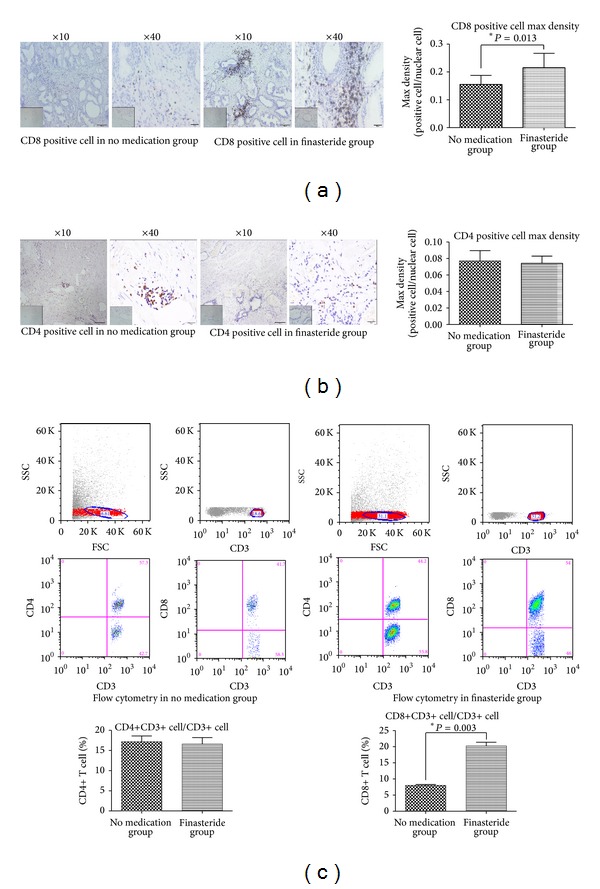
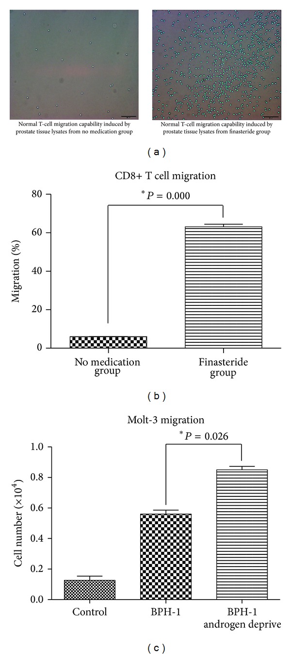
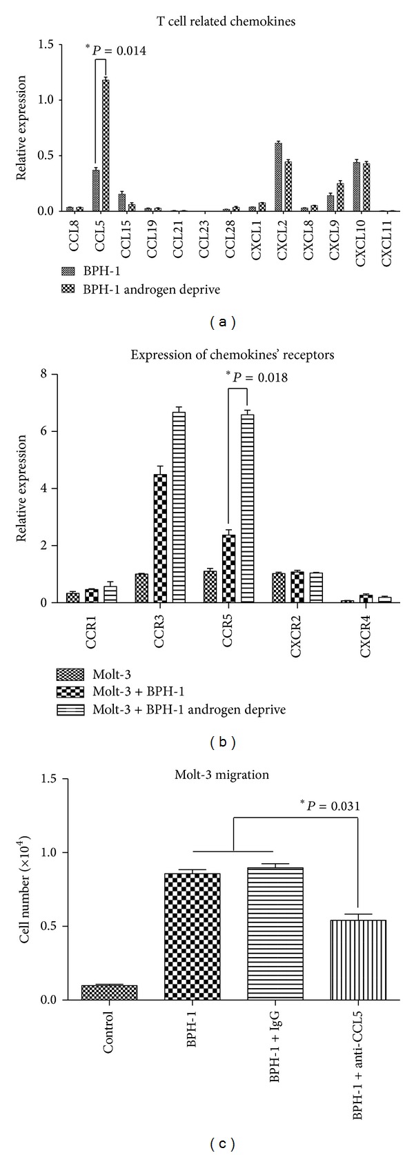
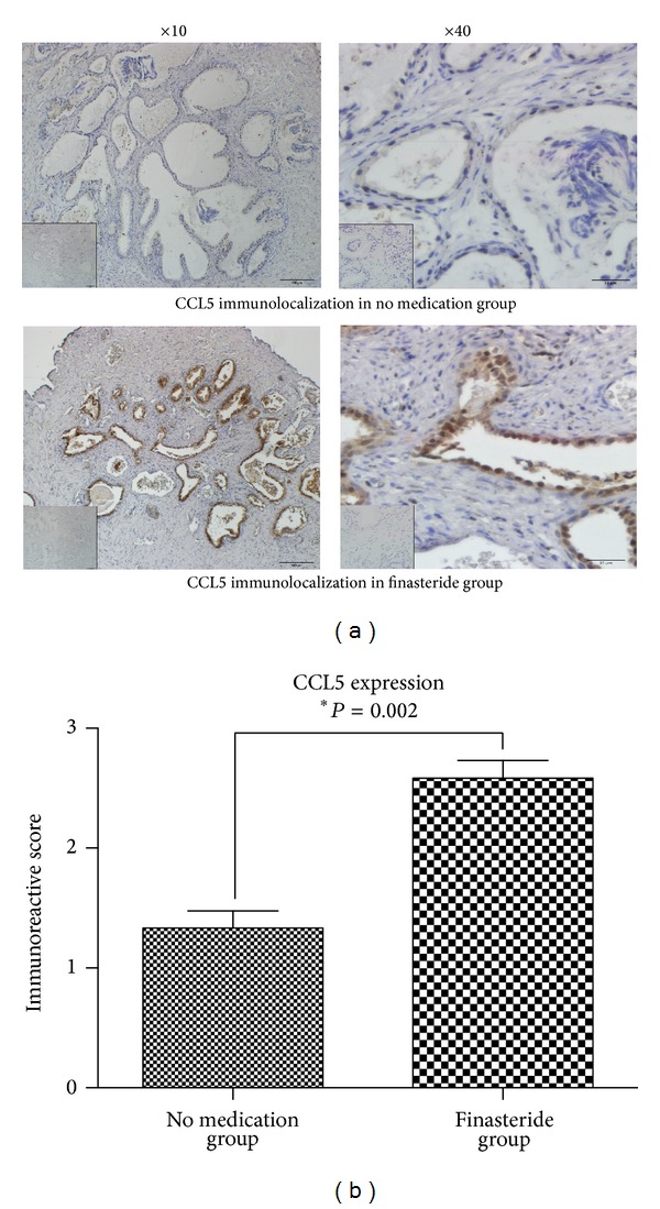
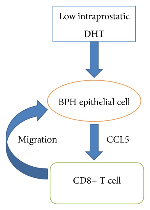
Clinical studies suggested thatandrogen might be associated with infiltrating T cells in prostate of benign prostatic hyperplasia (BPH) patients, but detail of T-cell subset and mechanism still remained unclear. The present study tested the hypothesis that intraprostatic 5 α -dihydrotestosterone (DHT) exerts effects on T cells recruitment by BPH epithelial cells. Prostate tissues from 64 cases of BPH patients after transurethral resection of prostate (TURP) were divided into 2 groups: (1) no medication history; (2) administration of 5 α -reductase type II inhibitor-finasteride 5 mg daily for at least 6 months before surgery. Group 2 presented significantly higher CD8+ T cells infiltration than group 1, but no changes in CD4+ T cells (immunohistochemistry and flow cytometry). In vitro study more CD8+ T cell migrated to the prostate tissue lysates from group 2 and BPH-1 cells in low DHT condition. Transcription of chemokine (C-C motif) Ligand 5 (CCL5) mRNA in BPH-1 cells and chemokine (C-C motif) receptor 5 (CCR5) mRNA in CD8+ T cells were upregulated in low DHT condition (q-PCR). CCL5 expression was also identified to be higher in group 2 prostate tissues by IHC. This study suggested that intraprostatic DHT may participate in regulating inflammatory response which was induced by human prostatic epithelial cell, via modulating CCL5 secretion.
Figures
Similar articles
-
Activation of cGMP/PKG/p65 signaling associated with PDE5-Is downregulates CCL5 secretion by CD8 + T cells in benign prostatic hyperplasia.Prostate. 2019 Jun;79(8):909-919. doi: 10.1002/pros.23801. Epub 2019 Apr 8. Prostate. 2019. PMID: 30958912 Free PMC article.
-
CD8+ T cells promote proliferation of benign prostatic hyperplasia epithelial cells under low androgen level via modulation of CCL5/STAT5/CCND1 signaling pathway.Sci Rep. 2017 Feb 20;7:42893. doi: 10.1038/srep42893. Sci Rep. 2017. PMID: 28216616 Free PMC article.
-
Differential effect of finasteride on the tissue androgen concentrations in benign prostatic hyperplasia.Clin Endocrinol (Oxf). 1997 Feb;46(2):137-44. doi: 10.1046/j.1365-2265.1997.950908.x. Clin Endocrinol (Oxf). 1997. PMID: 9135694 Clinical Trial.
-
Dihydrotestosterone and the concept of 5alpha-reductase inhibition in human benign prostatic hyperplasia.World J Urol. 2002 Apr;19(6):413-25. doi: 10.1007/s00345-002-0248-5. World J Urol. 2002. PMID: 12022710 Review.
-
Dihydrotestosterone and the concept of 5alpha-reductase inhibition in human benign prostatic hyperplasia.Eur Urol. 2000 Apr;37(4):367-80. doi: 10.1159/000020181. Eur Urol. 2000. PMID: 10765065 Review.
Cited by
-
Penile vascular abnormalities in young men with persistent side effects after finasteride use for the treatment of androgenic alopecia.Transl Androl Urol. 2020 Jun;9(3):1201-1209. doi: 10.21037/tau.2020.03.21. Transl Androl Urol. 2020. PMID: 32676403 Free PMC article.
-
Estrogen and G protein-coupled estrogen receptor accelerate the progression of benign prostatic hyperplasia by inducing prostatic fibrosis.Cell Death Dis. 2022 Jun 7;13(6):533. doi: 10.1038/s41419-022-04979-3. Cell Death Dis. 2022. PMID: 35672281 Free PMC article.
-
Tissue-derived extracellular vesicles in cancers and non-cancer diseases: Present and future.J Extracell Vesicles. 2021 Dec;10(14):e12175. doi: 10.1002/jev2.12175. J Extracell Vesicles. 2021. PMID: 34918479 Free PMC article. Review.
-
The Effects of Androgens on T Cells: Clues to Female Predominance in Autoimmune Liver Diseases?Front Immunol. 2020 Jul 29;11:1567. doi: 10.3389/fimmu.2020.01567. eCollection 2020. Front Immunol. 2020. PMID: 32849531 Free PMC article. Review.
-
Activation of cGMP/PKG/p65 signaling associated with PDE5-Is downregulates CCL5 secretion by CD8 + T cells in benign prostatic hyperplasia.Prostate. 2019 Jun;79(8):909-919. doi: 10.1002/pros.23801. Epub 2019 Apr 8. Prostate. 2019. PMID: 30958912 Free PMC article.
References
-
- Wei JT, Calhoun E, Jacobsen SJ. Urologic diseases in America project: benign prostatic hyperplasia. Journal of Urology. 2005;173(4):1256–1261. - PubMed
-
- Gandaglia G, Briganti A, Gontero P, et al. The role of chronic prostatic inflammation in the pathogenesis and progression of benign prostatic hyperplasia (BPH) BJU International. 2013;112(4):432–441. - PubMed
-
- Ho CKM, Habib FK. Estrogen and androgen signaling in the pathogenesis of BPH. Nature Reviews Urology. 2011;8(1):29–41. - PubMed
-
- Aggarwal S, Thareja S, Verma A, Bhardwaj TR, Kumar M. An overview on 5α-reductase inhibitors. Steroids. 2010;75(2):109–153. - PubMed
-
- McConnell JD, Roehrborn CG, Bautista OM, et al. The long-term effect of doxazosin, finasteride, and combination therapy on the clinical progression of benign prostatic hyperplasia. The New England Journal of Medicine. 2003;349(25):2387–2398. - PubMed
Publication types
MeSH terms
Substances
LinkOut - more resources
Full Text Sources
Other Literature Sources
Research Materials
