Cavernous hemangioma of the tongue: A rare case report
- PMID: 24808705
- PMCID: PMC4012128
- DOI: 10.4103/0976-237X.128680
Cavernous hemangioma of the tongue: A rare case report
Abstract
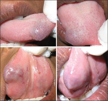
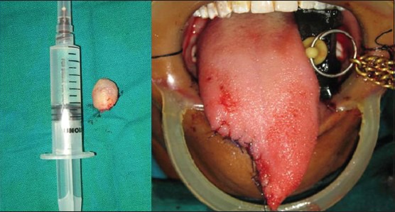
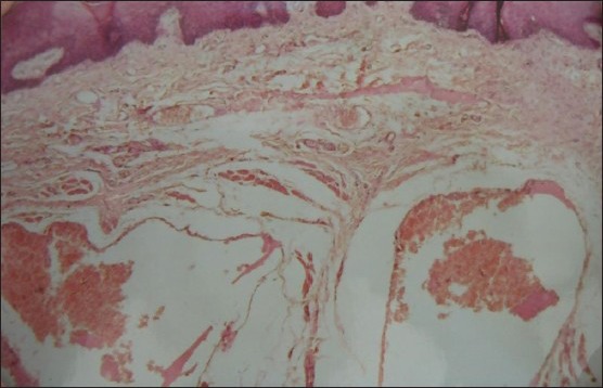
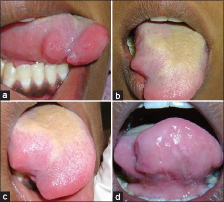
Hemangiomas are developmental vascular abnormalities and more than 50% of these lesions occur in the head and neck region, with the lips, tongue, buccal mucosa, and palate most commonly involved. They are considered as hamartomas rather than true neoplasms. Here we report a case of hemangioma of the body of the tongue, discussing the diagnostic aspects and treatment modalities of such lesion and emphasizing the role of the color Doppler ultrasonography, especially in the diagnosis and treatment. Factors such as patient's age, size and site of lesion and the proximity of lesion to vital structure are paramount in the determination of the therapeutic approach and surgical excision. Even though radiotherapy, cryotherapy, laser therapy, medical treatment, injection of sclerosing substances and the selective embolization of the lingual artery seem to have some efficacy, the author conclude that surgery is the therapy of choice in the isolated vascular lesions of the body of the tongue.
Keywords: Cavernous hemangioma; hemangioma; tongue hemangioma.
Conflict of interest statement
Figures
References
-
- Okoji VN, Alonge TO, Olusanya AA. Intra-tumoral ligation and the injection of sclerosant in the treatment of lingual cavernous hemangioma. Niger J Med. 2011;20:172–5. - PubMed
-
- Neville BW, Damm DD, Allen CM, Bouqot . 2nd ed. Philadelphia: WB Saunders; 2002. Oral and Maxillofacial Pathology.
-
- Avila ED, Molon RS, Conte Neto N, Gabrielli MA, Hochuli- Vieira E. Lip Cavernous hemangioma in a young child. Braz Dent J. 2010;21:370–4. - PubMed
-
- Bonet- Coloma C, Mínguez-Martínez I, Palma-Carrió C, Galan-Gil S, Penarroche-Diago M, Minguez-Sanz JM. Clinical characteristics, treatment and outcome of 28 oral hemangiomas in pediatric patients. Med Oral Patol Oral Cir Bucal. 2011;16:e19–22. - PubMed
-
- Slaba S, Braidy C, Sader RB, Hokayem N, Nassar J. Giant venous malformation of the tongue: The value of surgiflo. J Mal Vasc. 2010;35:197–201. - PubMed
Publication types
LinkOut - more resources
Full Text Sources
Other Literature Sources