Review
. 2014 Apr 1;41(2):135-43.
doi: 10.14503/THIJ-14-4172. eCollection 2014 Apr.

Cardiac magnetic resonance imaging for the investigation of cardiovascular disorders. Part 2: emerging applications

Affiliations
Review

Cardiac magnetic resonance imaging for the investigation of cardiovascular disorders. Part 2: emerging applications

Ajit H Goenka et al. Tex Heart Inst J. .

Abstract

Cardiac magnetic resonance imaging has emerged as a robust noninvasive technique for the investigation of cardiovascular disorders. The coming-of-age of cardiac magnetic resonance-and especially its widening span of applications-has generated both excitement and uncertainty in regard to its potential clinical use and its role vis-à-vis conventional imaging techniques. The purpose of this evidence-based review is to discuss some of these issues by highlighting the current (Part 1, previously published) and emerging (Part 2) applications of cardiac magnetic resonance. Familiarity with the versatile uses of cardiac magnetic resonance will facilitate its wider clinical acceptance for improving the management of patients with cardiovascular disorders.

Keywords: Cardiomyopathies/diagnosis; fibrosis; gadolinium DTPA/diagnostic use; hypertrophy, left ventricular/diagnosis; magnetic resonance angiography; magnetic resonance imaging/standards; myocarditis/diagnosis; pericarditis, constrictive/diagnosis; sarcoidosis; stroke volume; ventricular dysfunction, left/diagnosis.


Figures
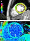
Fig. 1
Fig. 1
A) Parametric overlay of a T1 map on a short-axis slice of left ventricle (yellow ring) in a healthy volunteer. The color scale of yellow in this image corresponds to a mean T1 value of 563 ms. B) A 43-year-old woman with dilated nonischemic cardiomyopathy has diffusely abnormal T1 values throughout the left ventricular myocardium (arrows). The T1 values of the myocardium have a mean value of 174 ms, which corresponds to the green-blue portion of the parametric color scale (seen on the left border of the image).
Fig. 2
Fig. 2
A) Parametric overlay of a T2 map on a short-axis slice of left ventricle (greenish ring) in a patient without myocardial abnormalities. The color scale of green in this image (arrows) corresponds to a normal mean T2 value of 45 ms. B) A 26-year-old man with myocarditis involving the inferolateral wall has normal T2 values throughout the septum and anterior wall but has abnormal T2 values with a mean of 71 ms in the inferolateral wall (arrows). C) The corresponding delayed-enhancement image shows enhancement in the same region of the inferolateral wall (arrows) in a pattern that is compatible with myocarditis.
Fig. 3
Fig. 3
Short-axis cine steady-state free-precession dataset obtained A) with standard breath-hold technique and B) during free breathing in the same patient. Given the comparable contrast and the spatiotemporal resolution of the free-breathing technique, free breathing might be a viable alternative for the evaluation of left ventricular function in patients with impaired breath-holding capacity. Supplemental motion images are available for Figures 3A and 3B.
Fig. 4
Fig. 4
A 22-year-old man with a history of repaired tetralogy of Fallot underwent cardiac magnetic resonance imaging for evaluation and quantification of pulmonary insufficiency. Axial reformatted images from a navigator-gated, electrocardiogram-triggered, free-breathing, 3-dimensional steady-state free-precession volumetric dataset optimally show ostial and proximal courses of A) the left anterior descending coronary artery (double arrows), the left circumflex coronary artery (single arrow), and B) the right coronary artery (arrow).
Fig. 5
Fig. 5
A 24-year-old woman with a history of epidermolysis bullosa and recent stroke underwent cardiac magnetic resonance imaging for evaluation of newly diagnosed systolic dysfunction on echocardiography. A) Short-axis and B) 4-chamber delayed-enhancement images show mild dilation of the left ventricle, moderate (predominantly mid-myocardial) enhancement in the septum, and predominantly epicardial enhancement in the anterior, lateral, and inferior walls (arrows). The noncoronary distribution and nonischemic pattern of delayed enhancement were consistent with myocarditis as the underlying cause of the patient's systolic dysfunction.
Fig. 6
Fig. 6
A 59-year-old woman with a history of left atrial myxoma resection presented with worsening heart failure shortly after that surgery. Right- and left-sided heart catheterization results were normal. Cine steady-state free-precession images (not shown) showed hypokinesia of the inferior wall. A) Short-axis and B) vertical long-axis delayed-enhancement images show subendocardial enhancement extending variably into the mid-myocardium (arrows) in the inferior wall in a coronary distribution (right coronary artery territory). The most likely cause of the patient's heart failure was an embolic phenomenon in the right coronary artery during the perioperative period, which resulted in ischemic insult.

References

    1. Goenka AH, Flamm SD. Cardiac magnetic resonance imaging for the investigation of cardiovascular disorders. Part 1: current applications. Tex Heart Inst J. 2014;41(1):7–20. - PMC - PubMed
    1. Piechnik SK, Ferreira VM, Dall'Armellina E, Cochlin LE, Greiser A, Neubauer S, Robson MD. Shortened Modified Look-Locker Inversion recovery (ShMOLLI) for clinical myocardial T1-mapping at 1.5 and 3 T within a 9 heartbeat breathhold. J Cardiovasc Magn Reson. 2010;12:69. - PMC - PubMed
    1. Messroghli DR, Walters K, Plein S, Sparrow P, Friedrich MG, Ridgway JP, Sivananthan MU. Myocardial T1 mapping: application to patients with acute and chronic myocardial infarction. Magn Reson Med. 2007;58(1):34–40. - PubMed
    1. Messroghli DR, Radjenovic A, Kozerke S, Higgins DM, Sivananthan MU, Ridgway JP. Modified Look-Locker inversion recovery (MOLLI) for high-resolution T1 mapping of the heart. Magn Reson Med. 2004;52(1):141–6. - PubMed
    1. Goenka AH, Das CJ, Sharma R. Nephrogenic systemic fibrosis: a review of the new conundrum. Natl Med J India. 2009;22(6):302–6. - PubMed

MeSH terms