Review
. 2014 Apr 29:5:166.
doi: 10.3389/fpls.2014.00166. eCollection 2014.

The intriguing plant nuclear lamina

Affiliations
Review

The intriguing plant nuclear lamina

Malgorzata Ciska et al. Front Plant Sci. .

Abstract

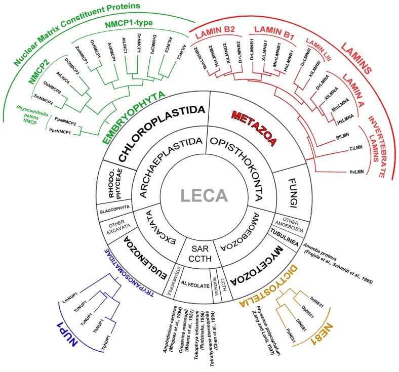
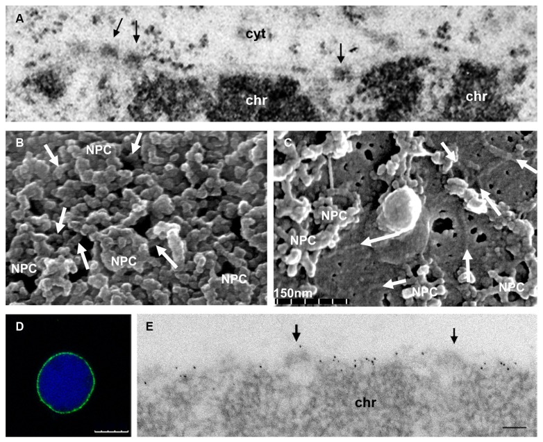
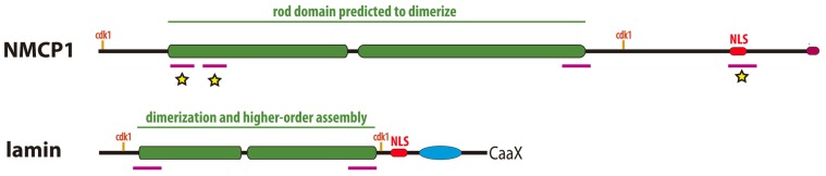
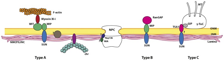
The nuclear lamina is a complex protein mesh attached to the inner nuclear membrane (INM), which is also associated with nuclear pore complexes. It provides mechanical support to the nucleus and nuclear envelope, and as well as facilitating the connection of the nucleoskeleton to the cytoskeleton, it is also involved in chromatin organization, gene regulation, and signaling. In metazoans, the nuclear lamina consists of a polymeric layer of lamins and other interacting proteins responsible for its association with the INM and chromatin. In plants, field emission scanning electron microscopy of nuclei, and thin section transmission electron microscopy of isolated nucleoskeletons, reveals the lamina to have a similar structure to that of metazoans. Moreover, although plants lack lamin genes and the genes encoding most lamin-binding proteins, the main functions of the lamina are fulfilled in plants. Hence, it would appear that the plant lamina is not based on lamins and that other proteins substitute for lamins in plant cells. The nuclear matrix constituent proteins are the best characterized structural proteins in the plant lamina. Although these proteins do not display strong sequence similarity to lamins, their predicted secondary structure and sub-nuclear distribution, as well as their influence on nuclear size and shape, and on heterochromatin organization, suggest they could be functional lamin analogs. In this review we shall summarize what is currently known about the organization and composition of the plant nuclear lamina and its interacting complexes, and we will discuss the activity of this structure in the plant cell and its nucleus.

Keywords: CRWN proteins; LINC proteins; NMCP proteins; Nup136; SUN proteins; plant nuclear envelope; plant nuclear lamina; plant nucleocytoplasmic linkers.


Figures

FIGURE 1
FIGURE 1
Presence of the lamina in Eukaryota and classification and phylogenetic relationships between the lamina proteins in the different groups. The lamina has been described in different Eukaryotic groups: Metazoan, Embryophyta, Dictyostelia, Trypanosomatidae, Alveolate and Tubulinea, although its constituent proteins differ in different groups. The main components of the metazoan lamina are lamins. Most invertebrates express a single lamin while vertebrates contain four genes encoding lamin B1, lamin B2, lamin A, and lamin LIII (lost in mammals; Peter and Stick, 2012). In Dictyostelia the lamina is made up of nuclear envelope 81 (NE81) protein, which is considered an ancestor of lamins (Batsios et al., 2012; Kruger et al., 2012). The lamina was also reported in the nucleus of various Alveolate species: Amphidinium carterae, Gregarina melanopli, Tokophrya infusionum, Tetrahymena thermophila, and in Amoeba proteus (Tubulinea) and Physarum polycephalum (Dictyostelia) that do not contain a gene encoding the NE81 protein. The composition of the lamina in these species is not known. In Trypanosomatidae it is made up of a single nuclear periphery 1 (NUP1) protein (Rout and Field, 2001; Dubois et al., 2012). In Embryophyta the lamina is made up of nuclear matrix constituent proteins (NMCPs; Masuda et al., 1997; Ciska et al., 2013). NMCPs are classified in flowering plants into NMCP1-type proteins and NMCP2. Monocots have one NMCP1 and one NMCP2 proteins while dicots contain one NMCP2 and two or three NMCP1-type proteins. The moss Physcomitrella patens contains two NMCP proteins (Ciska et al., 2013). Selected species for the representation of the NMCP protein family: Allium cepa (Ac), Arabidopsis thaliana (At), Daucus carota (Dc), Oryza sativa (Os), Physcomitrella patens (Ppa), and Zea mays (Zm); NE81 proteins: Dictyostelium discoideum (Dd), Dictyostelium fasciculatum (Df), Dictyostelium purpureum (Dp), and Polysphondylium pallidum (Pp); NUP1 proteins: Trypanosoma brucei (Tb), Trypanosoma gambiense (Tg), Trypanosoma cruzi (Tc), Trypanosoma vivax (Tv), and Leishmania major (Lm); and lamins: Hydra vulgaris (Hv), Ciona intestinalis (Ci), Branchiostoma lanceolatum (Bl), Danio rerio (Dr), Xenopus laevis (Xl), Mus musculus (Mm), and Homo sapiens (Hs). LECA, the last eukaryotic common ancestor; SAR, stramenopile, alveolate, Rhizaria; CCTH, cryptomonads, centrohelids, telonemids, haptophytes.
FIGURE 2
FIGURE 2
Ultrastructure of the plant nuclear lamina and localization of the NMCP proteins. (A) Conventional thin section TEM image of the nuclear periphery of an onion meristematic root cell, showing a portion of the NE with its two membranes, and the dense NPCs (arrows) that traverse it. The heterochromatin (chr) is tightly attached to the INM but the thin lamina is not conspicuous with this technique. Cytoplasm (cyt). (B) Cytoplasmic face of the NE of a tobacco BY-2 cell nucleus extracted with Triton X-100 to remove the membranes and visualized by feSEM. The filaments of the lamina interconnecting the NPCs are evident (arrows). (C) feSEM image of the nucleoplasmic face of the NE of a BY-2 nucleus that has been fractured but not extracted with Triton X-100. Arrows indicate the filaments of the lamina in the membrane. (D,E) Detection of NMCP1 in the lamina of isolated meristematic onion nuclei extracted with Triton X-100 after immunofluorescence and DAPI staining (D), or TEM immunogold labeling (E). After removing the membrane, the lamina with a lower electron density than chromatin and containing NMCP1 proteins is evident at the nuclear periphery. The association with NPCs (arrows) and the tight attachment to the condensed chromatin masses (chr) can be seen. (B,C courtesy of Drs J. Fiserova and M. W. Goldberg). Bars in D = 10 μm and in E = 100 nm.
FIGURE 3
FIGURE 3
Comparison of the structure of plant NMCP proteins and metazoan lamins. Both proteins have a similar tripartite structure with a central coiled–coil domain (green boxes) flanked by a short head and a long tail domains. The rod domain which is responsible for dimerization and higher order assembly in lamins, presents highly conserved regions at both ends (magenta bars) involved in head to tail association of dimers in the case of lamins. The rod domain is flanked by conserved cdk1 phosphorylation sites in both cases. In the tail domain both have a NLS (red boxes) and a conserved C-terminus (magenta box in NMCP1 and CAAX box in lamins). NMCPs lack the Ig fold for partner protein binding typical of lamins (blue oval). The conserved regions marked with a yellow star are involved in NMCP1 association to the nuclear periphery (Kimura et al., 2014).
FIGURE 4
FIGURE 4
Proposed model of the nuclear lamina organization and its main interacting partners in plants. The plant lamina is made up of NMCP proteins and it is attached to the INM of the NE through its interaction with INM proteins not yet identified. The lamina is also attached to NPCs, probably through its interaction with Nup136 and NUA. The factors involved in chromatin association to the lamina in plants remain unknown. The lamina associates to plant-specific nucleocytoplasmic linkers probably by interaction of NMCPs with SUN proteins, which are currently divided into three different types. The type A linker can be considered as a plant LINC complex because it connects the lamina with the actin cytoskeleton. The core organization of this complex is similar to that of type B linkers. The linker element that interacts with the cytoskeleton is myosin Xl-i, which binds to both the perinuclear actin filaments and the WIT protein in the ONM, that in turn interacts with the SUN–WIP bridge (Tamura et al., 2013). The type B linker is formed by SUN proteins anchored to the INM that form a bridge with the WIPs in the ONM, which may in turn complex with WIT proteins in some cases. This type of complex is necessary for RanGAP to associate with the NE (Zhou et al., 2012; Zhou and Meier, 2013). Recently a model for the attachment of γ-TuCs (γ-tubulin complexes) to the NE has been proposed (type C linker). In this complex, the interaction of a small component, GIP (GCP3-interacting protein), with TSA1, an ONM protein that contains a VIPt motif similar to the φ-VPT motif of WIPs, would facilitate an interaction with SUNs (Batzenschlager et al., 2013). Proteins in the different complexes are represented as monomers for simplification. Non-proven interactions are indicated by question marks.

References

    1. Aaronson R. P., Blobel G. (1975). Isolation of nuclear pore complexes in association with a lamina. Proc. Natl. Acad. Sci. U.S.A. 72 1007–1011 10.1073/pnas.72.3.1007 - DOI - PMC - PubMed
    1. Aebi U., Cohn J., Buhle L., Gerace L. (1986). The nuclear lamina is a meshwork of intermediate-type filaments. Nature 323 560–564 10.1038/323560a0 - DOI - PubMed
    1. Al-Haboubi T., Shumaker D. K., Koser J., Wehnert M., Fahrenkrog B. (2011). Distinct association of the nuclear pore protein Nup153 with A- and B-type lamins. Nucleus 2 500–509 10.4161/nucl.2.5.17913 - DOI - PubMed
    1. Batsios P., Peter T., Baumann O., Stick R., Meyer I., Graf R. (2012). A lamin in lower eukaryotes? Nucleus 3 237–243 10.4161/nucl.20149 - DOI - PMC - PubMed
    1. Batzenschlager M., Masoud K., Janski N., Houlne G., Herzog E., Evrard J. L., et al. (2013). The GIP gamma-tubulin complex-associated proteins are involved in nuclear architecture in Arabidopsis thaliana. Front. Plant Sci.1 4:480 10.3389/fpls.2013.00480 - DOI - PMC - PubMed