Review
. 2014 Dec;14(6):380-90.
doi: 10.1136/practneurol-2014-000821. Epub 2014 May 8.

A practical approach to, diagnosis, assessment and management of idiopathic intracranial hypertension

Affiliations
Free PMC article
Review

A practical approach to, diagnosis, assessment and management of idiopathic intracranial hypertension

Susan P Mollan et al. Pract Neurol. 2014 Dec.
Free PMC article

Abstract

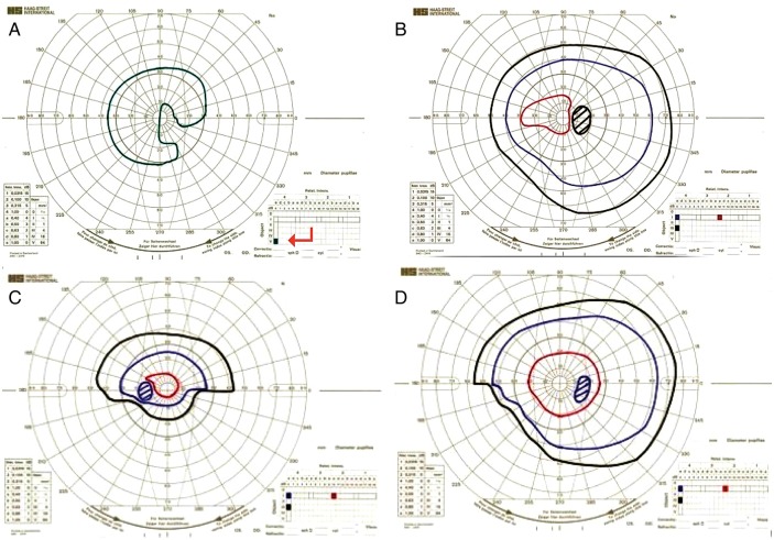
Adult patients who present with papilloedema and symptoms of raised intracranial pressure need urgent multidisciplinary assessment including neuroimaging, to exclude life-threatening causes. Where there is no apparent underlying cause for the raised intracranial pressure, patients are considered to have idiopathic intracranial hypertension (IIH). The incidence of IIH is increasing in line with the global epidemic of obesity. There are controversial issues in its diagnosis and management. This paper gives a practical approach to assessing patients with papilloedema, its investigation and the subsequent management of patients with IIH.

Keywords: HEADACHE; NEUROOPHTHALMOLOGY.


Figures

Figure 1
Figure 1
Single colour fundus photographs of patients with disc swelling secondary to raised intracranial pressure (papilloedema). (A) Mild papilloedema with burring and elevation of nasal disc margin (arrow). (B) Moderate papilloedema with obscuration of vessels by oedematous nerve fibre layer. (C, D, E) Severe papilloedema with cotton wool spots, nerve fibre layer haemorrhage (arrows C and D) and venous engorgement and tortuosity (arrow E). (F) Papilloedema with secondary optic atrophy. Note, as atrophy progresses, fewer nerve fibres can swell.
Figure 2
Figure 2
Single colour fundus photographs of pseudopapilloedema in patients initially thought to have IIH. (A) Elevated, lumpy disc with anomalous vascular pattern including trifurcation of central retinal artery (arrow) seen in optic nerve drusen. (B) Small disc height (arrow) leads to a crowded appearance of the optic nerve without a physiological cup. (C and D) High magnification photographs of right and left eyes of a patient with anomalous discs which show the indistinct nasal disc margin (arrow C) and absent physiological cup (arrow D).
Figure 3
Figure 3
Case one: (A and B) Right and left colour fundus photographs of the optic nerve head showing small crowded discs with anomalous branching of the blood vessels. (C and D) Left and right grey-scale plot taken from the Humphrey visual field test printout, showing he ‘clover leaf’ pattern of an unreliable test performance.
Figure 4
Figure 4
Surface and buried optic nerve head drusen. The disc is elevated and lumpy with visible yellowish deposits (drusen) within the optic nerve head. Note the absence of the physiological cup and anomalous vascular branching (arrow).
Figure 5
Figure 5
Colour fundus photograph of buried drusen. Elevated disc with absent physiological cup (arrow).
Figure 6
Figure 6
B-scan ultrasound of the right optic nerve, showing optic nerve head drusen. Ovoid echogenic foci in optic nerve (arrow).
Figure 7
Figure 7
Fundus fluorescein angiography of the left eye with papilloedema. A rapid series of fundus photographs follow the intravenous injection of a fluorescent contrast agent. In true disc swelling, the frames (A–E) show progressively increased intensity and area of fluorescence at the disc. This shows fluorescein leakage from the oedematous disc.
Figure 8
Figure 8
(A) MRI T1-weighted sagittal image showing a partially empty sella. (B) MRI T2-weighted coronal imaging showing increased fluid in the optic nerve sheath complex bilaterally. (C) MRI T1-weighted axial image showing flattening of the posterior globes, and dilated optic nerve sheaths in patient with raised intracranial pressure.
Figure 9
Figure 9
Case 2—Urgent CSF divergence surgery restored visual function. (A, B) Goldmann visual fields before and (C, D) after lumbar peritoneal shunt (left and right). (A) The left eye has an extensive visual field deficit and visual acuity of 1/60, the field is plotted (in green) only to the largest, brightest target called V4e (arrow). The patient was not able to perceive the smaller/ dimmer targets plotted in red, blue and black in figures 9B-D. (B) The right eye has an enlarged blind spot and reduced sensitivity of the visual field and visual acuity 6/18. (C and D) Left and right eye following the shunt show improved visual field and vision to 6/9 in both eyes.

References

    1. Caballero B. The global epidemic of obesity: an overview. Epidemiol Rev 2007;29:1–5. - PubMed
    1. Corbett JJ, Savino PJ, Thompson HS, et al. Visual loss in pseudotumor cerebri. Follow-up of 57 patients from five to 41 years and a profile of 14 patients with permanent severe visual loss. Arch Neurol 1982;39:461–74. - PubMed
    1. Friedman DI, Liu GT, Digre KB. Revised diagnostic criteria for the pseudotumor cerebri syndrome in adults and children. Neurology 2013;81:1159–65. - PubMed
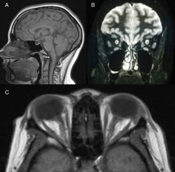
    1. Brodsky MC, Vaphiades M. Magnetic resonance imaging in pseudotumor cerebri. Ophthalmology 1998;105:1686–93. - PubMed
    1. Wright BL, Lai JT, Sinclair AJ. Cerebrospinal fluid and lumbar puncture: a practical review. J Neurol 2012;259:1530–45. - PubMed

Publication types

MeSH terms