. 2014 May 8;10(5):e1004115.
doi: 10.1371/journal.ppat.1004115. eCollection 2014 May.

Phosphorylation of KasB regulates virulence and acid-fastness in Mycobacterium tuberculosis

Affiliations

Phosphorylation of KasB regulates virulence and acid-fastness in Mycobacterium tuberculosis

Catherine Vilchèze et al. PLoS Pathog. .

Abstract

Mycobacterium tuberculosis bacilli display two signature features: acid-fast staining and the capacity to induce long-term latent infections in humans. However, the mechanisms governing these two important processes remain largely unknown. Ser/Thr phosphorylation has recently emerged as an important regulatory mechanism allowing mycobacteria to adapt their cell wall structure/composition in response to their environment. Herein, we evaluated whether phosphorylation of KasB, a crucial mycolic acid biosynthetic enzyme, could modulate acid-fast staining and virulence. Tandem mass spectrometry and site-directed mutagenesis revealed that phosphorylation of KasB occurred at Thr334 and Thr336 both in vitro and in mycobacteria. Isogenic strains of M. tuberculosis with either a deletion of the kasB gene or a kasB_T334D/T336D allele, mimicking constitutive phosphorylation of KasB, were constructed by specialized linkage transduction. Biochemical and structural analyses comparing these mutants to the parental strain revealed that both mutant strains had mycolic acids that were shortened by 4-6 carbon atoms and lacked trans-cyclopropanation. Together, these results suggested that in M. tuberculosis, phosphorylation profoundly decreases the condensing activity of KasB. Structural/modeling analyses reveal that Thr334 and Thr336 are located in the vicinity of the catalytic triad, which indicates that phosphorylation of these amino acids would result in loss of enzyme activity. Importantly, the kasB_T334D/T336D phosphomimetic and deletion alleles, in contrast to the kasB_T334A/T336A phosphoablative allele, completely lost acid-fast staining. Moreover, assessing the virulence of these strains indicated that the KasB phosphomimetic mutant was attenuated in both immunodeficient and immunocompetent mice following aerosol infection. This attenuation was characterized by the absence of lung pathology. Overall, these results highlight for the first time the role of Ser/Thr kinase-dependent KasB phosphorylation in regulating the later stages of mycolic acid elongation, with important consequences in terms of acid-fast staining and pathogenicity.


Conflict of interest statement

The authors have declared that no competing interests exist.

Figures
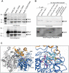
Figure 1
Figure 1. M. tuberculosis KasB is phosphorylated on Thr334 and Thr336. (A) In vitro phosphorylation of KasB with PknF.
The PknF kinase encoded by the Mtb genome was expressed and purified as a GST fusion and incubated with purified His-tagged KasB_WT, KasB_T334A, KasB_T336A and KasB_T334A/T336A in the presence of radiolabeled [γ-33]ATP. Samples were separated by SDS-PAGE, stained with Coomassie Blue and visualized by autoradiography after overnight exposure to a film as indicated. Upper bands reflect the autophosphorylation activity of PknF whereas the lower bands correspond to the phosphorylation signal of KasB. (B) In vivo phosphorylation of KasB. A kasB deletion mutant of M. bovis BCG was transformed with either pVV16_kasB_WT or pVV16_kasB_T334A/T336A and grown in Sauton medium. Exponential (expo) or stationary (stat) phase cells were harvested, lyzed and processed for KasB purification by affinity chromatography on Ni2+-containing beads. The BCG-derived KasB_WT and KasB_T334A/T336A proteins were separated by SDS-PAGE, either stained with Coomassie Blue (upper panel) or subjected to Western blot analysis after probing the membrane with anti-phosphoThreonine antibodies (lower panel). Specificity of phosphothreonine recognition was checked by probing the antibodies against recombinant KasB produced in E. coli strains carrying either pETPhos_kasB (non phosphorylated KasB) or pETDuet_kasB that co-expresses KasB with PknF (phosphorylated KasB). (C) Localization of Thr334 and Thr336 phospho-sites in the three-dimensional structure of KasB. Overall view (left panel) showing the KasB dimeric structure (; PDB entry 2GP6) in ribbon representation with the core domain in marine and the cap domain in orange. The second chain of the dimer is in light gray. The Cys-His-His catalytic triad and the two phospho-sites are displayed as ball-and-stick with carbon atoms in magenta and green, respectively. Also shown with carbon atoms in yellow are the TLM inhibitor and the PEG molecule as observed in the structure of the KasA C171Q acyl enzyme mimic (; PDB entry 2WGG). The PEG molecule is thought to delineate the acyl-binding channel . Nitrogens are in blue, oxygens in red, and sulfur in gold. Close-up view (right panel) after a 45° rotation of the left panel along a vertical axis. Side-chains of residues delineating the active site hydrophobic tunnel and of aspartic residues at positions 334 and 336 were also represented (carbons in cyan and white, respectively).
Figure 2
Figure 2. Construction of isogenic M. tuberculosis CDC1551 strains bearing the phosphoablative or phosphomimetic kasB alleles.
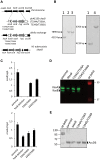
(A) Schematic representation of the specialized transduction phage. A replicating shuttle phasmid derivative of phAE159 containing kasB carrying the mutations T336A/T336A or T334D/T336D, sacB, a hyg resistance cassette, and the first 959 bp of accD6 was used to transduce Mtb CDC1551. If recombination occurs before the point mutation in kasB, this results in recombinant strains carrying the T334A/T336A or T334D/T336D mutations. The transductants were selected on hygromycin and screened by PCR amplification of kasB and presence of the desired mutations was confirmed by sequencing. (B) Southern blot analysis of M. tuberculosis kasB mutant strains. Genomic DNA from each strain was extracted, digested with BglII and analyzed: left panel, Southern for kasB point mutants (probe kasB); right panel, Southern for ΔkasB (probe kasA). The expected size of each band was: wild-type, 4219 bp; kasB point mutants, 7898 bp; ΔkasB, 6764 bp. Lane 1, CDC1551 wild-type strain; lane 2, CDC1551 KasB T334A/T336A; lane 3, CDC1551 KasB T334D/T336D; lane 4, CDC1551 ΔkasB. (C) kasB and cmaA2 expression levels in the different isogenic mutants. Analysis of kasB and cmaA2 mRNA levels of Mtb strains as determined by quantitative RT-PCR. The mean ± standard deviation of three real-time RT-PCR experiments is shown for each strain. The values were normalized to sigA mRNA levels. (D) KasA/KasB immunoblotting. Western blotting showing the expression level of KasB in the crude lysates of the parental strain and the various KasB mutant strains. The membrane was probed with rat anti-KasA antibodies which cross-react with KasB and the proteins revealed using secondary antibodies labeled with IRDye infrared dyes. (E) AccD6 immunoblotting. Western blot analysis showing the expression level of AccD6 in the crude lysates of the parental strain and the various KasB mutant strains. The membrane was probed with rabbit anti-AccD6 antibodies and the incubated with anti-rabbit antibodies conjugated to alkaline phosphatase.
Figure 3
Figure 3. Acid-fast staining of M. tuberculosis kasB isogenic mutant strains.
(A) Cultures were fixed on glass slides and acid-fast staining was performed on the fixed smears using either the BD TB Auramine Kit or the BD Carbolfuchsin kit. The left panels show phase contrast microscopy images of Mtb CDC1551 parental strain and the phosphomimetic and phosphoablative Mtb isogenic strains. (B) Restoration of the acid-fast staining phenotype in the ΔkasB and phosphomimetic KasB mutant strains complemented with pMV261::kasB. Magnification = 100×.
Figure 4
Figure 4. Structural analysis of mycolic acids in the phosphomimetic and phosphoablative kasB mutants. (A) Mycolic acid profile of the various KasB mutants and complemented strains.
Culture were grown at 37°C, harvested, and FAMEs and MAMEs were extracted and analyzed by one-dimensional TLC using hexane/ethyl acetate (19∶1, v/v; 3 runs). α-, methoxy- and keto-mycolic acids were revealed by spraying the plate with molybdophosphoric acid followed by charring. Mycolates migrating slightly faster in the Asp mutants than in the parental control strain can be observed. “-C” indicates complemented strain (with pMV261::kasB). (B) Relative proportions of cis - and trans -cyclopropanes in mycolates established from 1H NMR spectra. The relative quantification of specific signals associated to trans- and cis-cyclopropanes revealed that oxygenated mycolates synthesized by the KasB T334D/T336D and ΔkasB strains exclusively contain cis-cyclopropane rings. The % of trans-cyclopropanes for each mycolic acid sub-species and for each strain is indicated. (C) MALDI-MS analysis. Mass spectrometry analysis revealed that all three families of mycolates isolated from the KasB T334D/T336D and ΔkasB strains display reduced sizes compared to the parental or phosphoablative strains.
Figure 5
Figure 5. Infection of immunocompromised SCID mice with M. tuberculosis kasB isogenic mutants. (A) Survival curves in infected SCID mice.
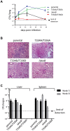
Low-dose aerosol infection of SCID mice (as in B) was performed with the following Mtb strains: parental, KasB_T334A/T336A, KasB_T334D/T336D and ΔkasB. (B) Growth of Mtb kasB strains in the lungs of SCID mice. At 1, 7, 21 and 56 days post-infection, one lung from each infected SCID mouse was harvested, homogenized and serial dilutions were plated on Middlebrook 7H10 supplemented with 10% OADC and 0.2% glycerol. (C) Pathology of lungs from infected SCID mice. One lung from each infected SCID mouse was harvested and fixed in 10% paraformaldehyde for a month prior to photography. (D) CFU plots in the liver and spleen three weeks (black bars) and eight weeks (grey bars) post-infection.
Figure 6
Figure 6. Infection of immunocompetent C57Bl/6 mice with M. tuberculosis kasB isogenic strains. (A) Growth of Mtb kasB strains in the lungs.
Low-dose aerosol infection was performed with the following Mtb strains: parental, KasB_T334A/T336A, KasB_T334D/T336D, and ΔkasB. Lungs from infected mice were harvested at 1, 7, 21 and 57 days post-infection, homogenized and serial dilutions were plated onto Middlebrook 7H10 plates supplemented with 10% OADC and 0.2% glycerol. (B) Pathology slides of lungs from infected mice at 21 days post-infection. Lung tissue sections from mice infected with the Mtb CDC1551 parental, KasB_T334A/T336A, KasB_T334D/T336D, and ΔkasB were stained with hematoxylin/eosin and observed. Magnification = ×20. (C) CFU plots in the liver and spleen three weeks (black bars) and eight weeks (grey bars) post-infection.
Figure 7
Figure 7. Infection of C57BL/6 bone marrow-derived macrophages with M. tuberculosis kasB variants. (A) Growth of the KasB mutants and parental strain in BMDM.
An MOI of 1 was used to infect the BMDM with the strains. Macrophages were lysed after 0, 1, 3 and 6 days post-infection and viable bacteria were counted by plating dilutions of the lysates on agar plates. Three independent experiments were performed and each experiment was done in triplicate. (B) Uptake of KasB strains by BMDM. The ratio between the bacterial titer after a 4 h infection and the inoculum titer is shown. A T-test (two-tailed distribution, unequal variance) was performed and showed a statistical difference between the uptake of the KasB phosphomimetic strain compared to deletion strain (left panel). Complementation with a functional KasB restores the uptake of the phosphomimetic mutant (right panel). Blue bar, parental strain; green bar, KasB_T334A/T336A; black bar ΔkasB, black bar with white stripes, ΔkasB pMV261-kasB; red bar, KasB_T334D/T336D; and red bar with white stripes, KasB_T334D/T336D pMV261-kasB.
Figure 8
Figure 8. Expression of PAT in the M. tuberculosis kasB variants.
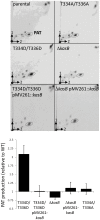
Left panel: autoradiographs of thin layer chromatograms of apolar lipids derived from [1-14C]-propionate labeling in the various Mtb KasB strains and complemented strains carrying pMV261-kasB. Total lipids (6,000 counts) were loaded on TLC plates and developed thrice in petroleum ether/acetone (92∶8, v/v) in the first direction and once in toluene/acetone (95∶5, v/v) in the second direction. Right panel: quantification of the PAT production (corresponding to the circled spots in left panel). Results are expressed in fold increase relative to the parental strain. Results are representative of two independent experiments.
Figure 9
Figure 9. Representation of the in vivo consequences of STPK-dependent phosphorylation of KasB.
Changes in cell wall and mycolic acid composition to various environmental stimuli are central to Mtb adaptation during infection. In response to external cues, STPKs undergo autophosphorylation, which in turn induces phosphorylation of KasB on Thr334 and Thr336. This presumably results in inactivation of KasB activity, thus directly affecting the activity of the elongation 2 FASII complex (E2-FASII) catalyzing the addition of the last carbon atoms required for full-length meromycolic acids. This leads to the production of shorter mycolic acids which is associated to dramatic phenotype changes, such as loss of acid-fastness, decreased cell wall permeability, severe attenuation in infected mice and defect in macrophage colonization.

Similar articles

Cited by

References

    1. Zhou P, Long Q, Zhou Y, Wang H, Xie J (2012) Mycobacterium tuberculosis two-component systems and implications in novel vaccines and drugs. Crit Rev Eukaryot Gene Expr 22: 37–52. - PubMed
    1. Cole ST, Brosch R, Parkhill J, Garnier T, Churcher C, et al. (1998) Deciphering the biology of Mycobacterium tuberculosis from the complete genome sequence. Nature 393: 537–544. - PubMed
    1. Av-Gay Y, Everett M (2000) The eukaryotic-like Ser/Thr protein kinases of Mycobacterium tuberculosis. Trends Microbiol 8: 238–244. - PubMed
    1. Prisic S, Dankwa S, Schwartz D, Chou MF, Locasale JW, et al. (2010) Extensive phosphorylation with overlapping specificity by Mycobacterium tuberculosis serine/threonine protein kinases. Proc Natl Acad Sci U S A 107: 7521–7526. - PMC - PubMed
    1. Chao J, Wong D, Zheng X, Poirier V, Bach H, et al. (2010) Protein kinase and phosphatase signaling in Mycobacterium tuberculosis physiology and pathogenesis. Biochim Biophys Acta 1804: 620–627. - PubMed

Publication types

MeSH terms

Substances