. 2014 Aug 15;23(16):1844-57.
doi: 10.1089/scd.2014.0124. Epub 2014 Jun 12.

Periostin secreted by mesenchymal stem cells supports tendon formation in an ectopic mouse model

Affiliations

Periostin secreted by mesenchymal stem cells supports tendon formation in an ectopic mouse model

Sandra Noack et al. Stem Cells Dev. .

Abstract

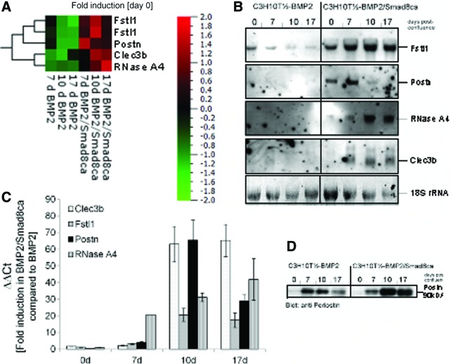
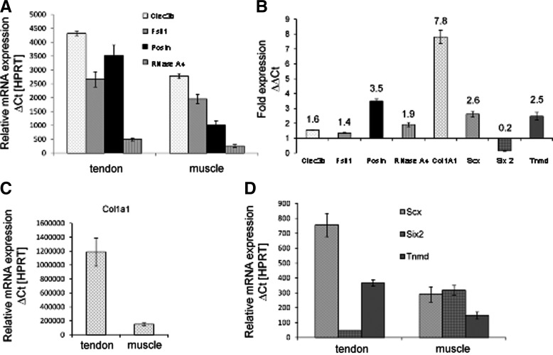
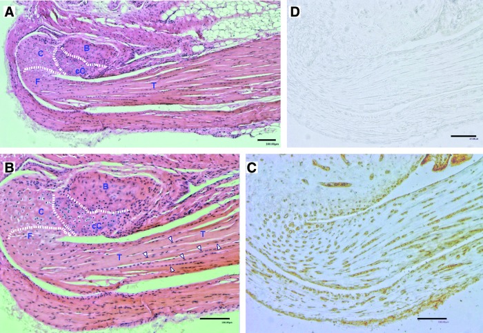
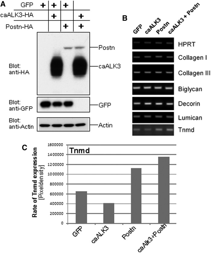
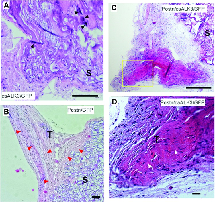
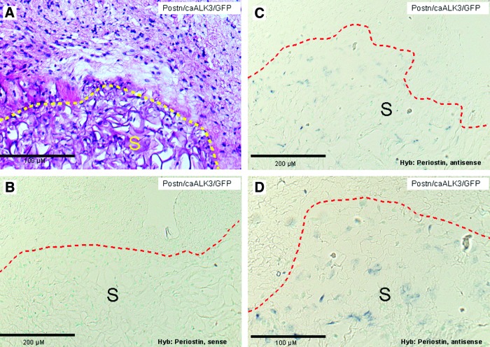
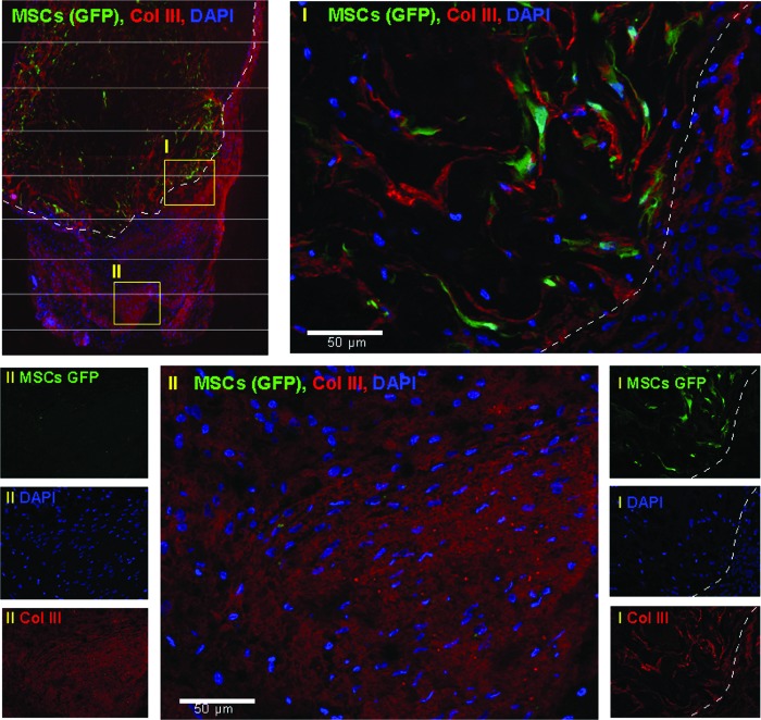
True tendon regeneration in human patients remains a vision of musculoskeletal therapies. In comparison to other mesenchymal lineages the biology of tenogenic differentiation is barely understood. Specifically, easy and efficient protocols are lacking that might enable tendon cell and tissue differentiation based on adult (stem) cell sources. In the murine mesenchymal progenitor cell line C3H10T½, overexpression of the growth factor bone morphogenetic protein 2 (BMP2) and a constitutively active transcription factor, Smad8 L+MH2, mediates tendon cell differentiation in vitro and the formation of tendon-like tissue in vivo. We hypothesized that during this differentiation secreted factors involved in extracellular matrix formation exert a major impact on tendon development. Gene expression analyses revealed four genes encoding secreted factors that are notably upregulated: periostin, C-type lectin domain family 3 (member b), RNase A4, and follistatin-like 1. These factors have not previously been implicated in tendon biology. Among these, periostin showed a specific expression in tenocytes of adult mouse Achilles tendon and in chondrocytes within the nonmineralized fibrocartilage zone of the enthesis with the calcaneus. Overexpression of periostin alone or in combination with constitutively active BMP receptor type in human mesenchymal stem cells and subsequent implantation into ectopic sites in mice demonstrated a reproducible moderate tenogenic capacity that has not been described before. Therefore, periostin may belong to the factors contributing to the development of tenogenic tissue.


Figures

<b>FIG. 1.</b>
FIG. 1.
Identification of secreted factors during tenogenic differentiation in C3H10T½-BMP2/Smad8ca in comparison with nontenogenic C3H10T½-BMP2 cells in a 20,000 clone mouse cDNA microarray. (A) Heat map of microarray data according to the results of the microarray analyses. Color-coded expression levels of differentially expressed genes in BMP2- or BMP2/Smad8 cell lines are shown. Normalization (day 0 of each cell line) was applied to accommodate for cell-line-specific effects. Qlucore Omics Explorer 2.3 Software was used to generate a heat map. High-expressed genes are depicted in red and low-expressed genes in green. For the Fstl1 gene, two different probes were present on the arrays. The results obtained with both probes are comparable. Periostin (Postn): ID 15044; Clec3b: ID 17142; RNase A4: ID 2987; and Fstl1: ID 807, ID 10435. (B) Northern blot analyses of genes shown in (A). RNA from in-vitro-cultivated C3H10T½ cells was blotted and subjected to nonradioactive hybridization as described in “Materials and Methods” section. Staining for 18S-rRNA was used as loading control. (C) The quantitative real-time PCR data from both cell lines at the four time points are presented as ΔΔCt using the BMP2 cell line as reference. Supplementary Figure S1 shows the corresponding ΔCt values (using the housekeeping gene HPRT as a reference). (D) Periostin expression in cultivated C3H10T½ cells as assessed by western blot analyses. Proteins were isolated as described in “Materials and Methods” section, subjected to sodium dodecyl sulfate gel electrophoresis, and blotted. Periostin was detected with an anti-periostin antibody (Acris, Herford, Germany). Periostin shows a higher expression rate in cells undergoing tenogenic differentiation than in nontenogenic mesenchymal progenitor cells C3H10T½-BMP2. BMP, bone morphogenetic protein; Clec3b, C-type lectin domain family 3, member b (previously called tetranectin); Fstl1, follistatin-like 1; HPRT, hypoxanthine phosphoribosyl transferase; PCR, polymerase chain reaction. Color images available online at www.liebertpub.com/scd
<b>FIG. 2.</b>
FIG. 2.
Expression rates of secreted factors upregulated in adult murine tendon and muscle tissue. RNA was isolated from murine thigh muscle and Achilles tendon tissue (mouse age: 6 weeks) as described in “Materials and Methods” section. The primers are indicated in “Materials and Methods” section. (A) Relative mRNA expression (ΔCt values normalized to HPRT) for Clec3b (tetranectin), follistatin-like 1 (Fstl1), periostin (Postn), and RNase A4. (B) Fold expression of the same four genes as well as of collagen 1a1 (Col1a1), scleraxis (Scx), sine oculis-related homeobox 2 (Six2), and tenomodulin (Tnmd) in tendon compared to muscle (ΔΔCt). (C) Relative mRNA expression (ΔCt values normalized to HPRT) for Col1a1, and (D) for Scx, Six2, and Tnmd. Apart from collagen 1a1, periostin is the gene with the most prominent expression in tendon compared to muscle.
<b>FIG. 3.</b>
FIG. 3.
Histology and periostin immunohistology of mouse Achilles tendon and its insertion into the calcaneus. The entire anatomical structure was removed from the animal, fixed with 4% paraformaldehyde overnight at 4°C, and subsequently decalcified with 16.8% EDTA for 7 days and then stored in 70% ethanol. After paraffin embedding, 5-μm sections were cut and parallel sections were stained. (A) HE stain, overview. An enlarged view is shown in (B). (B) Detailed view of tendon approaching the calcaneus. Dotted lines delineate bone from mineralized, calcified cartilage (cC) and from nonmineralized cartilage (C). Some representative tenocytes are indicated by white arrowheads. (C) Periostin staining. Positively stained, round cartilage cells and tenocytes are clearly visible. (D) Negative control without primary antibody. B, bone; cC, calcified cartilage; C, nonmineralized (fibro)cartilage; F, fibrocartilage; T, tendon. HE, hematoxylin and eosin. Color images available online at www.liebertpub.com/scd
<b>FIG. 4.</b>
FIG. 4.
Overexpression of caALK3, periostin, or caALK3 plus periostin in vitro induces tenomodulin transcription but does not result in formation of tenocyte-like cells. Human MSCs were lentivirally modified to overexpress GFP (control), caALK3, periostin, or caALK3 plus periostin. The cells were cultured under conditions favoring tenogenic differentiation (cf. “Materials and Methods” section) for 21 days after obtaining confluence. (A) Western blot analysis demonstrates expression of caALK3 and periostin with an HA antibody since both constructs are C-terminally HA tagged. Analysis was performed at day 0, that is, upon reaching of confluence and start of tenogenic differentiation. (B) Semiquantitative RT-PCR was performed to analyze expression of genes relevant for tenocytes and fibroblasts. Matrix proteins: collagen type I and collagen type III. Proteoglycans: biglycan, decorin, and lumican. Transmembrane protein: tenomodulin (Tnmd). No amplificates were obtained for the transcription factor scleraxis. (C) Rate of Tnmd upregulation by periostin. The periostin-dependent upregulation of Tnmd transcription was assessed by determining the pixel density of the RT-PCR bands of (B). Evaluation was performed with the ImageJ 1.41o software (NIH). caALK3, constitutively active human BMP receptor IA; GFP, green fluorescent protein; MSCs, mesenchymal stromal cells; RT-PCR, reverse transcription–polymerase chain reaction.
<b>FIG. 5.</b>
FIG. 5.
Periostin-dependent development of ordered collagen structures in human MSCs subjected to ectopic transplantations. Human MSCs were modified with lentiviruses encoding periostin, GFP, and caALK3 as described in “Materials and Methods” section. Modified cells were applied on a Duragen sponge and ectopically subcutaneously or intramuscularly implanted in female nude mice (6 weeks of age). After 4 weeks of implantation, mice were sacrificed and implants were explanted and subjected to histological analyses (HE staining). One representative implant of each group is shown in (A–D). (A) Implantation of human MSCs modified by lentiviral expression of caALK3 and GFP. The formation of bony elements may be assessed by the regions of intense HE staining (black arrowheads). (B) Human MSCs overexpressing periostin and GFP. The expression of periostin leads to the formation of ordered tendon-like tissue in the neighborhood of the implanted sponge and is indicated by the red arrowheads. (C) Overview of an implant harboring periostin- and caALK3-modified MSCs and surrounding tissue. The implantation of caALK3/periostin-modified MSCs favors the development of tendon-like tissue. (D) Detailed view of the yellow rectangle from (C). This magnification documents the ordered tendon-like organization of fibroblastic tissue and is pinpointed by white arrowheads. (A–D): scale bar, 100 μm. S, sponge; T, tendon-like tissue organization. Color images available online at www.liebertpub.com/scd
<b>FIG. 6.</b>
FIG. 6.
Nonradioactive in situ hybridizations with periostin-specific sense and antisense probes detect periostin-expressing MSCs predominantly in the implanted collagenous sponge. (A) Implant of lentivirally modified human MSCs expressing periostin, caALK3, and GFP. The border of the implant is indicated by the yellow dashed line. HE staining. (B–D) In situ hybridization with nonradioactively labeled periostin probes. Labeling and hybridization was as described in “Materials and Methods” section. The border of the implanted sponge is indicated with a red dashed line. Counter staining was performed with methyl green. (B) Control hybridization with a periostin-specific sense probe does not result in a hybridization signal. (C, D) Hybridization with a periostin-specific sense probe. Periostin-expressing MSCs are detected in the sponge only. S, sponge. Color images available online at www.liebertpub.com/scd
<b>FIG. 7.</b>
FIG. 7.
Modified MSCs are located in the implanted sponge and the tendon-like tissue surrounding the implant expresses collagen III, an early marker of fibroblastic tendon-like tissue formation. Immunofluorescence analysis was performed as described in “Materials and Methods” section. Red fluorescence: collagen III. Green fluorescence: GFP. Blue fluorescence: 4′,6-diamidino-2-phenylindole (DAPI). Left upper panel: overview of implant and its surrounding tissue. The border of the implant is indicated with a dashed white line. Two yellow rectangles (I, II) indicate regions shown in more detail. Right lower panels: individual immunofluorescence analyses show GFP and collagen III expression and DAPI staining of the implant and its immediate neighborhood (region I). GFP-expressing cells are only found in the implant. Upper right panel: merging of the individual fluorescence analyses (region I) indicates that MSCs remain in the sponge and are not directly involved in tendon differentiation. Left lower panels: individual immunofluorescence analyses do not show GFP-expressing MSCs and indicate collagen III expression and DAPI staining in a tendon-like tissue (region II). Lower middle panel: merging of the individual fluorescence analyses (region II). Tenogenic collagen III is detected in tendon-like tissue. The HE staining of this region is seen in Fig. 5C. Color images available online at www.liebertpub.com/scd

Similar articles

Cited by

References

    1. Schweitzer R, Zelzer E. and Volk T. (2010). Connecting muscles to tendons: tendons and musculoskeletal development in flies and vertebrates. Development 137:2807–2817 - PMC - PubMed
    1. Favata M, Beredjiklian PK, Zgonis MH, Beason DP, Crombleholme TM, Jawad AF. and Soslowsky LJ. (2006). Regenerative properties of fetal sheep tendon are not adversely affected by transplantation into an adult environment. J Orthop Res 24:2124–2132 - PubMed
    1. Hoffmann A. and Gross G. (2009). Innovative strategies for treatment of soft tissue injuries in human and animal athletes. Med Sport Sci 54:150–165 - PubMed
    1. Chen X, Song XH, Yin Z, Zou XH, Wang LL, Hu H, Cao T, Zheng M. and Ouyang HW. (2009). Stepwise differentiation of human embryonic stem cells promotes tendon regeneration by secreting fetal tendon matrix and differentiation factors. Stem Cells 27:1276–1287 - PubMed
    1. Hoffmann A, Pelled G, Turgeman G, Eberle P, Zilberman Y, Shinar H, Keinan-Adamsky K, Winkel A, Shahab S, et al. (2006). Neotendon formation induced by manipulation of the Smad8 signalling pathway in mesenchymal stem cells. J Clin Invest 116:940–952 - PMC - PubMed

Publication types

MeSH terms

LinkOut - more resources