. 2014 May 8;10(5):e1004359.
doi: 10.1371/journal.pgen.1004359. eCollection 2014 May.

A dominant-negative mutation of mouse Lmx1b causes glaucoma and is semi-lethal via LDB1-mediated dimerization [corrected]

Affiliations

A dominant-negative mutation of mouse Lmx1b causes glaucoma and is semi-lethal via LDB1-mediated dimerization [corrected]

Sally H Cross et al. PLoS Genet. .

Erratum in

  • PLoS Genet. 2014 Dec;10(12):e1004917

Abstract

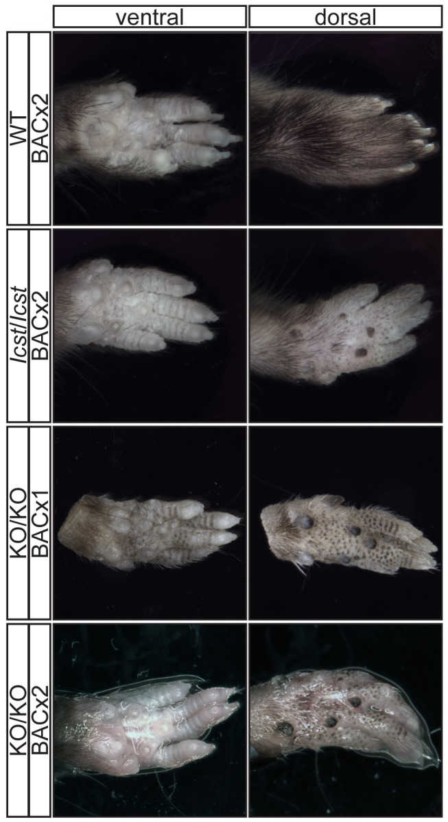
Mutations in the LIM-homeodomain transcription factor LMX1B cause nail-patella syndrome, an autosomal dominant pleiotrophic human disorder in which nail, patella and elbow dysplasia is associated with other skeletal abnormalities and variably nephropathy and glaucoma. It is thought to be a haploinsufficient disorder. Studies in the mouse have shown that during development Lmx1b controls limb dorsal-ventral patterning and is also required for kidney and eye development, midbrain-hindbrain boundary establishment and the specification of specific neuronal subtypes. Mice completely deficient for Lmx1b die at birth. In contrast to the situation in humans, heterozygous null mice do not have a mutant phenotype. Here we report a novel mouse mutant Icst, an N-ethyl-N-nitrosourea-induced missense substitution, V265D, in the homeodomain of LMX1B that abolishes DNA binding and thereby the ability to transactivate other genes. Although the homozygous phenotypic consequences of Icst and the null allele of Lmx1b are the same, heterozygous Icst elicits a phenotype whilst the null allele does not. Heterozygous Icst causes glaucomatous eye defects and is semi-lethal, probably due to kidney failure. We show that the null phenotype is rescued more effectively by an Lmx1b transgene than is Icst. Co-immunoprecipitation experiments show that both wild-type and Icst LMX1B are found in complexes with LIM domain binding protein 1 (LDB1), resulting in lower levels of functional LMX1B in Icst heterozygotes than null heterozygotes. We conclude that Icst is a dominant-negative allele of Lmx1b. These findings indicate a reassessment of whether nail-patella syndrome is always haploinsufficient. Furthermore, Icst is a rare example of a model of human glaucoma caused by mutation of the same gene in humans and mice.


Conflict of interest statement

The authors have declared that no competing interests exist.

Figures

Figure 1
Figure 1. Icst is a homeodomain missense mutation of Lmx1b that abolishes protein function.
(A) Genomic DNA sequence traces from Lmx1b exon 5 from wild-type (WT), heterozygous mutant (Icst/+) and homozygous mutant (Icst/Icst) embryonic samples. The position of the T-to-A transversion at position 725 (725T>A) in the Lmx1b gene, numbering from Genbank entry AF078166, is highlighted. This transversion causes a V265D mutation in the homeodomain numbering from entry O88609 in http://www.uniprot.org. (B) The Icst mutation impairs binding of LMX1B. Bandshift analysis using a fixed amount of 32P-labelled FLAT LMX1B binding element with increasing amounts (as indicated by the triangles) of His-tagged fusion proteins containing the homeodomain or full-length LMX1B either wild-type (WT) or containing the Icst mutation (ICST). In lane 1 no protein was added (-). Complexes as indicated by the arrows were formed with the wild-type proteins but not the proteins containing the Icst mutation. The position of the probe alone is also indicated by an arrow. (C) The Icst mutation abolishes transcriptional activity. A luciferase reporter plasmid, 6×FLAT-C2P-Luc4, containing six copies of the LMX1B binding element FLAT upstream of the Col2a1 promoter (, kind gift of Brendan Lee), was either transfected alone (-) or co-transfected with empty pcDNA-3.1 vector (V) or pcDNA-3.1 containing the wild-type (WT-L) or Icst (Icst-L) long form of LMX1B or wild-type (WT-S) or Icst (Icst-S) short form of LMX1B. Each experiment was performed in triplicate, and mean firefly luciferase expression normalized to control renilla luciferase expression is shown+standard error. Both WT-L and WT-S elicit robust induction of luciferase expression that reduced to background levels by the Icst mutation (t-test; * = P<0.005).
Figure 2
Figure 2. Icst causes defects in the anterior segment of the eye.
(A) Lmx1bKO/+ (KO/+) eye that is of normal appearance. (B) Lmx1bIcst/+ (Icst/+) eye where there is a fine strand between the pupillary margin and the cornea (arrowed). (C) This Lmx1bIcst/+ eye is enlarged with severe corneal vascularisation and central opacity. (D–F). Histological appearance of corneas. (D) Wild-type normal cornea (WT). (E) Lmx1bIcst/+ cornea that has stromal vascularisation (barbed arrows). Descemet's membrane is wrinkled (dashed arrows) and a membrane is present on the anterior surface of the iris (arrowheads). (F) Lmx1bIcst/+ cornea with basal epithelial oedema (dashed arrow). A stromal blood vessel with red blood cells is indicated by a barbed arrow. (G) Normal wild-type iridocorneal angle. (H–J) Iridocorneal angle morphology of Lmx1bIcst heterozygotes display a spectrum of phenotypes ranging from open-angle (H) to closed-angle morphology where the iris can be seen adhering to the cornea and blocking the iridocorneal angle as indicated by bars (I, J). (K–L) Higher magnification of wild-type (K) and Lmx1bIcst/+ (L) iridocorneal angles. (K) The wild-type has an open-angle with robust drainage structures ([). Schlemm's canal (arrowheads) is clearly identifiable and the trabecular meshwork (asterisks) has well-formed trabecular beams. (L) The Lmx1bIcst/+ iridocorneal angle is open but narrow. Iridocorneal strands (IS) are present. These are a normal feature of the mouse angle but are not present at all locations and do not prevent aqueous humor drainage. The drainage structures are not as robust as in the WT ([). Schlemm's canal is present but compressed (arrowheads) and the trabecular meshwork is hypoplastic (asterisks). Original magnification ×40 (D and F) and ×20 (E). Scale bar = 50 µm.
Figure 3
Figure 3. The IOP is elevated in Lmx1bIcst/+ mice.
(A) Box plot of IOP measurements of wild-type (WT) and Lmx1bIcst/+ (Het) mice taken at different ages. There is significantly elevated IOP in the Lmx1bIcst/+ mice up to 6 months (P = 0.006 at 2–3 months and P = 0.009 at 5–6 months). The IOP measurements are not significantly different in older mice owing to low readings of <10 mmHg in increasing numbers of the Lmx1bIcst/+ mice. (B) Frequency histogram summarizing the data from panel A showing increased incidence of high IOP measurements in Lmx1bIcst/+ mice.
Figure 4
Figure 4. Icst causes defects of the optic nerve and the ganglion cell layer of the retina.
(A) Sections through the optic nerve of adult mice. The optic nerve is variably affected in Lmx1bIcst/+ mice. Wild-type (WT) is normal (upper panel, left). In some Lmx1bIcst/+ (Icst/+) mice the optic nerve appears normal (lower panel, left) but in others there is evidence of optic nerve damage and cupping, indicated by asterisks (upper and lower panels, right). Scale bar = 50 µm. (B) Box plot of ganglion cell number present in Lmx1bIcst/+ (Icst/+), Lmx1b KO/+ (KO/+) and wild-type (WT) mice. Ganglion cells were stained using an anti-BRN3 antibody and the number present in each of four areas from around the optic disc from one eye were counted (n = 3 for Lmx1bIcst/+ and n = 2 for the other two genotypes). Compared to wild-type and Lmx1b KO/+ mice which show no difference (P = 0.95) the number of ganglion cells is greatly reduced in the Lmx1bIcst /+ mice compared to wild-type (P = 0.017) and to Lmx1b KO/+ mice (P = 0.034). (C) Optic nerve damage in Lmx1bIcst/+ mice. Optic nerve damage was assessed in wild-type (WT) and Lmx1bIcst /+ (Icst/+) mice at 8 months (WT, n = 23 and Icst/+, n = 16) and at 10–11 months (WT, n = 26 and Icst/+, n = 36). Nerves with no glaucoma have axon counts that match controls (green bar). Nerves with moderate nerve damage have an average of 30% axon (yellow bar). This degree of damage occurs in a low percentage of wild-type of this strain background at this age . All severely affected nerves have extensive axon damage throughout the optic nerve with obvious axon loss ranging from 50% to almost complete axon loss (red bar).
Figure 5
Figure 5. Ultrastructural analysis of E17.5 and E18.5 kidneys shows that Icst causes glomerular defects when heterozygous.
(A–B) Wild-type (WT) and Lmx1bKO/+ (KO/+) podocytes are normal with normal foot process formation (arrowheads). (C–D) Lmx1bKO/KO (KO/KO) and Lmx1bIcst/Icst (Icst/Icst) podocytes fail to form foot processes and appear immature. (E–L) Lmx1bIcst heterozygous podocytes (Icst/+). (E) Normal foot process formation (arrowheads). (F) The bar indicates an area of the GBM that appears fragmented. (G–H) Foot processes (arrowheads) abut GBM that is split (arrows). (I) The podocytes appear cuboidal (as indicated by asterisks) and immature. (J) The GBM is split as indicated by the bar and foot processes are rudimentary (arrows). (K) There is a large split in the GBM that is filled with fibrillar material (as indicated by an asterisk). (L) Podocytes are positioned flush against a split GBM. P, podocyte; endo = endothelial cell. Scale bar = 2 µm.
Figure 6
Figure 6. Forepaw phenotype of transgenic rescue mice.
Pictures of the ventral and dorsal sides of forepaws from wild-type (WT), Lmx1bIcst /Icst (Icst/Icst) and Lmx1bKO/KO (KO/KO) mice are shown and whether the BAC transgene is hemizygous or homozygous is indicated (BACx1 or BACx2). The ventral side of the paw is normal for all the rescue mice. The dorsal surface of all the homozygous mutant paws appears ventralised with pigmented footpads and no hair. The ages of the mice shown are as follows. Top two rows, P14; third row P26 and bottom row P35.
Figure 7
Figure 7. Forepaw skeletal phenotype of transgenic rescue mice.
µCT scans of the ventral and dorsal sides from wild-type (WT), Lmx1bIcst /Icst (Icst/Icst) and Lmx1bKO/KO (KO/KO) forepaws are shown and whether the BAC transgene is hemizygous or homozygous is indicated (BACx1 or BACx2). The ventral side of the skeleton appears normal for all the rescue mice (top panels). The dorsal surface appears completely ventralised for the Lmx1bIcst /Icst rescue mice homozygous for the transgene and for the Lmx1bKO/KO rescue hemizygous for the BAC transgene. Arrows point to sesamoid bones that are a feature of the ventral surface. In the case of the Lmx1bKO/KO homozygous for the BAC transgene the dorsal surface appears more normal although there are still aspects of ventral morphology seen, for example a sesamoid bone (arrowed). The images shown are from mice between four and five weeks of age except for Lmx1bKO/KO hemizygous for the BAC transgene which was P26.
Figure 8
Figure 8. Patella and scapula phenotype of transgenic rescue mice.
All mice shown are homozygous for the transgene. µCT scans of the patella and scapula are shown. (A) Lmx1bIcst /+ (Icst/+) with normal patella (arrowed). (B) Lmx1bIcst /Icst rescue (Icst/Icst) knee where the patella is absent (arrow indicates its expected location). (C) Lmx1bKO/KO rescue (KO/KO) knee where the patella is present (arrowed). (D) Normal scapula in Lmx1bIcst /+ (arrowed). (E) The scapula is very underdeveloped and small in Lmx1bIcst /Icst rescue (arrowed). (F) The scapula appears more normal in Lmx1bKO/KO rescue (arrowed). The ages of the mice shown are as follows; P23 (A, B, D, E), three months (C) and five weeks (F).
Figure 9
Figure 9. Wild-type and Icst LMX1B are in a complex with LDB1.
Western blot analysis of co-immunoprecipitation experiments using anti-Myc (α-Myc), anti-FLAG (α-FLAG) and anti-LDB1 (α-LDB1) antibodies. (A) Protein lysates (INPUT) of HEK 293T cells transfected with WT-Myc and ICST-FLAG as indicated were immunoprecipitated using anti-c-Myc antibody coupled to agarose and the bound fraction (BOUND) eluted. WT-Myc is present in the immunoprecipitated bound fraction but Icst-FLAG is not. (B) Protein lysates (INPUT) of HEK 293T cells transfected with pcDNA-LDB1 along with WT-Myc and ICST-FLAG as indicated were immunoprecipitated using anti-c-Myc agarose and the bound fraction (BOUND) eluted. Both LDB1 and ICST-FLAG are in a complex with the WT-Myc and found in the immunoprecipitated bound fraction.

Similar articles

Cited by

  • AP-2β is required for formation of the murine trabecular meshwork and Schlemm's canal.
    Akula M, Taiyab A, Deschamps P, Yee S, Ball AK, Williams T, West-Mays JA. Akula M, et al. Exp Eye Res. 2020 Jun;195:108042. doi: 10.1016/j.exer.2020.108042. Epub 2020 Apr 27. Exp Eye Res. 2020. PMID: 32353428 Free PMC article.
  • A multiethnic genome-wide association study of primary open-angle glaucoma identifies novel risk loci.
    Choquet H, Paylakhi S, Kneeland SC, Thai KK, Hoffmann TJ, Yin J, Kvale MN, Banda Y, Tolman NG, Williams PA, Schaefer C, Melles RB, Risch N, John SWM, Nair KS, Jorgenson E. Choquet H, et al. Nat Commun. 2018 Jun 11;9(1):2278. doi: 10.1038/s41467-018-04555-4. Nat Commun. 2018. PMID: 29891935 Free PMC article.
  • Analysis combining correlated glaucoma traits identifies five new risk loci for open-angle glaucoma.
    Gharahkhani P, Burdon KP, Cooke Bailey JN, Hewitt AW, Law MH, Pasquale LR, Kang JH, Haines JL, Souzeau E, Zhou T, Siggs OM, Landers J, Awadalla M, Sharma S, Mills RA, Ridge B, Lynn D, Casson R, Graham SL, Goldberg I, White A, Healey PR, Grigg J, Lawlor M, Mitchell P, Ruddle J, Coote M, Walland M, Best S, Vincent A, Gale J, RadfordSmith G, Whiteman DC, Montgomery GW, Martin NG, Mackey DA, Wiggs JL, MacGregor S, Craig JE; NEIGHBORHOOD consortium. Gharahkhani P, et al. Sci Rep. 2018 Feb 15;8(1):3124. doi: 10.1038/s41598-018-20435-9. Sci Rep. 2018. PMID: 29449654 Free PMC article.
  • Consensus Recommendation for Mouse Models of Ocular Hypertension to Study Aqueous Humor Outflow and Its Mechanisms.
    McDowell CM, Kizhatil K, Elliott MH, Overby DR, van Batenburg-Sherwood J, Millar JC, Kuehn MH, Zode G, Acott TS, Anderson MG, Bhattacharya SK, Bertrand JA, Borras T, Bovenkamp DE, Cheng L, Danias J, De Ieso ML, Du Y, Faralli JA, Fuchshofer R, Ganapathy PS, Gong H, Herberg S, Hernandez H, Humphries P, John SWM, Kaufman PL, Keller KE, Kelley MJ, Kelly RA, Krizaj D, Kumar A, Leonard BC, Lieberman RL, Liton P, Liu Y, Liu KC, Lopez NN, Mao W, Mavlyutov T, McDonnell F, McLellan GJ, Mzyk P, Nartey A, Pasquale LR, Patel GC, Pattabiraman PP, Peters DM, Raghunathan V, Rao PV, Rayana N, Raychaudhuri U, Reina-Torres E, Ren R, Rhee D, Chowdhury UR, Samples JR, Samples EG, Sharif N, Schuman JS, Sheffield VC, Stevenson CH, Soundararajan A, Subramanian P, Sugali CK, Sun Y, Toris CB, Torrejon KY, Vahabikashi A, Vranka JA, Wang T, Willoughby CE, Xin C, Yun H, Zhang HF, Fautsch MP, Tamm ER, Clark AF, Ethier CR, Stamer WD. McDowell CM, et al. Invest Ophthalmol Vis Sci. 2022 Feb 1;63(2):12. doi: 10.1167/iovs.63.2.12. Invest Ophthalmol Vis Sci. 2022. PMID: 35129590 Free PMC article. Review.
  • A long noncoding RNA cluster-based genomic locus maintains proper development and visual function.
    Wang F, Ren D, Liang X, Ke S, Zhang B, Hu B, Song X, Wang X. Wang F, et al. Nucleic Acids Res. 2019 Jul 9;47(12):6315-6329. doi: 10.1093/nar/gkz444. Nucleic Acids Res. 2019. PMID: 31127312 Free PMC article.

References

    1. Sweeney E, Fryer A, Mountford R, Green A, McIntosh I (2003) Nail patella syndrome: a review of the phenotype aided by developmental biology. J Med Genet 40: 153–162. - PMC - PubMed
    1. Mimiwati Z, Mackey DA, Craig JE, Mackinnon JR, Rait JL, et al. (2006) Nail-patella syndrome and its association with glaucoma: a review of eight families. Br J Ophthalmol 90: 1505–1509. - PMC - PubMed
    1. Dreyer SD, Zhou G, Baldini A, Winterpacht A, Zabel B, et al. (1998) Mutations in LMX1B cause abnormal skeletal patterning and renal dysplasia in nail patella syndrome. Nat Genet 19: 47–50. - PubMed
    1. Vollrath D, Jaramillo-Babb VL, Clough MV, McIntosh I, Scott KM, et al. (1998) Loss-of-function mutations in the LIM-homeodomain gene, LMX1B, in nail-patella syndrome. Hum Mol Genet 7: 1091–1098. - PubMed
    1. McIntosh I, Dreyer SD, Clough MV, Dunston JA, Eyaid W, et al. (1998) Mutation analysis of LMX1B gene in nail-patella syndrome patients. Am J Hum Genet 63: 1651–1658. - PMC - PubMed

Publication types

Substances