Comparison of radiological and clinical features of temporal lobe necrosis in nasopharyngeal carcinoma patients treated with 2D radiotherapy or intensity-modulated radiotherapy
- PMID: 24809780
- PMCID: PMC4037838
- DOI: 10.1038/bjc.2014.243
Comparison of radiological and clinical features of temporal lobe necrosis in nasopharyngeal carcinoma patients treated with 2D radiotherapy or intensity-modulated radiotherapy
Abstract
Background: To compare the imaging and clinical features of temporal lobe necrosis (TLN) in nasopharyngeal carcinoma (NPC) patients treated with two-dimensional radiotherapy (2D-RT) or those with intensity-modulated radiotherapy (IMRT).
Methods: We retrospectively analysed NPC patients who underwent 2D-RT (72 patients, 128 temporal lobes) or IMRT (36 patients, 50 lobes) and developed radiation-induced, MRI-confirmed TLN.
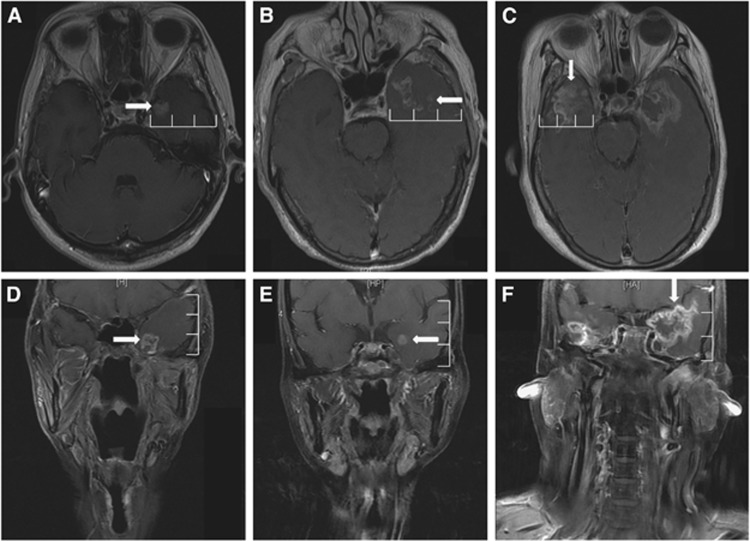
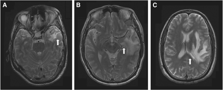
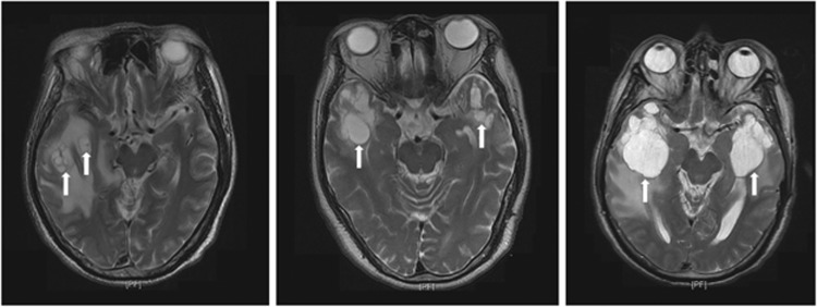
Results: White-matter lesions (WMLs), contrast-enhanced lesions, cysts and local mass effects were present in 128 out of 128 vs 48 out of 50 (P=0.078), 123 out of 128 vs 47 out of 50 (P=0.688), 10 out of 128 vs 1 out of 50 (P=0.185) and 57 out of 128 vs 13 out of 50 (P=0.023) temporal lobes, respectively, in the 2D-RT and IMRT groups. The WMLs were more extensive in the 2D-RT group (P<0.001). The maximum diameter of contrast-enhanced lesions was greater in the 2D-RT group (P<0.001), and these lesions tended to extend far away from the nasopharynx. The WMLs and enhancement had no impact on cyst development (both P=1). Local mass effects were always accompanied with contrast-enhanced lesions (P=0.024) but were not correlated with WMLs or cysts (P=0.523 and 0.341, respectively). There were no between-group differences in clinical features (all P-values>0.05), whereas the difference in the incidence of severe debility was of marginal significance (18.1% vs 5.6%, P=0.077).
Conclusions: The IMRT-induced TLN was less extensive and milder than 2D-RT-induced TLN, but both had similar clinical features.
Figures


Similar articles
-
Radiation induced temporal lobe necrosis in nasopharyngeal cancer patients after radical external beam radiotherapy.Radiat Oncol. 2020 May 15;15(1):112. doi: 10.1186/s13014-020-01560-0. Radiat Oncol. 2020. PMID: 32414378 Free PMC article. Review.
-
Analysis of dosimetric factors associated with temporal lobe necrosis (TLN) in patients with nasopharyngeal carcinoma (NPC) after intensity modulated radiotherapy.Radiat Oncol. 2013 Jan 22;8:17. doi: 10.1186/1748-717X-8-17. Radiat Oncol. 2013. PMID: 23336282 Free PMC article.
-
Tumor-Infiltrating T Cell Receptor-Beta Repertoires are Linked to the Risk of Late Chemoradiation-Induced Temporal Lobe Necrosis in Locally Advanced Nasopharyngeal Carcinoma.Int J Radiat Oncol Biol Phys. 2019 May 1;104(1):165-176. doi: 10.1016/j.ijrobp.2019.01.002. Epub 2019 Jan 14. Int J Radiat Oncol Biol Phys. 2019. PMID: 30654091
-
Effect of dosimetric factors on occurrence and volume of temporal lobe necrosis following intensity modulated radiation therapy for nasopharyngeal carcinoma: a case-control study.Int J Radiat Oncol Biol Phys. 2014 Oct 1;90(2):261-9. doi: 10.1016/j.ijrobp.2014.05.036. Epub 2014 Jul 24. Int J Radiat Oncol Biol Phys. 2014. PMID: 25066214
-
Dosimetric parameters predict radiation-induced temporal lobe necrosis in nasopharyngeal carcinoma patients: A systematic review and meta-analysis.Radiother Oncol. 2024 Jun;195:110258. doi: 10.1016/j.radonc.2024.110258. Epub 2024 Mar 26. Radiother Oncol. 2024. PMID: 38537680
Cited by
-
Evolution of radiation-induced temporal lobe injury after intensity-modulated radiation therapy in nasopharyngeal carcinoma: a large cohort retrospective study.Radiat Oncol. 2024 Jan 19;19(1):9. doi: 10.1186/s13014-024-02400-1. Radiat Oncol. 2024. PMID: 38243277 Free PMC article.
-
Radiation induced temporal lobe necrosis in nasopharyngeal cancer patients after radical external beam radiotherapy.Radiat Oncol. 2020 May 15;15(1):112. doi: 10.1186/s13014-020-01560-0. Radiat Oncol. 2020. PMID: 32414378 Free PMC article. Review.
-
Temporal lobe microstructural abnormalities in patients with nasopharyngeal carcinoma quantitatively evaluated by high-resolution DWI and DKI after concurrent chemoradiotherapy.Clin Transl Radiat Oncol. 2019 Dec 25;21:36-43. doi: 10.1016/j.ctro.2019.12.003. eCollection 2020 Mar. Clin Transl Radiat Oncol. 2019. PMID: 31970294 Free PMC article.
-
A retrospective study comparing the outcomes and toxicities of intensity-modulated radiotherapy versus two-dimensional conventional radiotherapy for the treatment of children and adolescent nasopharyngeal carcinoma.J Cancer Res Clin Oncol. 2017 Aug;143(8):1563-1572. doi: 10.1007/s00432-017-2401-y. Epub 2017 Mar 25. J Cancer Res Clin Oncol. 2017. PMID: 28342002 Free PMC article.
-
Altered properties of brain white matter structural networks in patients with nasopharyngeal carcinoma after radiotherapy.Brain Imaging Behav. 2020 Dec;14(6):2745-2761. doi: 10.1007/s11682-019-00224-2. Brain Imaging Behav. 2020. PMID: 31900892
References
-
- Butler CR, Zeman AZ. Recent insights into the impairment of memory in epilepsy: transient epileptic amnesia, accelerated long-term forgetting and remote memory impairment. Brain. 2008;131 (Pt 9:2243–2263. - PubMed
-
- Coderre JA, Morris GM, Micca PL, Hopewell JW, Verhagen I, Kleiboer BJ, van der Kogel AJ. Late effects of radiation on the central nervous system: role of vascular endothelial damage and glial stem cell survival. Radiat Res. 2006;166 (3:495–503. - PubMed
-
- Edge SB, Byrd DR, Compton CC, Fritz AG, Greene FL, Trotti A, American Joint Committee on Cancer 2010AJCC Cancer Staging Manual7th edn.Springer: New York
-
- Hsiao KY, Yeh SA, Chang CC, Tsai PC, Wu JM, Gau JS. Cognitive function before and after intensity-modulated radiation therapy in patients with nasopharyngeal carcinoma: a prospective study. Int J Radiat Oncol Biol Phys. 2010;77 (3:722–726. - PubMed
Publication types
MeSH terms
LinkOut - more resources
Full Text Sources
Other Literature Sources
Miscellaneous