. 2014 Jul 15;391(2):241-50.
doi: 10.1016/j.ydbio.2014.04.017. Epub 2014 May 6.

Sema3A maintains corneal avascularity during development by inhibiting Vegf induced angioblast migration

Affiliations

Sema3A maintains corneal avascularity during development by inhibiting Vegf induced angioblast migration

Chelsey C McKenna et al. Dev Biol. .

Abstract

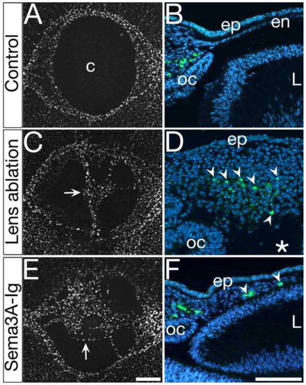
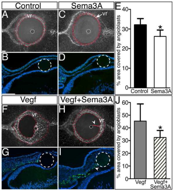
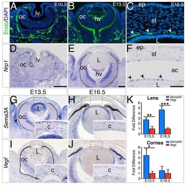
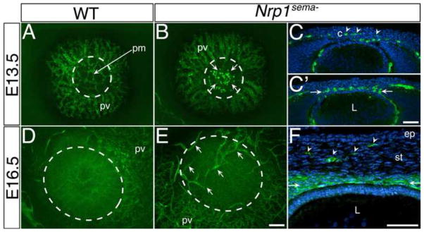
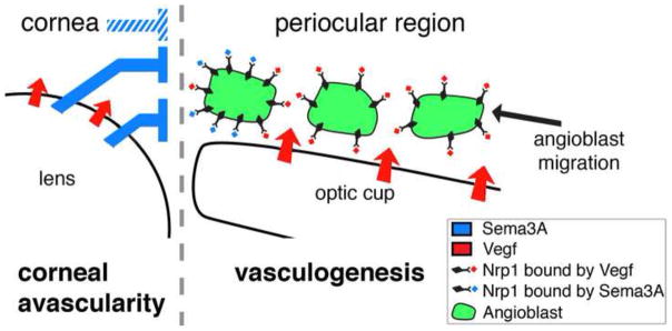
Corneal avascularity is important for optical clarity and normal vision. However, the molecular mechanisms that prevent angioblast migration and vascularization of the developing cornea are not clear. Previously we showed that periocular angioblasts and forming ocular blood vessels avoid the presumptive cornea despite dynamic ingression of neural crest cells. In the current study, we investigate the role of Semaphorin3A (Sema3A), a cell guidance chemorepellent, on angioblast migration and corneal avascularity during development. We show that Sema3A, Vegf, and Nrp1 are expressed in the anterior eye during cornea development. Sema3A mRNA transcripts are expressed at significantly higher levels than Vegf in the lens that is positioned adjacent to the presumptive cornea. Blockade of Sema3A signaling via lens removal or injection of a synthetic Sema3A inhibitor causes ectopic migration of angioblasts into the cornea and results in its subsequent vascularization. In addition, using bead implantation, we demonstrate that exogenous Sema3A protein inhibits Vegf-induced vascularization of the cornea. In agreement with these findings, loss of Sema/Nrp1 signaling in Nrp1(Sema-) mutant mice results in ectopic angioblasts and vascularization of the embryonic mouse corneas. Altogether, our results reveal Sema3A signaling as an important cue during the establishment of corneal avascularity in both chick and mouse embryos. Our study introduces cornea development as a new model for studying the mechanisms involved in vascular patterning during embryogenesis and it also provides new insights into therapeutic potential for Sema3A in neovascular diseases.

Keywords: Angioblast; Cornea; Eye development; Nrp1; Sema3A; Vasculogenesis; Vegf.


Figures

Figure 1
Figure 1. Expression of Vegf, Sema3A, and Nrp1 during vasculogenesis of the avian anterior eye
Transverse sections through the anterior eye at E3 showing the localization of (A) Vegf in the lens and optic cup, and (B) Sema3A in the lens. (C) Quantification of Sema3A and Vegf mRNA levels in the lens at E3, E5, and E7 by qRT-PCR. Each time point is an average of triplicate reactions and expressed as fold change relative to Vegf. Student’s t-test, *P < 0.05 and **P < 0.01; error bars indicate s.d. (D and E) GFP-positive angioblasts and primitive blood vessels in the periocular region express Nrp1. Insets in D and E are higher magnifications of outlined regions. (F and G) Binding of Sema3A with Tg(tie1:H2B:eYFP) endothelial cells in vitro. Cells cultured (G) with media containing AP-Sema3A show a dark precipitate indicating binding of Sema3A, which is absent in (F) cells cultured in control media. Abbreviations: ec, ectoderm; en, endothelium; ep, epithelium; L, lens; oc; optic cup; ret, retina; vr, vascular ring; ir, iris. Scale bars: A, B 100 μm; D, E 100 μm; D, E (insets) 50 μm F, G 200 μm.
Figure 2
Figure 2. Removal of lens-derived Sema3A causes ectopic angioblast migration into the cornea
Wholemount (A, C, E) and transverse sections (B, D, F) of E5 Tg(tie1:H2B:eYFP) quail corneas showing localization of GFP-positive angioblasts. (A and B) Controls showing absence of angioblasts in the presumptive cornea. (C and D) Lensless, and (E and F) Sema3A-Ig peptide injected eyes showing ectopic angioblasts (arrows and arrowheads) in the presumptive cornea. Asterisk in D indicates absence of lens. Abbreviations: c, presumptive cornea; en, endothelium; ep, epithelium; L, lens; oc; optic cup. Scale bars: A, C, E, 200 μm; B, D, F, 100 μm.
Figure 3
Figure 3. Sema3A inhibits Vegf-induced angioblast migration into the cornea
Wholemount and transverse sections of (A–D) E5, and (E–F) E6 Tg(tie1:H2B:eYFP) eyes showing GFP-positive angioblasts. No ectopic angioblasts where observed in corneas after implantation of beads soaked in (A and B) Ringer’s solution or (C and D) Sema3A. Implantation of beads soaked in Vegf induced angioblast migration into the cornea (F and G), which was inhibited when beads were soaked in Vegf + Sema3A (H). In some cases angioblasts avoided the cornea but migrated in the anterior chamber towards the bead (I). Dotted white circles indicate location of the beads. (E and J) quantification of the area covered by angioblasts demarcated with red dotted lines: (E) Control (black bar, n = 4) against Sema3A (white bar, n = 4), and (J) Vegf (grey bar, n = 4) against Vegf + Sema3A (stripped bar, n = 4). Student’s t-test, *P < 0.05; error bars indicate s.d. Abbreviations: vr, vascular ring. Scale bars: ACFH, 100 μm; BDGI, 100 μm.
Figure 4
Figure 4. Ocular vasculogenesis during mouse eye development and the expression of Nrp1, Sema3A, and Vegf
Cross sections through E10.5 (A, D), E13.5 (B, E), and E16.5 (C, F) eyes. Immunostaining with endomucin (green) to show the localization of angioblasts and primitive vasculature (A–C). Section in situ hybridization showing expression patterns of Nrp1 (D–F). Arrowheads in C and F indicate the pupillary membrane. Expression patterns of Sema3A (G and H), and Vegf (I and J). Quantification of Sema3A and Vegf mRNA levels in the mouse lens and cornea at E13.5 and E16.5 by qRT-PCR (K). Each time point is an average of triplicate reactions and expressed as fold change relative to Vegf. Student’s t-test, *P < 0.05, **P < 0.01 and ***P < 0.001; error bars indicate s.d. Abbreviations: lpt, lens pit; oc, optic cup; c, presumptive cornea; L, lens; hv, hyaloid vasculature; ep, epithelium; st, stroma; ac, anterior chamber. Scale bars: AD and insets, 100 μm; BEGHIJ, 100 μm; CF, 50 μm.
Figure 5
Figure 5. Ectopic angioblast migration and vascularization of Nrp1Sema- mutant corneas
Wholemount images of wild type (A, D) and Nrp1sema- (B, E) anterior eyes, and sections through Nrp1sema- eyes (C, C′, F) showing localization of endomucin-positive (green) angioblasts and vasculature. Ectopic angioblasts and blood vessels are respectively indicated by arrows in the wholemount corneas (B and E), and by the arrowheads in (C and F). Arrows in C′ and F indicate the pupillary membrane. White dotted circles demarcates the cornea. Abbreviations: c, presumptive cornea; pv, periocular vasculature; pm, pupillary membrane; L, lens; ep, epithelium; st, stroma. Scale bars: ABDE, 100 μm; CC′F, 50 μm.
Figure 6
Figure 6. Model depicting the regulation of angioblast migration in the anterior eye by Sema3A and Vegf via Nrp1
Schematic of a cross section through the anterior eye showing the localization of Sema3A and Vegf in the anterior eye and the response to these cues by Nrp1-positive angioblasts. Angioblasts migrate to the periocular region in response to Vegf signaling from the optic cup. The lens secrets a higher concentration of Sema3A than Vegf into the region of the developing cornea. In the mouse, the presumptive cornea may also secrete Sema3A (inhibitory sign with white hatch lines). Therefore angioblasts encounter strong Sema3A repulsion at the interface between the cornea and periocular region that prevents their migration into the presumptive cornea. These unequal concentrations of Sema3A and Vegf signaling in the anterior eye allow for the formation of the limbal vasculature in the periocular region while maintaining avascularity in the presumptive cornea.

Similar articles

Cited by

References

    1. Abramoff MD, Magalhães PJ, Ram SJ. Image processing with ImageJ. Biophotonics Int. 2004;11:36–42.
    1. Amano S, Rohan R, Kuroki M, Tolentino M, Adamis AP. Requirement for vascular endothelial growth factor in wound- and inflammation-related corneal neovascularization. Invest Ophthalmol Vis Sci. 1998;39:18–22. - PubMed
    1. Ambati BK, Nozaki M, Singh N, Takeda A, Jani PD, Suthar T, Albuquerque RJC, Richter E, Sakurai E, Newcomb MT, et al. Corneal avascularity is due to soluble VEGF receptor-1. Nature. 2006;443:993–997. - PMC - PubMed
    1. Ash J, Overbeek P. Lens-specific VEGF-A expression induces angioblast migration and proliferation and stimulates angiogenic remodeling. Dev Biol. 2000;223:383–398. - PubMed
    1. Bagri A, Tessier-Lavigne M, Watts RJ. Neuropilins in tumor biology. Clin Cancer Res Off J Am Assoc Cancer Res. 2009;15:1860–1864. - PubMed

Publication types

MeSH terms

LinkOut - more resources