. 2014 May 8;5(5):e1219.
doi: 10.1038/cddis.2014.193.

Cab45S inhibits the ER stress-induced IRE1-JNK pathway and apoptosis via GRP78/BiP

Affiliations

Cab45S inhibits the ER stress-induced IRE1-JNK pathway and apoptosis via GRP78/BiP

L Chen et al. Cell Death Dis. .

Abstract

Disturbance of endoplasmic reticulum (ER) homeostasis causes ER stress and leads to activation of the unfolded protein response, which reduces the stress and promotes cell survival at the early stage of stress, or triggers cell death and apoptosis when homeostasis is not restored under prolonged ER stress. Here, we report that Cab45S, a member of the CREC family, inhibits ER stress-induced apoptosis. Depletion of Cab45S increases inositol-requiring kinase 1 (IRE1) activity, thus producing more spliced forms of X-box-binding protein 1 mRNA at the early stage of stress and leads to phosphorylation of c-Jun N-terminal kinase, which finally induces apoptosis. Furthermore, we find that Cab45S specifically interacts with 78-kDa glucose-regulated protein/immunoglobulin heavy chain binding protein (GRP78/BiP) on its nucleotide-binding domain. Cab45S enhances GRP78/BiP protein level and stabilizes the interaction of GRP78/BiP with IRE1 to inhibit ER stress-induced IRE1 activation and apoptosis. Together, Cab45S, a novel regulator of GRP78/BiP, suppresses ER stress-induced IRE1 activation and apoptosis by binding to and elevating GRP78/BiP, and has a role in the inhibition of ER stress-induced apoptosis.


Figures

Figure 1
Figure 1
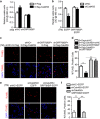
Cab45S promotes cell survival and protects cells from drug-induced apoptosis. (a and b) MTS assay of viable HeLa cells overexpressing 3 × Flag or 3 × Flag-Cab45S treated with different TG (a) or TM (b) concentrations for 48 h (n=3). (c and d) MTS assay of viable HeLa cells overexpressing scrambled shRNA (shNC) or Cab45S shRNA (shCab45S) treated with different concentrations of TG (c) or TM (d) for 48 h (n=3). (e) Representative photomicrographs from TUNEL assay of apoptotic HeLa cells transfected with vectors expressing 3 × Flag and 3 × Flag-Cab45S, treated with TG (2 μM) or TM (2 μg/ml) for 48 h. Scale bar, 100 μm. (f) Quantification of TUNEL-positive cells as in e (n=3; >100 cells per experiment). (g) Western blots of Cab45S from stable HeLa cell lines with Cab45S knockdown. Numbers represent different cell lines. (h) Representative photomicrographs from TUNEL assay of apoptotic cells labeled in Cab45S-knockdown HeLa cells treated with TG (2 μM) or TM (2 μg/ml) for 24 h. Scale bar, 100 μm. (i) Quantification of TUNEL-positive cells as in h (n=3; >100 cells per experiment). (j) Western blots of procaspase-3 and cleaved caspase-3 in Cab45S-knockdown HeLa cells after treatment with TM for the indicated times. For ad, data are presented as mean±S.E.M. *P<0.05, ***P<0.001, as determined by two-way ANOVA. For f and i, data are presented as mean±S.E.M. ***P<0.001, determined by unpaired two-tailed Student's t-test
Figure 2
Figure 2
The IRE1-JNK signaling is required for activating ER stress-induced apoptosis in Cab45S-depleted cells. (a) Quantitative real-time PCR of relative mRNA expression levels of XBP1S in control (shNC, scrambled shRNA) and Cab45S-knockdown HeLa cell lines treated with TM (2 μg/ml) for the indicated periods (n=3). (b) Western blots of CHOP, IRE1 and p-IRE1 in control and Cab45S-knockdown HeLa cell lines after treatment with TM (2 μg/ml) at the indicated time points. (c) Quantification of p-IRE1 in b. GAPDH was used as a loading control. (d) MTS assay of viable HeLa cells transfected with vectors expressing shNC, shCab45S and shNC, shCab45S and shPERK, and shCab45S and shIRE1 with TM (2 μg/ml) treatment (n=3). (e) Representative photomicrographs of apoptotic cells in transfected HeLa cells in which Cab45S or both Cab45S and IRE1 were depleted with TM (2 μg/ml, 48 h) treatment (TUNEL assay). Scale bar, 100 μm. (f) Quantification of TUNEL-positive cells as in e (n=3; >100 cells per experiment). (g) Western blots of cleaved caspase-3 and p-JNK in control and Cab45S-knockdown HeLa cell lines after treatment with TM (2 μg/ml) at the indicated time points. (h) Western blots of cleaved caspase-3 and p-JNK in control and Cab45S-knockdown HeLa cell lines after 24 h treatment with indicated drugs. TM, 2 μg/ml; sp600125, 20 μM. (i) Quantitative real-time PCR of relative mRNA expression levels of BAX in control and Cab45S-knockdown HeLa cell lines treated with TM (2 μg/ml) for the indicated periods (n=3). For a, c, d, f and i, data are presented as mean±S.E.M. *P<0.05, **P<0.01, ***P<0.001, as determined by unpaired two-tailed Student's t-test
Figure 3
Figure 3
Cab45S interacts with the nucleotide-binding domain (NBD) of GRP78/BiP. (a) Immunoprecipitation assay (IP) with anti-Flag antibody in HEK293T cell lysates expressing 3 × Flag-Cab45S. Immunoprecipitates were subjected to SDS-PAGE and then MS analysis. (b) Extracts of HEK293T cells co-transfected with GRP78/BiP-EGFP and 3 × Flag, 3 × Flag-Cab45S, 3 × Flag-Cab45G or 3 × Flag-RCN1 were immunoprecipitated using anti-Flag antibody. The immunoprecipitates were immunoblotted with anti-Flag or anti-GFP antibody. (c and d) Mapping the domain at which GRP78/BiP interacted with Cab45S. Schematics of GRP78/BiP truncates (c). Extracts of HEK293T cells overexpressing GFP-tagged GRP78/BiP truncates and 3 × Flag-Cab45S were immunoprecipitated with anti-GFP antibody, and the immunoprecipitates were immunoblotted with anti-GFP or anti-Flag antibody (d). SBD, substrate-binding domain. (e and f) Mapping the domain of Cab45S, which interacted with GRP78/BiP. Schematics of Cab45S truncates (e). Extracts of HEK293T cells overexpressing 3 × Flag-tagged Cab45S truncates and GRP78/BiP-EGFP were immunoprecipitated with anti-Flag antibody, and the immunoprecipitates were immunoblotted with anti-GFP and anti-Flag antibodies (f). Asterisks indicate 3 × Flag-tagged Cab45S truncates immunoprecipitated by anti-Flag antibody. SP, signal peptide; EFh, EF-hand
Figure 4
Figure 4
Cab45S increases the GRP78/BiP protein level. (a and d) Western blots of GRP78/BiP and two other ER molecular chaperones, PDI and calnexin, in stable Cab45S-knockdown (a) or Cab45S-overexpressed (d) PANC-1 cell lines. Numbers represent different cell lines. (b and c) Western blots (b) and quantification (c) of GRP78/BiP in Cab45S-knockdown and control (shNC, scrambled shRNA) HeLa cells treated with TM (2 μg/ml) for the indicated periods. GAPDH was used as a loading control. (e) Quantitative real-time PCR of the relative GRP78/BiP mRNA expression levels in Cab45S-knockdown and control HeLa cells treated with TM for the indicated times (n=3). (f) Western blots of GRP78/BiP in Cab45S-knockdown and control HeLa cells treated with TM (2 μg/ml, 4 h) followed by cycloheximide (Chx; 100 μM) for the indicated periods. For c and e, data are presented as mean±S.E.M. **P<0.01, ***P<0.001, as determined by unpaired two-tailed Student's t-test
Figure 5
Figure 5
Cab45S inhibits ER stress-induced apoptosis via GRP78/BiP. (a) MTS assay of viable HeLa cells transfected with vectors expressing 3 × Flag and shNC (scrambled shRNA), 3 × Flag-Cab45S and shNC, 3 × Flag and shGRP78/BiP, or 3 × Flag-Cab45S and shGRP78/BiP treated with TM (2 μg/ml, 48 h; n=3). (b) MTS assay of viable HeLa cells transfected with vectors expressing shNC and EGFP, shCab45S and EGFP, shNC and GRP78/BiP-EGFP, or shCab45S and GRP78/BiP-EGFP treated with TM (2 μg/ml, 48 h; n=3). (c) Representative photomicrographs from TUNEL assay of apoptotic HeLa cells transfected with vectors expressing 3 × Flag and shNC, 3 × Flag-Cab45S and shNC, 3 × Flag and shGRP78/BiP, or 3 × Flag-Cab45S and shGRP78/BiP treated with TM (2 μg/ml, 48 h). Scale bar, 100 μm. (d) Quantification of TUNEL-positive cells as in c (n=3; >100 cells per experiment). (e) Representative photomicrographs from TUNEL assay of apoptotic HeLa cells transfected with vectors expressing shNC and EGFP, shCab45S and EGFP, or shCab45S and GRP78/BiP-EGFP treated with TM (2 μg/ml, 48 h). Scale bar, 100 μm. (f) Quantification of TUNEL-positive cells as in e (n=3; >100 cells per experiment). For a, b, d and f, data are presented as mean±S.E.M. **P<0.01, ***P<0.001, as determined by unpaired two-tailed Student's t-test
Figure 6
Figure 6
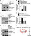
Cab45S inhibits IRE1 activation via GRP78/BiP. (a and b) Western blots (a) and quantification (b) of p-IRE1 in HeLa cells transfected with vectors expressing 3 × Flag and shNC (scrambled shRNA), 3 × Flag-Cab45S and shNC, 3 × Flag and shGRP78/BiP, or 3 × Flag-Cab45S and shGRP78/BiP after TM treatment (2 μg/ml, 48 h). GAPDH was used as a loading control. (c and d) Western blots (c) and quantification (d) of p-IRE1 in HeLa cells expressing shNC and EGFP, shCab45S and EGFP, shNC and GRP78/BiP-EGFP, or shCab45S and GRP78/BiP-EGFP after TM treatment (2 μg/ml, 48 h). GAPDH was used as a loading control. (e) Extracts of HEK293T cells overexpressing the indicated vectors treated with TM (1 μg/ml, 24 h) were immunoprecipitated with anti-Flag antibody. The immunoprecipitates were immunoblotted with anti-Flag, anti-EGFP or anti-Cab45S antibody. (f) Working model of the mechanism by which Cab45S controls ER stress-induced apoptosis. Cab45S interacts with the NBD of GRP78/BiP, which prevents its disassociation from IRE1 and increases the protein level of GRP78/BiP. These effects lead to inhibition of the IRE1-JNK pathway and ER stress-induced apoptosis. For b and d, data are presented as mean±S.E.M. **P<0.01, ***P<0.001, as determined by unpaired two-tailed Student's t-test

Similar articles

Cited by

References

    1. Anelli T, Sitia R. Protein quality control in the early secretory pathway. EMBO J. 2008;27:315–327. - PMC - PubMed
    1. Pizzo P, Pozzan T. Mitochondria-endoplasmic reticulum choreography: structure and signaling dynamics. Trends Cell Biol. 2007;17:511–517. - PubMed
    1. Kim I, Xu W, Reed JC. Cell death and endoplasmic reticulum stress: disease relevance and therapeutic opportunities. Nat Rev Drug Discov. 2008;7:1013–1030. - PubMed
    1. Ron D, Walter P. Signal integration in the endoplasmic reticulum unfolded protein response. Nat Rev Mol Cell Biol. 2007;8:519–529. - PubMed
    1. Hetz C. The unfolded protein response: controlling cell fate decisions under ER stress and beyond. Nat Rev Mol Cell Biol. 2012;13:89–102. - PubMed

Publication types

MeSH terms