. 2014 Jul;24(7):796-808.
doi: 10.1038/cr.2014.64. Epub 2014 May 9.

Redefining the modular organization of the core Mediator complex

Affiliations

Redefining the modular organization of the core Mediator complex

Xuejuan Wang et al. Cell Res. 2014 Jul.

Abstract

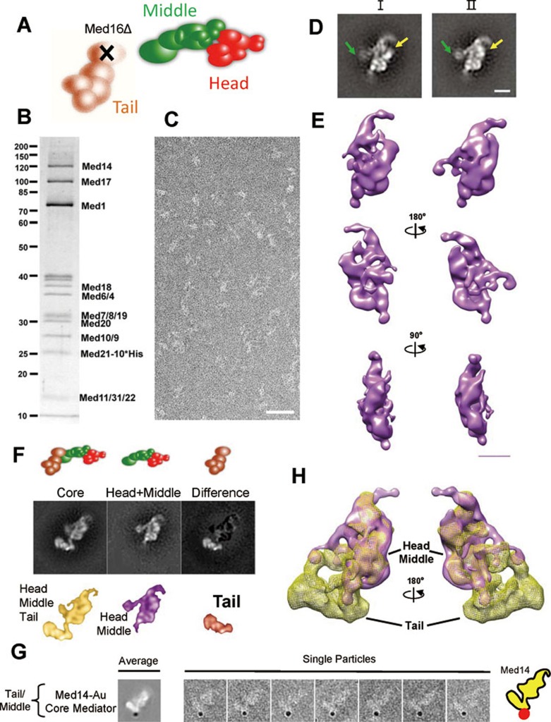
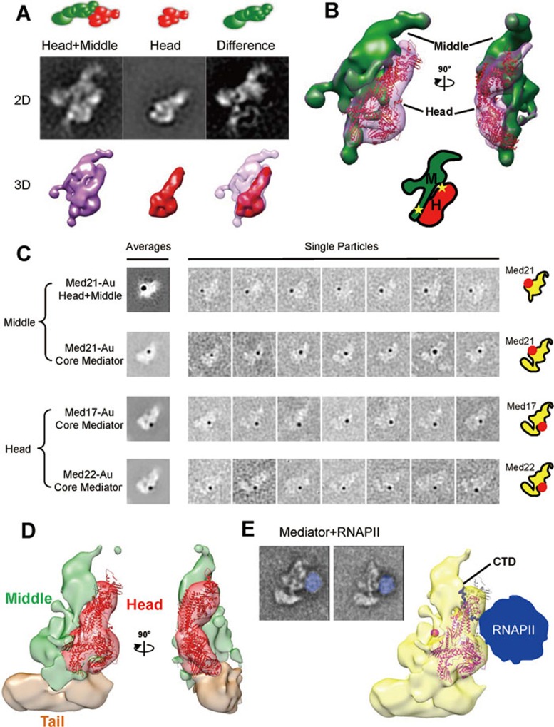
The Mediator complex plays an essential role in the regulation of eukaryotic transcription. The Saccharomyces cerevisiae core Mediator comprises 21 subunits, which are organized into Head, Middle and Tail modules. Previously, the Head module was assigned to a distinct dense domain at the base, and the Middle and Tail modules were identified to form a tight structure above the Head module, which apparently contradicted findings from many biochemical and functional studies. Here, we compared the structures of the core Mediator and its subcomplexes, especially the first 3D structure of the Head + Middle modules, which permitted an unambiguous assignment of the three modules. Furthermore, nanogold labeling pinpointing four Mediator subunits from different modules conclusively validated the modular assignment, in which the Head and Middle modules fold back on one another and form the upper portion of the core Mediator, while the Tail module forms a distinct dense domain at the base. The new modular model of the core Mediator has reconciled the previous inconsistencies between the structurally and functionally defined Mediator modules. Collectively, these analyses completely redefine the modular organization of the core Mediator, which allow us to integrate the structural and functional information into a coherent mechanism for the Mediator's modularity and regulation in transcription initiation.


Figures

Figure 1
Figure 1
Structure of the core Mediator complex. (A) Schematic view of subunit and modular organization of the core Mediator from S. cerevisiae. (B) SDS-PAGE analysis of the native core Mediator purified from S. cerevisiae. The purifications were separated by SDS-PAGE, followed by Ruby staining. (C) A typical micrograph of the well-preserved particles under negative staining. Scale bar, 50 nm. (D) Two-dimensional (2D) EM analysis of the core Mediator. Three different conformations were identified through reference-free alignment and classification of EM images, showing variability in the position of a dense domain at the base. Scale bar, 100 Å. (E) Three-dimensional (3D) reconstructions of the core Mediator in three conformations. Scale bar, 100 Å. (F) Different views of the 3D reconstructions of the core Mediator in closed conformation. (G) Comparison of the 2D structures of the core Mediator complex from S. pombe and S. cerevisiae and difference mapping establishes that the dense domain at the base of S. cerevisiae Mediator most likely corresponds to the Tail module, which is absent in S. pombe Mediator. Contour plots calculated from 2D structures of the core Mediators from S. pombe and S. cerevisiae are color-coded to highlight structural conservation and the approximate boundary of the Tail module.
Figure 2
Figure 2
Structure of the Head + Middle modules and boundary of the Tail module. (A) Schematic view of obtaining the Tail-less core Mediator by deletion of the med16 gene. (B) SDS-PAGE analysis of the native Head + Middle modules. (C) A typical micrograph of the Head + Middle particles preserved under negative staining. Scale bar, 50 nm. (D) Representative 2D structures of the Head + Middle modules. The yellow and green arrows indicate the regions showing high degree of structural flexibility. Scale bar, 100 Å. (E) Different views of the 3D reconstructions of the Head + Middle modules in two different conformations. Scale bar, 100 Å. (F) Comparison of the structures of the core Mediator and the Head + Middle modules and difference mapping establishes that the dense domain at the base of the core Mediator corresponds to the Tail module. Contour plots calculated from 2D structures are color-coded to highlight the modular correspondence and the boundary of the Tail module. (G) Localization of the C terminus of the Med14 subunit revealed by Ni-NTA-Nanogold labeling. Single particles of nanogold-labeled core Mediator complex (middle), a corresponding class average (left), and a diagram representing the localization of the Med14 C terminus (right) are shown. (H) Fitting of the structure of the Head + Middle modules (semitransparent purple surface) into the core Mediator structure (semitransparent yellow mesh). Comparison of the structures of the core Mediator and the Head + Middle modules combined with the nanogold labeling of the Med14 subunit firmly establishes the boundary of the Tail module.
Figure 3
Figure 3
Boundaries of the Head and Middle modules. (A) Comparison of the structures of the Head + Middle modules and the Head module, combined with difference mapping, establishes that the Middle module corresponds to the extended domain at the left and the Head module forms the right portion of the structure. (B) Two views of the 3D reconstructions of the Head + Middle modules unambiguously fitted into the crystal structural model of the Head module (PDBID: 4GWP). The additional density related to the Middle module is represented as a green solid surface. A diagram (inset) revealing the boundary and connectors (yellow stars) between the Head and Middle modules is shown. (C) Localization of Mediator subunits from the Middle and Head modules revealed by using Ni-NTA nanogold probes against the His10 tag incorporated into the C-termini of Med21 (Middle), Med17 (Head) and Med22 (Head). Single particles of the nanogold-labeled core Mediator (middle), corresponding class averages (left) and diagrams representing the localization of the C terminus of each subunit (right) are shown. (D) Fitting the crystal structural model of the Head module (PDBID: 4GWP) into the core Mediator structure (semitransparent colors according to the module segmentation). Comparison of the structures of the core Mediator and module assemblages, combined with the nanogold labeling of several subunits of different modules, completely redefines the modular organization of the core Mediator. (E) Representative class averages of the core Mediator interacting with RNAPII (left) and a figure representing the interaction modes of Mediator with RNAPII (right). The CTD of RNAPII deduced from the crystal structure (PDBID: 4GWQ) is highlighted as the blue sphere. The X-ray structures of the four Srb subunits (Med17/Srb4, Med18/Srb5, Med20/Srb2, and Med22/Srb6) in the Head module are shown in purple ribbon, and the only Srb subunit in the Middle module (Med21/Srb7) is represented by a purple ball.
Figure 4
Figure 4
Redefined modular organization of the core Mediator complex and its functional implications. (A) Modular organization of the yeast Mediator complex. Previously defined modular architecture (left),,, which was solely based on comparing low-resolution EM structures of the core Mediator in RNAPII-associated and free forms. The redefined modular organization (right) is derived from detailed comparison of the structures of the core Mediator and module assemblages, combined with the nanogold labeling of several subunits of different modules. The inserted histogram lists the theoretical MW of each module and the calculated MW obtained from the number of voxels enclosed by each segmented density from both the previous and redefined modular model. (B) Multipartite interactions between Mediator and RNAPII and general transcription factors. The redefined Mediator modular structure fitted with the X-ray structure of the Head module (left, PDBID: 4GWQ) and the surface representation of the RNAPII transcription PIC model (right),. The figure of the PIC model is adapted from a previous study, and the position of TFIIH is inferred from structural information on the human PIC. The documented regions of the Mediator Head module that could be responsible for interacting with RNAPII (CTD,, Rpb3 subunit, and Rpb4/7 sub-complex,) and general transcription factors (TBP, and TFIIH) are labeled with ovals in different colors. The three putative interaction surfaces with Mediator on the PIC are centered on RNAPII (CTD,, Rpb3 subunit, and Rpb4/7 sub-complex,), TBP,, TFIIB, and TFIIH (the Rad3 subunit), which are highlighted with red dashed ovals.

Comment in

  • Mediator redefines itself.
    Sennett NC, Taatjes DJ. Sennett NC, et al. Cell Res. 2014 Jul;24(7):775-6. doi: 10.1038/cr.2014.76. Epub 2014 Jun 10. Cell Res. 2014. PMID: 24913192 Free PMC article.

References

    1. Lee TI, Young RA. Transcription of eukaryotic protein-coding genes. Annu Rev Genet. 2000;34:77–137. - PubMed
    1. Conaway RC, Conaway JW. Function and regulation of the Mediator complex. Curr Opin Genet Dev. 2011;21:225–230. - PMC - PubMed
    1. Kornberg RD. Mediator and the mechanism of transcriptional activation. Trends Biochem Sci. 2005;30:235–239. - PubMed
    1. Naar AM, Lemon BD, Tjian R. Transcriptional coactivator complexes. Annu Rev Biochem. 2001;70:475–501. - PubMed
    1. Malik S, Roeder RG. Transcriptional regulation through Mediator-like coactivators in yeast and metazoan cells. Trends Biochem Sci. 2000;25:277–283. - PubMed

Publication types

MeSH terms

Substances

LinkOut - more resources