. 2014 May 9:4:4914.
doi: 10.1038/srep04914.

Berberine exposure triggers developmental effects on planarian regeneration

Affiliations

Berberine exposure triggers developmental effects on planarian regeneration

Linda Balestrini et al. Sci Rep. .

Abstract

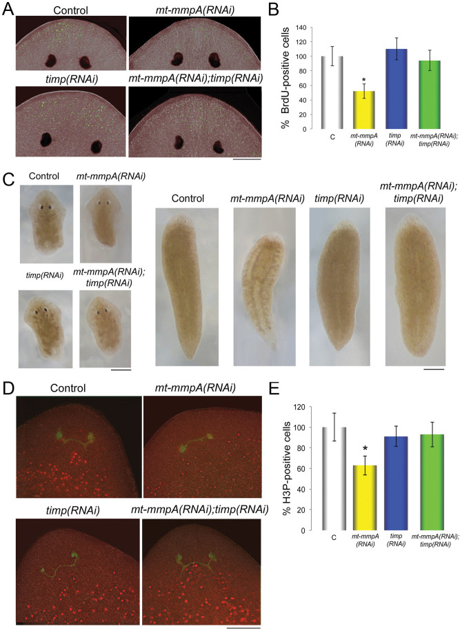
The mechanisms of action underlying the pharmacological properties of the natural alkaloid berberine still need investigation. Planarian regeneration is instrumental in deciphering developmental responses following drug exposure. Here we report the effects of berberine on regeneration in the planarian Dugesia japonica. Our findings demonstrate that this compound perturbs the regenerative pattern. By real-time PCR screening for the effects of berberine exposure on gene expression, we identified alterations in the transcriptional profile of genes representative of different tissues, as well as of genes involved in extracellular matrix (ECM) remodeling. Although berberine does not influence cell proliferation/apoptosis, our experiments prove that this compound causes abnormal regeneration of the planarian visual system. Potential berberine-induced cytotoxic effects were noticed in the intestine. Although we were unable to detect abnormalities in other structures, our findings, sustained by RNAi-based investigations, support the possibility that berberine effects are critically linked to anomalous ECM remodeling in treated planarians.


Figures

Figure 1
Figure 1. Berberine absorption in planarians.
(A) Dorsal view of a live planarian. The highly branched morphology of the intestine, consisting of a single anterior and two posterior primary branches, is visualized by red food dye. Anterior is up. Black box indicates the region magnified in (B–E). (B–E) Head of an intact planarian, as visualized under UV light. Berberine shows a wide range of distribution and is mainly accumulated in the intestinal branches (g). (B) 400 μM berberine. (C) Control. The planarian body and eyes are highlighted by white lines. e: eye; g: gut branches. (D,E) Magnification of two regions in (B), localized at the gut branch level (D) and near the epidermis (E). (F–J) 600 μM berberine visualization in dissociated cells. (F) A gastrodermal cell showing a berberine-positive nucleus. The cytoplasm is filled with droplets and vacuoles. (G) A big elongated hypothetical muscle cell with a berberine-positive nucleus. (H) An oval, middle-sized cell, probably an acidophilic cell, shows both cytoplasmic and nuclear localization of berberine. (I) A middle-sized cell, probably a cell in the process of differentiation, shows a berberine-positive nucleus. (J) berberine stain in a neoblast-like cell. (K) Chemical structure of berberine. (L) Graph representing the concentration of berberine absorbed by planarians versus the berberine concentration in the soaking medium. Intact: intact planarians. Regenerating: regenerating planarians. Scale bar: 500 μm in (A); 150 μm in (B) and (C); 75 μm in (D); 30 μm in (E); 10 μm in (F–J).
Figure 2
Figure 2. Berberine treatment affects visual-system regeneration.
(A) Schematic of the experimental procedure used to obtain regenerating fragments. (B–E) Representative brighfield images of planarians regenerating a head following berberine treatments (regeneration: 7 days). (B′–E′) eyes and the optic chiasm are visualized by immunostaining with anti VC-1 antibody. (B) Control. (C) B50: berberine 50 μM. (D) B400: berberine 400 μM. (E) B600: berberine 600 μM. (F) The graph shows the percentage of the different phenotypes. A number of 300 animals was analyzed for each berberine concentration. Light grey: normal eyes; grey: asymmetric and smaller eyes; dark grey: cyclopic eyes; black: no eyes. (G) Schematic of the phototactic test used to assess the behavior of regenerating planarians following 600 μM berberine treatment (regeneration: 7 days). (H) The graph indicates the time spent in the target area by tail fragments regenerating a head. (I) The graph indicates the time spent in the target area by head fragments regenerating a tail. Each graph represents the mean ± s.d. from 10 independent specimens. H2O: control; B600: berberine 600 μM. Scale bar: 100 μm in (B–E) and 50 μm in (B′-E′).
Figure 3
Figure 3. Effect of berberine treatment on cell proliferation and apoptosis in regenerating planarians.
(A) Expression level of Dj-mcm2 and Dj-bruno in berberine-treated planarians and controls, analyzed by Real Time RT-PCR. H2O: controls; B50: berberine 50 μM; B400: berberine 400 μM; B600: berberine 600 μM. In Real Time RT-PCR experiments, the expression levels are indicated in relative units, assuming as unitary the value of the controls. Each value is the mean ± s.d. of 3 independent experiments, carried out in 6 replicates (B) Morphometric analysis of blastema size in planarians regenerating a head followig berberine 600 μM (B600) treatment and in controls. Values are average ± s.d. from 30 samples. (C) Effects of berberine on activation of caspase-3 in planarians after 7 days of regeneration. Berberine treatments: B50: 50 μM berberine-treated planarians; B400: 400 μM berberine-treated planarians; B600: 600 μM berberine-treated planarians; control: planarians regenerating in water. Caspase-3 activation was monitored for 4 hours (h). A405: absorbance at 405 nm. Data represent mean values ± s.d. from 3 independent samples.
Figure 4
Figure 4. Effects of berberine on the transcriptional profile of representative markers of nerve cells.
(A) Comparison of mRNA levels of representative markers of the visual system: Dj-six1, Dj-eya, Dj-pax6A, Dj-ops and Dj-netB in berberine-treated planarians and in controls. H2O: controls; B50: berberine 50 μM; B400: berberine 400 μM; B600: berberine 600 μM. (B) Expression of Dj-syt is used to detect the CNS in a control planarian (H2O) and following berberine 600 μM (B600) treatment. (C) The graph shows quantification of the size of the regenerating cephalic ganglia in berberine 600 μM (B600)-treated planarians and controls (H2O), as measured after in situ hybridization with Dj-syt. Values are average ± s.d. from 30 samples. (D) Comparison of mRNA levels of the neuronal markers Dj-gad, Dj-rax, Dj-syt and Dj-otp in berberine-treated planarians and in controls. H2O: controls; B50: berberine 50 μM; B400: berberine 400 μM; B600: berberine 600 μM. In Real Time RT-PCR experiments, the expression levels are indicated in relative units, assuming as unitary the value of the controls. Each value is the mean ± s.d. of 3 independent experiments, carried out in 6 replicates. (E) Expression of Dj-otp is used to detect regeneration of the branches localized on the outer side of each lobe of the brain in a control planarian (H2O) and following berberine 600 μM (B600) treatment. (F) Expression of Dj-inx3 is used to detect regeneration of the branches in a control planarian (H2O) and following berberine 600 μM (B600) treatment. Scale bars: 100 μm in (B) and 200 μm in (E,F).
Figure 5
Figure 5. Effects of berberine on the transcriptional profile of representative markers of other differentiated cell types.
(A) Comparison of mRNA levels of representative markers expressed in different tissues: Dj-ahnak, Dj-inx1, Dj-mhca; Dj-inx10; Dj-mmp1; Dj-mt-mmpA, Dj-ast4 and Dj-timp in berberine-treated planarians and in controls. H2O: controls; B50: berberine 50 μM; B400: berberine 400 μM; B600: berberine 600 μM. In Real Time RT-PCR experiments, the expression levels are indicated in relative units, assuming as unitary the value of the controls. Each value is the mean ± s.d. of 3 independent experiments, carried out in 6 replicates. (B) Whole mount in situ hybridizations reveal the pattern of representative markers expressed in different tissues: Dj-ahnak, Dj-inx1, Dj-mhca, Dj-inx10; Dj-mmp1;Dj-mt-mmpA, Dj-ast4 and Dj-timp in 600 μM berberine-treated planarians (B600) and in controls (H2O). Scale bar: 400 μm.
Figure 6
Figure 6. Analysis of long-term effects of Dj-mt-mmpA, Dj-timp and simultaneous Dj-mt-mmpA;Dj-timp knockdown in intact and regenerating planarians.
(A) Representative images of the region anterior to the photoreceptors in β-gal(RNAi) control, mt-mmp(RNAi), timp(RNAi) and mt mmpA(RNAi) planarians. BrdU-labeling, used to monitor cell migration, is visualized in green and superposed upon a bright-field image (in dark red). Anterior is up. (B) The graph shows the significant decrease in the percentage of BrdU-positive cells in the region anterior to eyes of mt-mmpA(RNAi) phenotypes, compared to β-gal(RNAi) controls. No significant variation in the number of BrdU-positive cells with respect to the controls is detected after timp(RNAi) or simultaneous mt-mmpA(RNAi);timp(RNAi). Results represent average ± s.d. from 15 specimens assuming as 100% the value of control planarians. (unpaired t-test) *P < 0.05. C: β-gal(RNAi); mt-mmpA(RNAi): Dj-mt-mmpA(RNAi); timp(RNAi): Dj-timp(RNAi); mt-mmpA;timp(RNAi): simultaneous Dj-mt-mmpA;Dj-timp (RNAi). (C) Brightfield images of representative tail and head fragments: regeneration 5 days after 5 weeks of feeding RNAi. (D) Immunostaining with anti-H3P antibody (red spots): 5 days of head regeneration after 5 weeks of feeding RNAi. VC-1 antibody (in green). H3P-positive cells and VC-1-positive photoreceptors are visualized superposed upon a bright-field image (in brown). Delayed morphogenesis of the visual system and a lower number of H3P-positive cells in the blastema area can be observed in mt-mmpA(RNAi) fragments. Control: β-gal(RNAi); mt-mmpA(RNAi): Dj-mt-mmpA(RNAi); timp(RNAi): Dj-timp(RNAi); mt-mmpA;timp(RNAi): simultaneous Dj-mt-mmpA;Dj-timp (RNAi). (E) Graph shows the significant decrease in the percentage of H3P-positive cells in the blastema of mt-mmpA(RNAi) fragments compared to controls. No significant variation in the number of H3P-positive cells was detected in the blastema of timp(RNAi) and mt-mmpA(RNAi);Dj-timp(RNAi) fragments. Results represent average ± s.d. from 20 specimens assuming as 100% the value of control planarians. (unpaired t- test) *P < 0.05. (C): β-gal(RNAi); mt-mmpA(RNAi): Dj-mt-mmpA(RNAi); timp(RNAi): Dj-timp(RNAi); mt-mmpA;timp(RNAi): simultaneous Dj-mt-mmpA;Dj-timp (RNAi). Scale bars: 100 μm in (A,D); 500 μm in (C).

References

    1. Chen X. W. et al. Interaction of herbal compounds with biological targets: a case study with berberine. ScientificWorldJournal. 708292 (2012). - PMC - PubMed
    1. Gan R. Y. Bioactivities of Berberine: An Update. Int. J. Modern Biol. Med. 1, 48–81 (2012).
    1. Gilca M., Gaman L., Panait E., Stoian I. & Atanasiu V. Chelidonium majus: an integrative review: traditional knowledge versus modern findings. Forsch. Komplementmed. 17, 241–248 (2010). - PubMed
    1. Isolani M. E. et al. The in vivo effect of chelidonine on the stem cell system of planarians. Eur. J. Pharmacol. 686, 1–3 (2012). - PubMed
    1. Chignell C. F., Sik R. H., Watson M. A. & Wielgus A. R. Photochemistry and Photocytotoxicity of Alkaloids from Goldenseal (Hydrastis canadensis L.) 3. Effect on Human Lens and Retinal Pigment Epithelial Cells. Photochem. Photobiol. 83, 938–943 (2007). - PMC - PubMed

Publication types

LinkOut - more resources