. 2014 May 8;8(5):e2845.
doi: 10.1371/journal.pntd.0002845. eCollection 2014 May.

Field-evaluation of a new lateral flow assay for detection of cellular and humoral immunity against Mycobacterium leprae

Affiliations

Field-evaluation of a new lateral flow assay for detection of cellular and humoral immunity against Mycobacterium leprae

Kidist Bobosha et al. PLoS Negl Trop Dis. .

Abstract

Background: Field-applicable tests detecting asymptomatic Mycobacterium leprae (M. leprae) infection or predicting progression to leprosy, are urgently required. Since the outcome of M. leprae infection is determined by cellular- and humoral immunity, we aim to develop diagnostic tests detecting pro-/anti-inflammatory and regulatory cytokines as well as antibodies against M. leprae. Previously, we developed lateral flow assays (LFA) for detection of cytokines and anti-PGL-I antibodies. Here we evaluate progress of newly developed LFAs for applications in resource-poor settings.

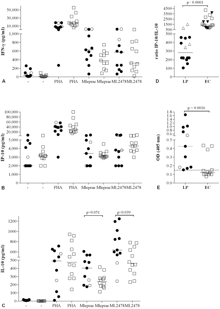
Methods: The combined diagnostic value of IP-10, IL-10 and anti-PGL-I antibodies was tested using M. leprae-stimulated blood of leprosy patients and endemic controls (EC). For reduction of the overall test-to-result time the minimal whole blood assay time required to detect distinctive responses was investigated. To accommodate LFAs for field settings, dry-format LFAs for IP-10 and anti-PGL-I antibodies were developed allowing storage and shipment at ambient temperatures. Additionally, a multiplex LFA-format was applied for simultaneous detection of anti-PGL-I antibodies and IP-10. For improved sensitivity and quantitation upconverting phosphor (UCP) reporter technology was applied in all LFAs.

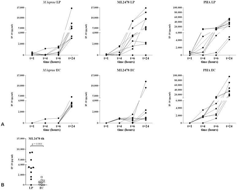
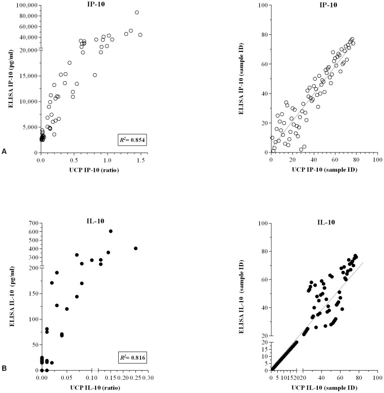
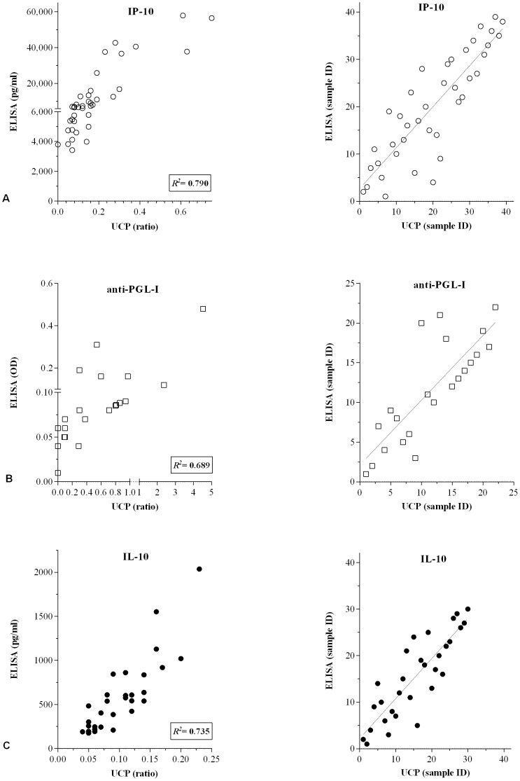
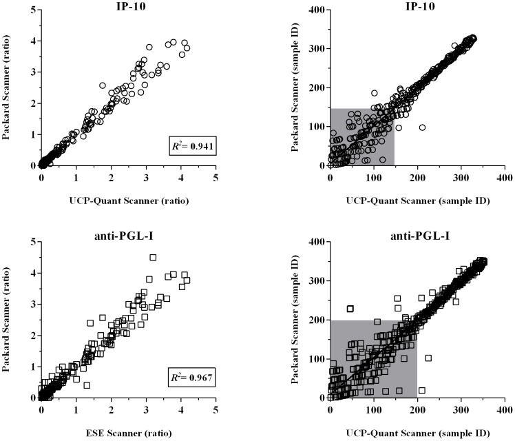
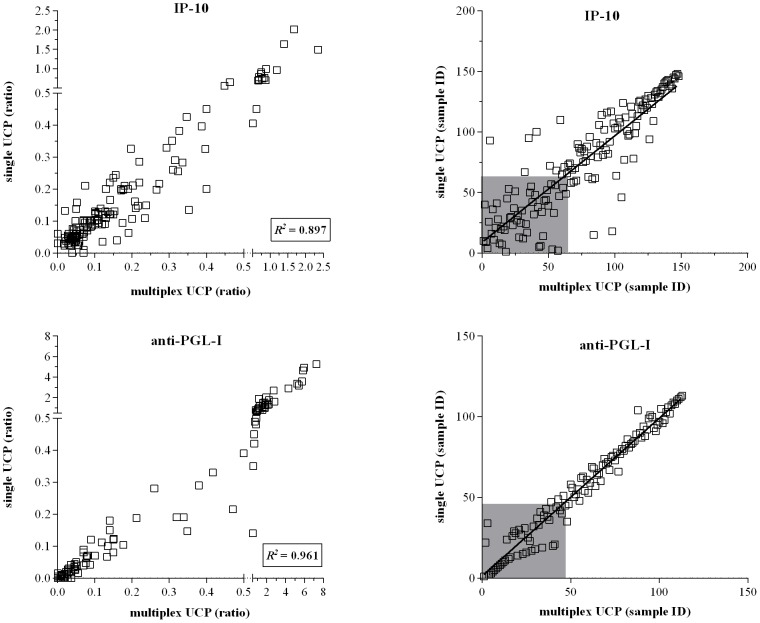
Results: Single and multiplex UCP-LFAs correlated well with ELISAs. The performance of dry reagent assays and portable, lightweight UCP-LF strip readers indicated excellent field-robustness. Notably, detection of IP-10 levels in stimulated samples allowed a reduction of the whole blood assay time from 24 h to 6 h. Moreover, IP-10/IL-10 ratios in unstimulated plasma differed significantly between patients and EC, indicating the feasibility to identify M. leprae infection in endemic areas.

Conclusions: Dry-format UCP-LFAs are low-tech, robust assays allowing detection of relevant cytokines and antibodies in response to M. leprae in the field. The high levels of IP-10 and the required shorter whole blood assay time, render this cytokine useful to discriminate between leprosy patients and EC.


Conflict of interest statement

The authors have declared that no competing interest exist.

Figures

Figure 1
Figure 1. Combined cytokine profiles in response to M. leprae.
Production of IFN-γ (A), IP-10 (B) and IL-10 (C) determined by ELISA, in response to medium (-), PHA, M. leprae WCS or the M. leprae-unique protein ML2478 in 24 h WBA for Ethiopian leprosy patients (n = 11: 2 BT (○) and 9 BL (•), and healthy endemic controls (EC; n = 12; □). For comparison between BT and BL, significant differences were found for M. leprae WCS (Mlep) induced IFN-γ responses (p = 0.036) and ML2478 induced IL-10 responses (p = 0.035). (D): IP-10/IL-10 ratios are depicted for unstimulated samples after 24 h {LP (•) and EC (□)} or after 1 h WBA {LP (▵) and EC (▾)}. (E): Anti-PGL-I antibodies for BL (○) and BT (•) patients were detected by ELISA using natural disaccharide of PGL-I linked to HSA (ND-O-HSA). Optical density (OD450) readings were performed using 1∶800 serum dilutions. Median values per group are indicated by horizontal lines. The cut-off for positivity is indicated by the dashed horizontal line.
Figure 2
Figure 2. Kinetics of IP-10 production in WBA.
(A): IP-10 concentrations produced in stimulated whole blood cultures of leprosy patients (upper panel; LP; n = 10: 5 BL (Ethiopia); 2 BT (Ethiopia); 3 BT (The Netherlands) and healthy endemic controls (lower panel; EC, n = 8) in response to M. leprae WCS (left panel; 10 µg/ml), M. leprae unique protein ML2478 (middle panel; 10 µg/ml) and PHA (right panel; 1 µg/ml). IP-10 concentrations were determined by ELISA after 1 h, 4 h, 6 h and 24 h antigen stimulation. Values on the y-axis are concentrations corrected for background values. (B): Comparison of IP-10 concentrations determined by ELISA after 6 h stimulation with ML2478 (10 µg/ml) of whole blood samples.
Figure 3
Figure 3. Correlation between ELISAs and UCP-LFAs.
Levels of IP-10 (A) and IL-10 (B) in 24 h whole blood samples of 77 M. leprae (antigen), LPS and PHA stimulated WBA samples of Dutch healthy controls were simultaneously determined by ELISAs and wet-format UCP-LFAs. Left panels: results for ELISAs are indicated in pg/ml (ELISA) or as the ratio of the relative fluorescence units (RFUs) measured at Test and Flow-Control lines (UCP-LFA). R2 equals the square of the Pearson correlation coefficient. Right panels: Spearman ranking.
Figure 4
Figure 4. Correlation between ELISAs and UCP-LFAs.
Levels of IP-10 (A; n = 40), anti-PGL-I antibodies (B; n = 22) or IL-10 (C; n = 40) in WBA samples were simultaneously determined by ELISAs and UCP-LFAs in Ethiopia using dry-format (A, B) or wet format (C) UCP-LFAs. For cytokine analysis (A and C), samples of Ethiopian leprosy patients (2 BT and 8 BL) that were unstimulated or stimulated with M. leprae WCS, ML2478 or PHA were used. For anti-PGL-I antibodies (B), samples of Ethiopian leprosy patients (2 BT and 8 BL) and healthy endemic controls (n = 12) were used. Left panels: results for ELISA are indicated in pg/ml (A, C) or OD450 (B) or as the ratio of the relative fluorescence units (RFUs) measured at Test and Flow-Control lines (UCP-LFA). R2 equals the square of the Pearson correlation coefficient. Correlation was calculated for samples with ELISA values higher than the cut-off threshold. Right panels: Spearman ranking.
Figure 5
Figure 5. Performance of the portable lightweight UCP-Quant LF strip reader.
Dry-format UCP-LFAs were performed for single detection of IP-10 and anti-PGL-I antibodies in an Ethiopian field setting (Figure 3). LF strips were analyzed using a portable reader (UCP-Quant). Subsequently, LF strips were shipped to The Netherlands and re-analysed using a dedicated lab-based FluoroCount microtiterplate reader (Packard) adapted for reading UCP-LF strips. Left panel: results are indicated as the ratio of the relative fluorescence units (RFUs) measured at Test and Flow-Control lines. R2 equals the square the Pearson correlation coefficient. Right panel: Spearman ranking. The grey box indicates samples scoring values below the specificity threshold.
Figure 6
Figure 6. Comparison between single and multiplex UCP-LFAs.
UCP-LFAs were performed for single or multiplex detection of IP-10 (upper panel; n = 149 samples) and anti-PGL-I (lower panel; n = 115 samples) using M. leprae antigen-stimulated WBA samples of Dutch and Ethiopian leprosy patients. Simultaneous detection of IP-10 and anti-PGL-I IgM was performed following the two phase protocol using the UCPαIP-10conjugate and the UCPαIgM conjugate. Left panel: Results for UCP-LFAs are displayed as the ratio of the relative fluorescence units (RFUs) measured at Test and Flow-Control lines. R2 equals the square of the Pearson correlation coefficient. Right panel: Spearman ranking. The grey box indicates samples scoring values below the specificity threshold.

References

    1. Global leprosy: update on the 2012 situation. Wkly Epidemiol Rec 88: 365–379. - PubMed
    1. Ridley DS, Jopling WH (1966) Classification of leprosy according to immunity. A five-group system. Int J Lepr Other Mycobact Dis 34: 255–273. - PubMed
    1. Burki T (2010) Fight against leprosy no longer about the numbers. Lancet Infect Dis 10: 74. - PubMed
    1. Niedbala RS, Feindt H, Kardos K, Vail T, Burton J, et al. (2001) Detection of analytes by immunoassay using up-converting phosphor technology. Anal Biochem 293: 22–30. - PubMed
    1. Qu Q, Zhu Z, Wang Y, Zhong Z, Zhao J, et al. (2009) Rapid and quantitative detection of Brucella by up-converting phosphor technology-based lateral-flow assay. J Microbiol Methods 79: 121–123 S0167-7012(09)00228-0 [pii];10.1016/j.mimet.2009.07.015 [doi] - PubMed

Publication types