

Bone marrow-derived mesenchymal stem cells migrate to healthy and damaged salivary glands following stem cell infusion
- PMID: 24810808
- PMCID: PMC4170149
- DOI: 10.1038/ijos.2014.23
Bone marrow-derived mesenchymal stem cells migrate to healthy and damaged salivary glands following stem cell infusion
Abstract
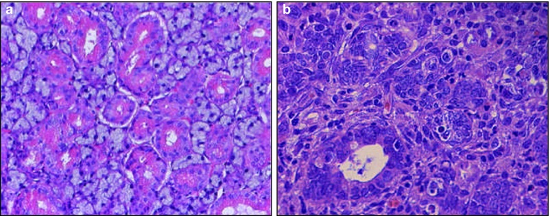
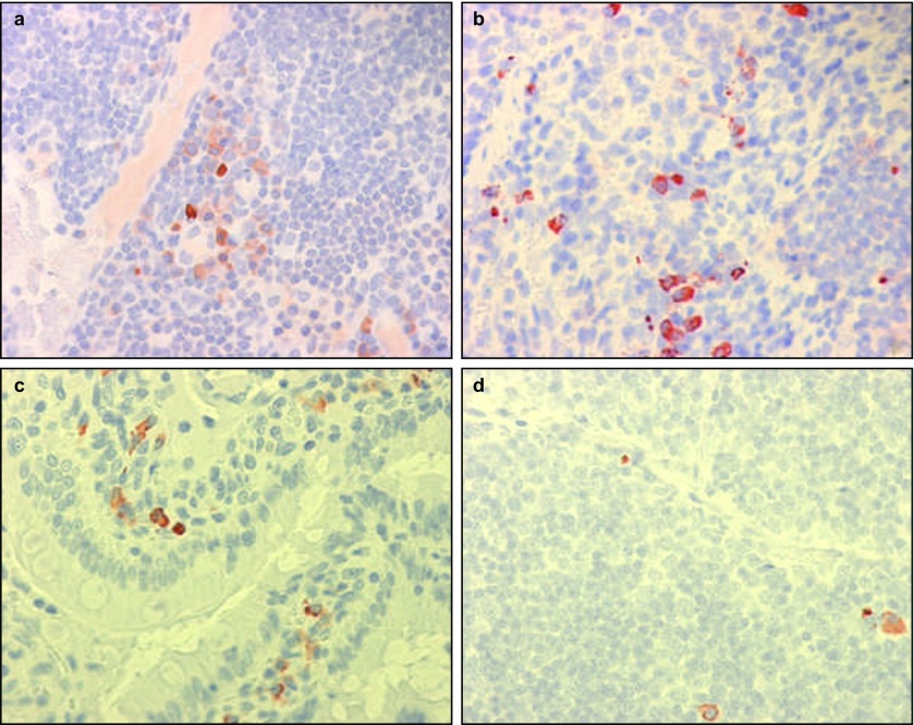
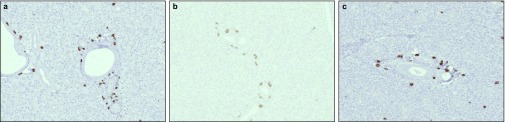
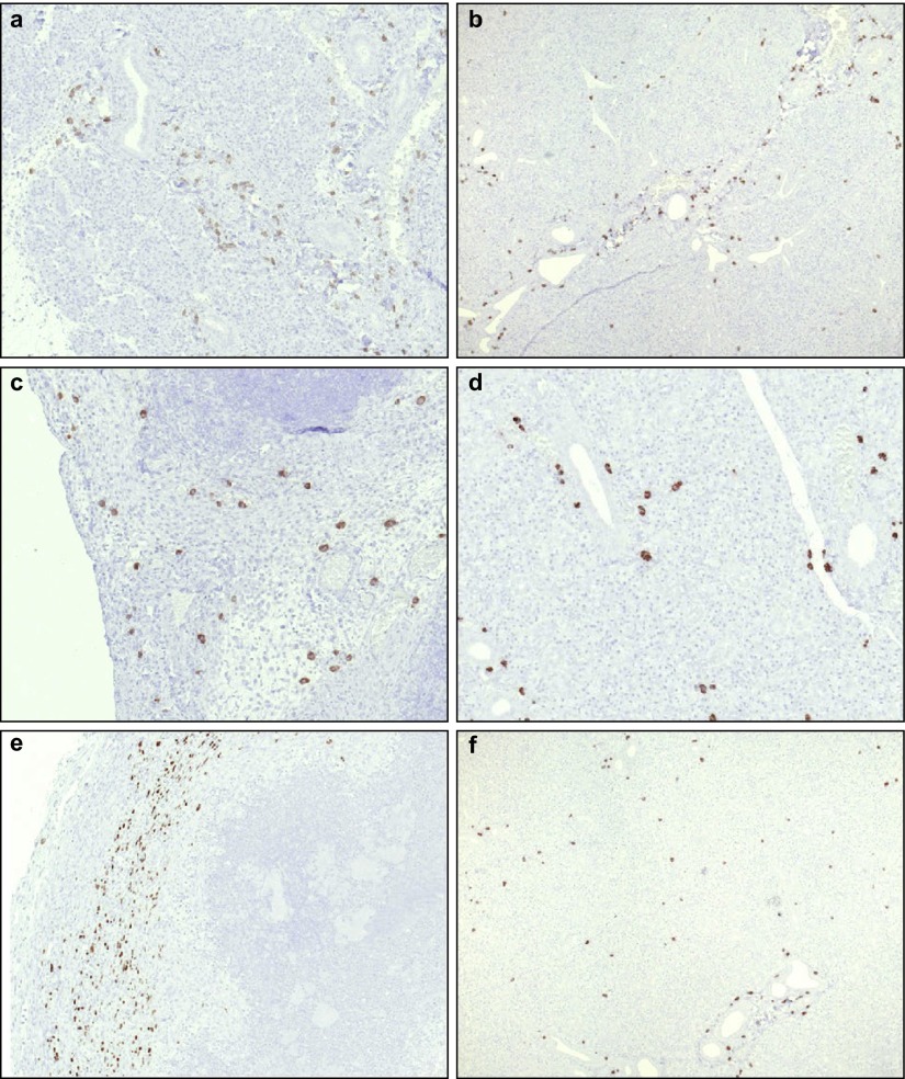
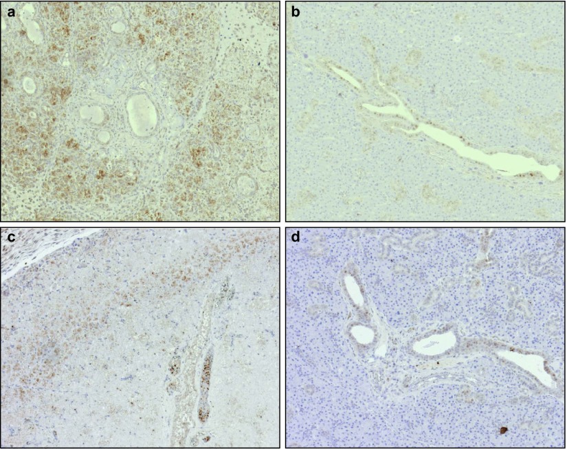
Xerostomia is a severe side effect of radiation therapy in head and neck cancer patients. To date, no satisfactory treatment option has been established. Because mesenchymal stem cells (MSCs) have been identified as a potential treatment modality, we aimed to evaluate stem cell distribution following intravenous and intraglandular injections using a surgical model of salivary gland damage and to analyse the effects of MSC injections on the recruitment of immune cells. The submandibular gland ducts of rats were surgically ligated. Syngeneic adult MSCs were isolated, immortalised by simian virus 40 (SV40) large T antigen and characterized by flow cytometry. MSCs were injected intravenously and intraglandularly. After 1, 3 and 7 days, the organs of interest were analysed for stem cell recruitment. Inflammation was analysed by immunohistochemical staining. We were able to demonstrate that, after intravenous injection, MSCs were recruited to normal and damaged submandibular glands on days 1, 3 and 7. Unexpectedly, stem cells were recruited to ligated and non-ligated glands in a comparable manner. After intraglandular injection of MSCs into ligated glands, the presence of MSCs, leucocytes and macrophages was enhanced, compared to intravenous injection of stem cells. Our data suggest that injected MSCs were retained within the inflamed glands, could become activated and subsequently recruited leucocytes to the sites of tissue damage.
Figures

Similar articles
-
Acute salivary gland hypofunction in the duct ligation model in the absence of inflammation.Oral Dis. 2008 Sep;14(6):520-8. doi: 10.1111/j.1601-0825.2007.01413.x. Epub 2008 Jan 22. Oral Dis. 2008. PMID: 18221457 Free PMC article.
-
Bone marrow derived mesenchymal stem cells restored GLUT1 expression in the submandibular salivary glands of ovariectomized rats.Arch Oral Biol. 2024 Oct;166:106048. doi: 10.1016/j.archoralbio.2024.106048. Epub 2024 Jul 13. Arch Oral Biol. 2024. PMID: 39002180
-
Sonographic analysis of rat submandibular glands in experimentally-induced sialadenitis.Dentomaxillofac Radiol. 2000 Mar;29(2):90-6. doi: 10.1038/sj/dmfr/4600508. Dentomaxillofac Radiol. 2000. PMID: 10808222
-
Mesenchymal Stromal/Stem Cell Therapy Improves Salivary Flow Rate in Radiation-Induced Salivary Gland Hypofunction in Preclinical in vivo Models: A Systematic Review and Meta-Analysis.Stem Cell Rev Rep. 2024 May;20(4):1078-1092. doi: 10.1007/s12015-024-10700-y. Epub 2024 Mar 2. Stem Cell Rev Rep. 2024. PMID: 38430363 Free PMC article.
-
Chronic sclerosing sialadenitis of the submandibular and parotid glands: a report of a case and review of the literature.Oral Surg Oral Med Oral Pathol Oral Radiol Endod. 2000 Jun;89(6):720-3. doi: 10.1067/moe.2000.102515. Oral Surg Oral Med Oral Pathol Oral Radiol Endod. 2000. PMID: 10846127 Review.
Cited by
-
Endogenous Mobilization of Mesenchymal Stromal Cells: A Pathway for Interorgan Communication?Front Cell Dev Biol. 2021 Jan 8;8:598520. doi: 10.3389/fcell.2020.598520. eCollection 2020. Front Cell Dev Biol. 2021. PMID: 33490065 Free PMC article. Review.
-
Drug Therapeutics Delivery to the Salivary Glands: Intraglandular and Intraductal Injections.Adv Exp Med Biol. 2023;1436:119-130. doi: 10.1007/5584_2023_765. Adv Exp Med Biol. 2023. PMID: 36809639 Review.
-
Adult stem cell transplantation combined with conventional therapy for the treatment of end-stage liver disease: a systematic review and meta-analysis.Stem Cell Res Ther. 2021 Oct 30;12(1):558. doi: 10.1186/s13287-021-02625-x. Stem Cell Res Ther. 2021. PMID: 34717737 Free PMC article.
-
First-in-man intraglandular implantation of stromal vascular fraction and adipose-derived stem cells plus platelet-rich plasma in irradiation-induced gland damage: a case study.Int Med Case Rep J. 2017 Aug 16;10:295-299. doi: 10.2147/IMCRJ.S142514. eCollection 2017. Int Med Case Rep J. 2017. PMID: 28860871 Free PMC article.
-
Regenerative rehabilitation: a novel multidisciplinary field to maximize patient outcomes.Med Rev (2021). 2024 Jun 14;4(5):413-434. doi: 10.1515/mr-2023-0060. eCollection 2024 Oct. Med Rev (2021). 2024. PMID: 39444794 Free PMC article. Review.
References
-
- Vokes EE, Weichselbaum RR, Lippman SM, et al. Head and neck cancer. N Engl J Med. 1993;328 (3:184–194. - PubMed
-
- Vergeer MR, Doornaert PA, Rietveld DH, et al. Intensity-modulated radiotherapy reduces radiation-induced morbidity and improves health-related quality of life: results of a nonrandomized prospective study using a standardized follow-up program. Int J Radiat Oncol Biol Phys. 2009;74 (1:1–8. - PubMed
-
- O'Connell AC. Natural history and prevention of radiation injury. Adv Dent Res. 2000;14:57–61. - PubMed
Publication types
MeSH terms
Substances
LinkOut - more resources
Full Text Sources
Other Literature Sources
