. 2014 May 8:4:4881.
doi: 10.1038/srep04881.

A PrP(C)-caveolin-Lyn complex negatively controls neuronal GSK3β and serotonin 1B receptor

Affiliations

A PrP(C)-caveolin-Lyn complex negatively controls neuronal GSK3β and serotonin 1B receptor

Julia Hernandez-Rapp et al. Sci Rep. .

Abstract

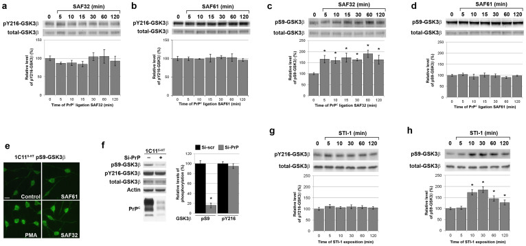
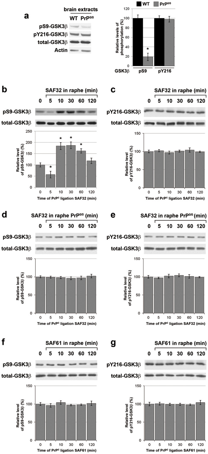
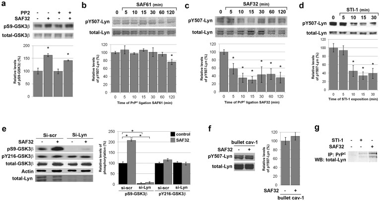
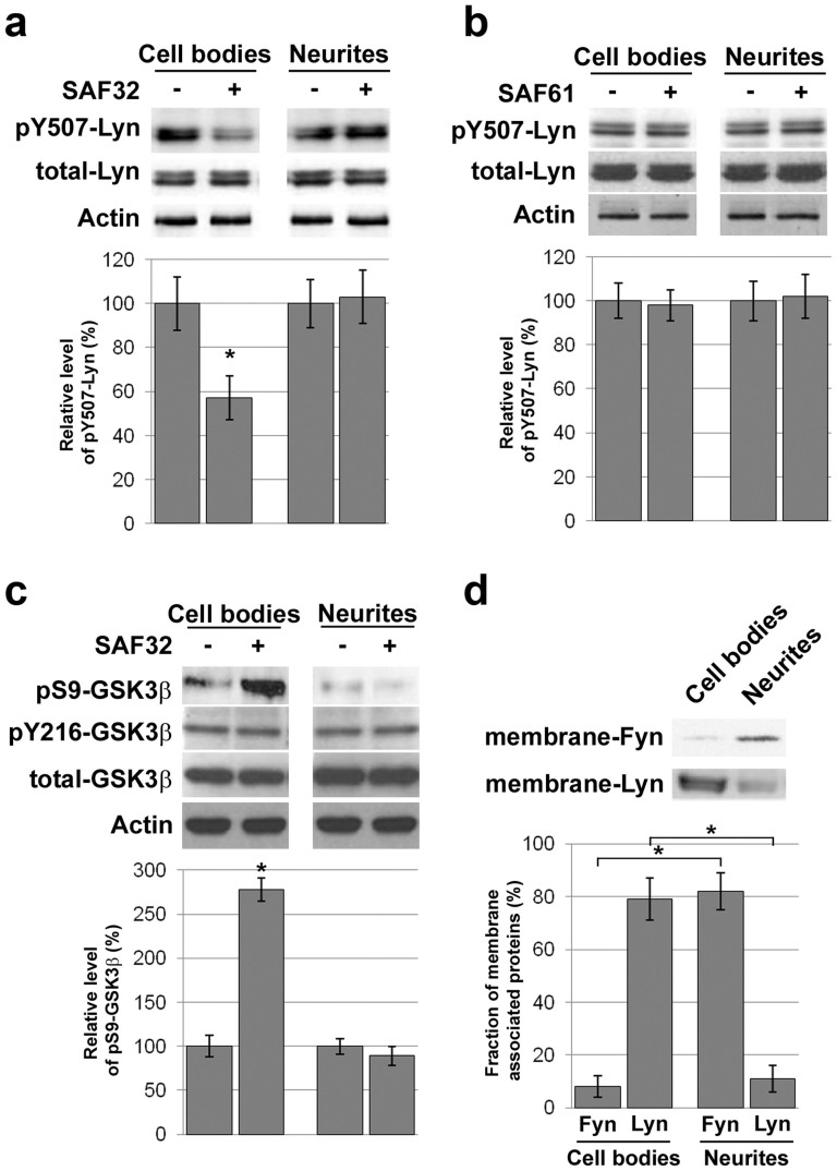
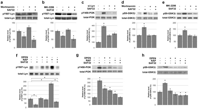
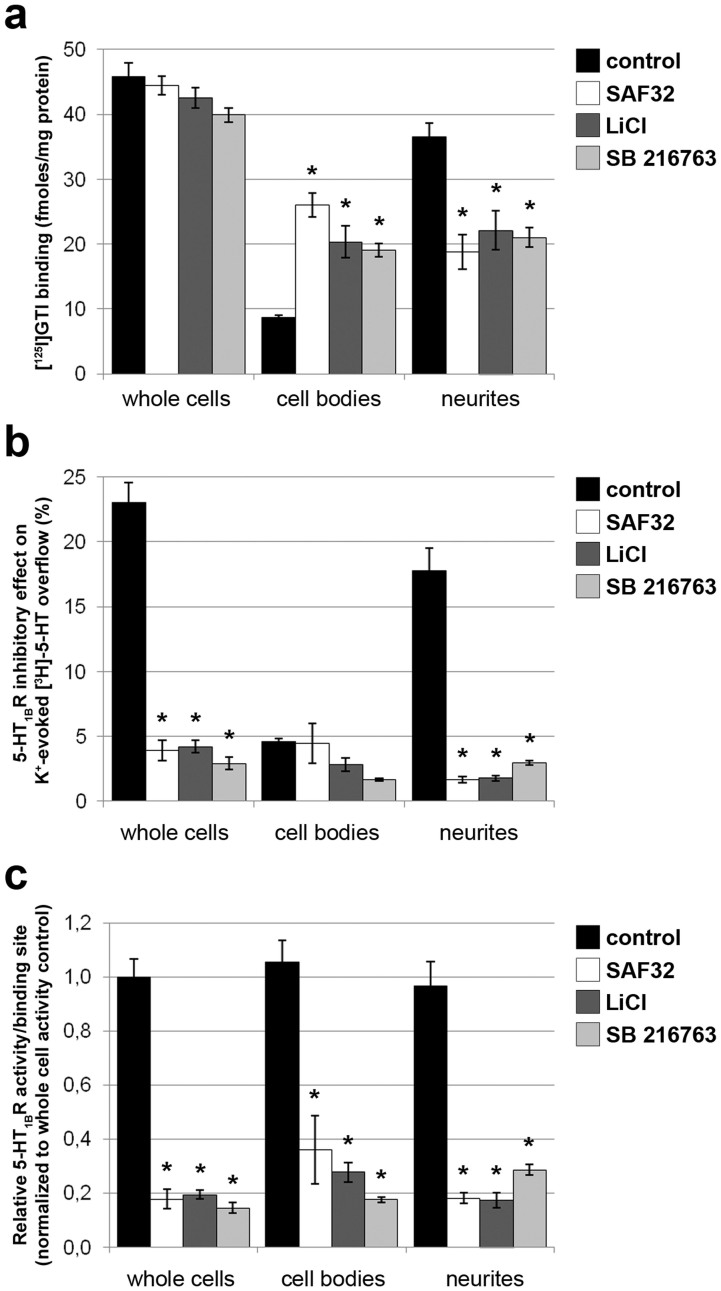
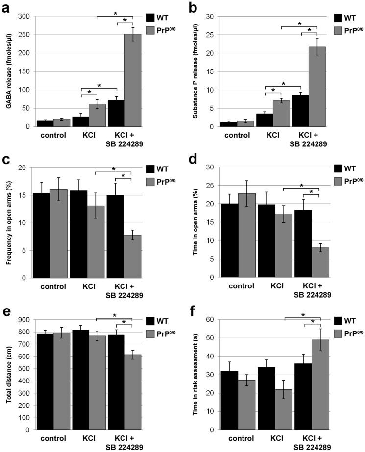
The cellular prion protein, PrP(C), is a glycosylphosphatidylinositol-anchored protein, abundant in lipid rafts and highly expressed in the brain. While PrP(C) is much studied for its involvement under its abnormal PrP(Sc) isoform in Transmissible Spongiform Encephalopathies, its physiological role remains unclear. Here, we report that GSK3β, a multifunctional kinase whose inhibition is neuroprotective, is a downstream target of PrP(C) signalling in serotonergic neuronal cells. We show that the PrP(C)-dependent inactivation of GSK3β is relayed by a caveolin-Lyn platform located on neuronal cell bodies. Furthermore, the coupling of PrP(C) to GSK3β potentiates serotonergic signalling by altering the distribution and activity of the serotonin 1B receptor (5-HT1BR), a receptor that limits neurotransmitter release. In vivo, our data reveal an increased GSK3β kinase activity in PrP-deficient mouse brain, as well as sustained 5-HT1BR activity, whose inhibition promotes an anxiogenic behavioural response. Collectively, our data unveil a new facet of PrP(C) signalling that strengthens neurotransmission.


Figures

Figure 1
Figure 1. Stimulation of native PrPC promotes GSK3β phosphorylation on S9.
(a–d) 1C115-HT neuronal cells were exposed to anti-PrPC antibodies targeting a N-terminal epitope (SAF32, 10 μg/ml) (a), (c) or a C-terminal epitope (SAF61, 10 μg/ml) (b), (d). Cell lysates were immunoblotted with antibodies targeting either pY216-GSK3β (a), (b) or pS9-GSK3β (c), (d). Levels of pY216-GSK3β (a), (b) or pS9-GSK3β (c), (d) were normalized to total GSK3β for quantification. (e) 1C115-HT neuronal cells exposed to various anti-PrPC antibodies (SAF32, SAF61, each 10 μg/ml) for 30 min were stained with anti pS9-GSK3β antibodies. Unstimulated cells and cells treated with the phorbol ester PMA (5 μM, 30 min), a known inducer of phosphorylation of GSK3β at S9, were included as negative and positive controls, respectively. Scale bar = 25 μm. (f) The levels of pS9-GSK3β, pY216-GSK3β and total GSK3β were assessed by immunoblotting in 1C115-HT cells transfected for 36 h with a siRNA targeted against PrP (Si-PrP) or a control scramble siRNA (Si-scr). immunoblotting with antibodies to PrPC and actin was carried out to check knockdown and as loading control, respectively. (g), (h) 1C115-HT neuronal cells were exposed to a peptide corresponding to the domain of STI-1 that binds PrPC (aa 230–245) (25 μM). Cell lysates were immunoblotted with antibodies targeting either pY216-GSK3β (g) or pS9-GSK3β (h). Levels of pY216-GSK3β (g) or pS9-GSK3β (h) were normalized to total GSK3β for quantification. Gels have been cropped for clarity and conciseness purposes; original images corresponding to (a–d) are shown in Supplemental Figure 4. All data are representative of a set of n = 4 to 6 independent experiments. Results are expressed as means ± S.E.M. *P < 0.05 vs. control, Kolmogorov-Smirnov test.
Figure 2
Figure 2. PrPC-negatively controls GSK3β in mouse brain.
(a) The levels of pS9-GSK3β, pY216-GSK3β and total GSK3β were assessed by immunoblotting in brain extracts from PrP0/0 mice versus WT mice. Actin was used as loading control. Data are representative of n = 4 animals. (b–g) SAF32 (b–e) or SAF61 (f–g) anti-PrPC antibodies (2 μl at 1 mg/ml) were stereotaxically injected in the raphe nuclei of WT (b), (c), (f), (g) or PrP0/0 mice (d), (e). Mice were sacrificed at the indicated time to collect raphe nuclei samples. The levels of pS9-GSK3β (b), (d), (f) or pY216-GSK3β (c), (e), (g) were assessed by immunoblotting of the raphe extracts and normalized to total GSK3β for quantification. Data are representative of n = 4 animals. Gels have been cropped for clarity and conciseness purposes and have been run under the same experimental conditions. Results are expressed as means ± S.E.M. *P < 0.05 vs. control, Kolmogorov-Smirnov test.
Figure 3
Figure 3. PrPC-mediated GSK3β phosphorylation on S9 is relayed by the Lyn kinase in 1C115-HT neuronal cells.
(a) 1C115-HT neuronal cells were pre-incubated with the Fyn inhibitor PP2 (50 pM, 1 h) prior to exposure to SAF32 anti-PrPC antibodies (10 μg/ml, 30 min), targeting native PrPC. Cell lysates were immunoblotted with antibodies targeting pS9-GSK3β and total GSK3β for normalization. (b–d) 1C115-HT neuronal cells were exposed to anti-PrPC antibodies targeting (b) a C-terminal epitope (SAF61, 10 μg/ml), (c) a N-terminal epitope (SAF32, 10 μg/ml) or (d) to a peptide corresponding to the domain of STI-1 that binds PrPC (aa 230–245) (25 μM). Cell lysates were immunoblotted with antibodies targeting pY507-Lyn and total Lyn for normalization. (e) 1C115-HT neuronal cells were transfected for 36 h with a siRNA targeted against Lyn (Si-Lyn) or a control scramble siRNA (Si-scr) prior to exposure to SAF32 anti-PrPC antibodies (30 min). Cell lysates were immunoblotted with antibodies targeting pS9-GSK3β, pY216-GSK3β, total GSK3β. Total Lyn was used to check knockdown and actin was used as loading control. (f) 1C115-HT neuronal cells were submitted to caveolin-1 immunosequestration prior to exposure to SAF32 antibodies (10 μg/ml, 15 min). Cell lysates were immunoblotted with antibodies against pY507-Lyn and total Lyn for normalization. (g) 1C115-HT neuronal cells were exposed to the STI-1 peptide (25 μM) or SAF32 antibodies (10 μg/ml) for 30 min. Cell lysates were immunoprecipitated with SAF61 anti-PrPC antibodies and immunoblotted with antibodies against Lyn. Gels have been cropped for clarity and conciseness purposes; original images corresponding to (b–c) are shown in Supplemental Figure 5. Data are expressed as means ± S.E.M of n = 4 to 6 independent analyses. *P < 0.05 vs. control, Kolmogorov-Smirnov test.
Figure 4
Figure 4. The PrPC-Lyn-GSK3β cascade involves PrPC molecules located at the cell bodies of 1C115-HT neuronal cells.
(a), (b) Cell bodies and neurites of 1C115-HT cells were separated prior exposure to SAF32 (a) or SAF61 (b) anti-PrPC antibodies (10 μg/ml, 15 min). Lysates were immunoblotted with antibodies targeting pY507-Lyn or total Lyn. Actin was used as loading control. Cell fractionation was verified through immunoblotting with antibodies against lamin A/C, as shown in Supplemental Figure 6a. (c) pS9-GSK3β, pY216-GSK3β or total GSK3β levels were measured in cell bodies and neurites of 1C115-HT cells exposed to SAF32 anti-PrPC antibodies (10 μg/ml, 30 min). Actin was used as loading control. (d) Membrane-associated Fyn and Lyn were quantified in cell bodies versus neurites of 1C115-HT cells. Membrane preparation was verified through immunoblotting with antibodies against NaK-ATPase, as shown in Supplemental Figure 6b. Gels have been cropped for clarity and conciseness purposes and have been run under the same experimental conditions. All data are representative of a set of n = 4 to 6 independent experiments. Results are expressed as means ± S.E.M. *P < 0.05 vs. control, Kolmogorov-Smirnov test.
Figure 5
Figure 5. The PrPC-Lyn-GSK3β cascade in 1C115-HT neuronal cells is relayed by PI3K and Akt and involves copper and LRP1.
(a), (b) 1C115-HT neuronal cells were pre-incubated for 1 h with the PI3K inhibitor Wortmannin (5 nM) (a) or the Akt Inhibitor MK-2206 (1 μM) (b), and then exposed to SAF32 anti-PrPC antibodies (10 μg/ml, 15 min). Cell lysates were immunoblotted with antibodies targeting pY507-Lyn or total Lyn for normalization. (c) 1C115-HT neuronal cells were transfected for 36 h with a siRNA targeted against Lyn (Si-Lyn) or a control scramble siRNA prior to exposure to SAF32 anti-PrPC antibodies. Cell lysates were immunoblotted with antibodies targeting pY458-p85 PI3K or total p85 PI3K. (d), (e) 1C115-HT neuronal cells were pre-incubated for 1 h with the PI3K inhibitor Wortmannin (5 nM) (d) or the Akt Inhibitor MK-2206 (1 μM) (e) prior to exposure to SAF32 anti-PrPC antibodies (10 μg/ml, 30 min). Cell lysates were immunoblotted with antibodies targeting pS9-GSK3β or total GSK3β for normalization. (f–h) 1C115-HT cells were incubated for 1 h with the copper chelator DPEN (0.1 μg/ml) or the LRP1 antagonist RAP (200 nM), prior to exposure to SAF32 antibodies. Cell lysates were immunoblotted with antibodies targeting pY507-Lyn (f), pY458-p85 PI3K (g), or pS9-GSK3β (h). Levels of phosphorylated proteins were normalized to total corresponding proteins. Gels have been cropped for clarity and conciseness purposes and have been run under the same experimental conditions. All data are representative of a set of n = 4 to 6 independent experiments. Results are expressed as means ± S.E.M. *P < 0.05 vs. control, Kolmogorov-Smirnov test.
Figure 6
Figure 6. The PrPC-GSK3β cascade affects the subcellular distribution of the serotonin 1B receptor and negatively regulates its activity.
(a) 1C115-HT neuronal cells were exposed to SAF32 (10 μg/ml) anti-PrPC antibodies or the GSK3β inhibitors LiCl (1 mM) or SB216763 (10 nM) for 30 min. 5-HT1BRs were quantified through [125I]-GTI binding in whole cells and the corresponding cell bodies and neuritic fractions. (b) The 5-HT1BR activity was determined by measuring the inhibitory effect of the 5-HT1BR agonist L694247 on K+-evoked [3H]-5-HT overflow in whole cells. Experiments were repeated after cell fractionation in the corresponding cell bodies and neuritic fractions. (c) The relative 5-HT1BR activity was calculated by normalizing the inhibitory effect on [3H]-5-HT overflow to [125I]-GTI binding for each pool of cells, both before and after cell fractionation. A total of n = 6 independent cultures were analyzed. Results are expressed as means ± S.E.M. *P < 0.05 vs. control, Kolmogorov-Smirnov test.
Figure 7
Figure 7. Inhibition of serotonin 1B receptors in the substantia nigra of PrP-null mice unleashes GABA and substance P release and promotes an anxiogenic behavioural response.
(a), (b) The effect of 5-HT1BR inhibition on KCl-evoked GABA (a) and substance P (b) release was measured by in vivo microdialysis in awake WT and PrP0/0 mice. In response to KCl alone, PrP0/0 mice exhibited stronger GABA (a) and substance P (b) release than WT mice. 5-HT1BR pharmacological inhibition (SB224289, 100 nM) exacerbated GABA (a) and substance P (b) release in WT animals. The potentiating effect of the 5-HT1BR antagonist on GABA (a) and substance P (b) release was dramatically increased in PrP0/0 mice. (c–f) The anxiety-like behaviour of PrP0/0 versus WT mice was probed in the elevated plus maze. Three groups of each type of mice were tested: one group served as controls, a second group received a KCl injection (30 mM) in the substantia nigra and the last group received SB224289 (100 nM) prior to KCl injection. Mice were placed in the elevated plus maze 30 min after KCl treatment. Each trial was recorded for 5 min and further analysed to quantify frequency in open arms (c), time spent in open arms (d), total distance traversed (e) and time spent in risk assessment (f). Data are expressed as means ± S.E.M of n = 6 animals. *P < 0.05 vs. control, Kolmogorov-Smirnov test.

References

    1. Aguzzi A. & Calella A. M. Prions: protein aggregation and infectious diseases. Physiol Rev. 89, 1105–1152 (2009). - PubMed
    1. Larson M. et al. The complex PrP(c)-Fyn couples human oligomeric Abeta with pathological tau changes in Alzheimer's disease. J Neurosci 32, 16857–16871a (2012). - PMC - PubMed
    1. Lauren J., Gimbel D. A., Nygaard H. B., Gilbert J. W. & Strittmatter S. M. Cellular prion protein mediates impairment of synaptic plasticity by amyloid-beta oligomers. Nature. 457, 1128–1132 (2009). - PMC - PubMed
    1. Rambold A. S. et al. Stress-protective signalling of prion protein is corrupted by scrapie prions. EMBO J. 27, 1974–1984 (2008). - PMC - PubMed
    1. Roffe M. et al. Prion protein interaction with stress-inducible protein 1 enhances neuronal protein synthesis via mTOR. Proc Natl Acad Sci U S A 107, 13147–13152 (2010). - PMC - PubMed

Publication types