. 2014 May 8;10(5):e1004326.
doi: 10.1371/journal.pgen.1004326. eCollection 2014 May.

Enhanced interaction between pseudokinase and kinase domains in Gcn2 stimulates eIF2α phosphorylation in starved cells

Affiliations

Enhanced interaction between pseudokinase and kinase domains in Gcn2 stimulates eIF2α phosphorylation in starved cells

Sebastien Lageix et al. PLoS Genet. .

Abstract

The stress-activated protein kinase Gcn2 regulates protein synthesis by phosphorylation of translation initiation factor eIF2α, from yeast to mammals. The Gcn2 kinase domain (KD) is inherently inactive and requires allosteric stimulation by adjoining regulatory domains. Gcn2 contains a pseudokinase domain (YKD) required for high-level eIF2α phosphorylation in amino acid starved yeast cells; however, the role of the YKD in KD activation was unknown. We isolated substitutions of evolutionarily conserved YKD amino acids that impair Gcn2 activation without reducing binding of the activating ligand, uncharged tRNA, to the histidyl-tRNA synthetase-related domain of Gcn2. Several such Gcn- substitutions cluster in predicted helices E and I (αE and αI) of the YKD. We also identified Gcd- substitutions, evoking constitutive activation of Gcn2, mapping in αI of the YKD. Interestingly, αI Gcd- substitutions enhance YKD-KD interactions in vitro, whereas Gcn- substitutions in αE and αI suppress both this effect and the constitutive activation of Gcn2 conferred by YKD Gcd- substitutions. These findings indicate that the YKD interacts directly with the KD for activation of kinase function and identify likely sites of direct YKD-KD contact. We propose that tRNA binding to the HisRS domain evokes a conformational change that increases access of the YKD to sites of allosteric activation in the adjoining KD.


Conflict of interest statement

The authors have declared that no competing interests exist.

Figures

Figure 1
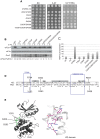
Figure 1. Structure-based sequence alignment of the YKD region of Gcn2.
The multiple sequence alignment of YKDs from 40 fungal Gcn2 sequences, identified on the far left by abbreviations of their species of origin, was built using the MUSCLE program. Residues are colored according to evolutionary sequence variation as analyzed with the CONSURF on-line server, with magenta corresponding to the most conserved residues, and teal indicating the most variable. Numbering corresponds to residue positions in full-length S. cerevisiae Gcn2 (residues 280–534). Regions of predicted α-helical and β-strand secondary structures within the YKD are denoted above the S. cerevisiae sequence, based on the alignment of Gcn2 YKD sequences with authentic KDs in Fig. S1A–G. Substitutions (described below) conferring Gcn phenotypes are shown in red, those conferring Gcd phenotypes are shown in green, and those preserving WT function are shown in black.
Figure 2
Figure 2. Predicted three-dimensional structure and sequence conservation of surface residues of the Gcn2 YKD.
The degree of sequence conservation of Gcn2 YKD residues, shown in Fig. 1, was projected onto the three-dimensional structure of the authentic Gcn2 KD monomer using the CONSURF program. Yellow indicates amino acids for which the data are insufficient to calculate a reliable conservation grade. Except for residues in β3, the most highly conserved residues (magenta and red) are largely clustered on one surface (view II), whereas most of the variable residues (shades of blue) are on the opposite face (I). The most conserved regions (β3, αC, αE, αI, the activation loop and the hinge) are circled in red for emphasis.
Figure 3
Figure 3. Substitutions of predicted surface-exposed residues of the Gcn2 YKD conferring Gcn phenotypes in vivo.
(A) Transformants of gcn2Δ strain H1149 containing derivatives of low-copy plasmid p722 with WT GCN2, gcn2-m2, GCN2c-M788V, or the indicated mutations in the YKD were replica-plated to synthetic complete medium lacking uracil (SC-Ura) and SC-Ura plus 30 mM 3-AT and incubated for 3 d at 30°C. (B) Cultures of strains from panel A were grown in liquid SC medium lacking uracil and histidine to saturation, diluted into fresh medium at A600 of ∼0.2, and grown 6 h at 30°C. 3-AT was added at 10 mM to one culture for 1 h before harvesting (even-numbered lanes). WCEs were resolved by SDS-PAGE and subjected to Western analysis using the indicated specific antibodies and enhanced chemiluminescence to detect immune complexes. (C) Localization of the Gcn substitutions on the predicted 3-D structure of the Gcn2 YKD domain. Residues in the authentic Gcn2 KD that align in Fig. S1A–G with Gcn substitutions in the YKD from (A) were colored red and labeled on the crystal structure of the Gcn2 KD monomer.
Figure 4
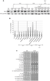
Figure 4. Mutations in the αI helix of the YKD constitutively activates Gcn2 in vivo.
(A) Transformants of gcn2Δ strain H1149 containing p722 derivatives with WT GCN2, gcn2-m2, GCN2c-M788V, or mutations affecting residues in helix αI of the YKD were replica-plated to SC-Ura, SC-Ura plus 30 mM 3-AT, or SD plus 0.5 mM 5-FT and 0.125 mM TRA (5FT/TRA) and incubated for 3 d at 30°C. (B) Cultures of strains from panel A were analyzed for levels of eIF2α-P as in Fig. 3B. Western signals on the upper panel (P-eIF2α) were quantified by scanning densitometry of exposed films using ImageJ software, normalized for the corresponding signals in the middle panel (total eIF2α), and the ratios of the two signals (eIF2α-P; eIF2α) are indicated below the lanes. Standard errors are less than 6.5% of the mean values shown. (C) Gcd phenotypes of the indicated mutants were quantified by measuring HIS4-lacZ expression. Strains from (A) were cultured in nonstarvation conditions as described in Materials and Methods and WCEs were prepared and assayed for β-galactosidase activities. Results are the means and S.E.M.s calculated from three transformants, with activity expressed as nanomoles of o-nitrophenyl-β-D-galactopyronoside hydrolyzed per minute per milligram of protein. (D) Locations on the predicted structure of the YKD domain of Gcd substitutions (from panel A; green) and a subset of Gcn substitutions in αE and αI (red) determined as in Fig. 3C.
Figure 5
Figure 5. Substitutions in the predicted hinge of the YKD constitutively activate Gcn2 in vivo.
(A) Transformants of gcn2Δ strain H1149 harboring the indicated GCN2 alleles were analyzed for resistance to 3-AT and 5-FT/TRA as in Fig. 4A. (B) Strains from (A) were analyzed for eIF2α-P as in Fig. 4B. Standard errors are less than 8.5% of the mean values shown. (C) Strains from (A) were analyzed for HIS4-lacZ expression in non-starvation conditions as in Fig. 4C. (D) Structure-based sequence alignment and conservation of the hinge regions of the Gcn2 KD and YKD domains. Regions of predicted α-helical and β-strand secondary structures are denoted schematically above sequence. The HRD catalytic motif in the KD domain is underlined. Mutations causing Gcd phenotypes in each domain are indicated. (E) Locations of Gcd substitutions on the predicted YKD structure (left) and KD (right, in boldface) along with the residue interactions that rigidify the hinge in the KD domain (right). (Image on the right reproduced from Fig. 3C of Padyana et al. (2005)).
Figure 6
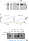
Figure 6. Effect of YKD substitutions on kinase activity and tRNA binding by purified Gcn2 in vitro.
(A) The indicated Gcn2 proteins (0.25 µg) were incubated with 3 µCi of [γ-32P]ATP (6000 Ci/mmol, Amersham), 1 µg of recombinant eIF2α−ΔC purified from E. coli, and 0.5 µg of bovine serum albumin in 20 µL of kinase assay buffer (20 mM Tris–HCl [pH 7.9], 50 mM NaCl, 10 mM MgCl2, 1 mM dithiothreitol, and 100 µM PMSF) for 5 to 15 min at 30°C. The samples were resolved by 8%–16% SDS–PAGE and subjected to autoradiography. Positions of autophosphorylated Gcn2 (Gcn2-P) and phosphorylated eIF2α−ΔC (eIF2α-P) are indicated. All results were cropped from the same autoradiogram except for those obtained for the R1325E variant, which was analyzed separately with the same WT protein. The results for WT in the latter autoradiogram were essentially identical to those shown here. (B) The extent of Gcn2 autophosphorylation and eIF2α phosphorylation at each time point in (A) was determined by quantifying the intensity of the relevant bands by phosphorimaging of the respective gel bands. Data obtained from 3 independent experiments was averaged and plotted with S.E.M.s as error bars. (C) Purified Gcn2 proteins were incubated at the indicated concentrations with [32P]-labeled total yeast tRNA in 20 µL of GMSA buffer. Gcn2-tRNA complexes were resolved by electrophoresis through a 1% agarose gel in 1×MOPS buffer (1.5 h, 100 V), transferred to a nitrocellulose membrane and visualized by autoradiography. Unbound [32P]-tRNA, which has a higher mobility, was present at essentially identical amounts in each lane at levels ∼15-fold higher than the WT Gcn2/tRNA complexes formed at 4 µM (data not shown). All results shown originate from the same gel except for those obtained for the R1325E variant, which was analyzed separately with the same WT protein. The results for WT in the latter gel were similar to those shown here.
Figure 7
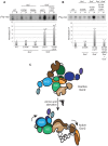
Figure 7. Certain Gcd substitutions in the YKD enhance coimmunoprecipitation of Gcn2 with LexA-HA-KD from yeast extracts.
(A) WCEs were prepared from transformants of gcn2Δ strain HQY132 bearing high-copy-number plasmid p2825 encoding LexA-HA-KD (720–999) and plasmids encoding wild-type Gcn2 (p630) or the indicated Gcn2 mutant. Aliquots of extracts containing 50 µg of protein were immunoprecipitated with anti-HA antibodies and the precipitates were resolved by SDS-PAGE and subjected to immunoblot analysis with anti-Gcn2 antibodies (upper panel) or anti-LexA antibodies (lower panel), and enhanced chemiluminescence was used to detect immune complexes. Input (I) lanes contain 5 µg of starting WCEs and pellet (P) lanes contain immune complexes recovered from 25 µg of WCEs. All results shown for each protein were cropped from the same immunoblot. (B) Densities of bands in I and P lanes of (A) were quantified by scanning densitometry of exposed films using ImageJ software, and P∶I ratios of Gcn2 signals were normalized to the LexA-HA-KD signals in the corresponding P lanes. The normalized ratios were calculated from three independent experiments and the average and S.E.M.s were plotted for each Gcn2 variant. (C) Transformants of gcn2Δ strain HQY132 containing high-copy-number plasmids encoding WT Gcn2 (p630) or the indicated YKD mutants were replica-plated to SC-Ura and SC-Ura plus 30 mM 3-AT and incubated for 3 d at 30°C.
Figure 8
Figure 8. Gcd YKD substitutions in αI enhance direct binding between YKD and KD segments in a manner suppressed by Gcn YKD substitutions in αE.
(A–B) YKD Gcn2 segments (WT or the indicated mutants) were translated in vitro with [35S]methionine and incubated with 50 µg of WCEs from transformants of gcn2Δ strain HQY132 expressing LexA-HA-KD(720–999). Reactions were immunoprecipitated with anti-HA antibodies and immune complexes were resolved by SDS-PAGE and visualized by fluorography. Input (I) lanes contain aliquots of reticulocyte lysates containing the input [35S]-labeled YKD fragments and pellet (P) lanes contain immune complexes recovered from reaction aliquots corresponding to 1/2 of the starting lysates. (B) Densities of bands in I and P lanes of (A) were quantified using a PhosphorImager Storm Scanner and ImageQuant software and P∶I ratios were calculated and normalized to that determined for the WT YKD fragment. Normalized ratios were calculated from three independent experiments and the average and S.E.M.s were plotted (C) Model of allosteric activation of the KD via direct interaction with the YKD, regulated by competing CTD∶KD association and tRNA binding to the HisRS-like domain. Inactive WT Gcn2 in nonstarved cells contains the CTD and HisRS-like domains engaged with the KD. These interactions could contribute directly to latency of the KD, but we propose here that the CTD acts indirectly to block the stimulatory YKD-KD interaction. YKD-CTD interaction helps to stabilize the inhibitory CTD-KD association in the inactive state. Uncharged tRNA binding to the HisRS domain and possibly also the CTD would activate Gcn2 by dissociating the CTD from the KD to enable the stimulatory YKD-KD interaction uncovered in this study. YKD Gcn and Gcd- substitutions would weaken or strengthen, respectively, the stimulatory YKD-KD interaction.

Similar articles

Cited by

References

    1. Lu PD, Harding HP, Ron D (2004) Translation reinitiation at alternative open reading frames regulates gene expression in an integrated stress response. J Cell Biol 167: 27–33. - PMC - PubMed
    1. Vattem KM, Wek RC (2004) Reinitiation involving upstream ORFs regulates ATF4 mRNA translation in mammalian cells. Proc Natl Acad Sci U S A 101: 11269–11274. - PMC - PubMed
    1. Hinnebusch AG (2005) Translational regulation of GCN4 and the general amino acid control of yeast. Annu Rev Microbiol 59: 407–450. - PubMed
    1. Guo F, Cavener DR (2007) The GCN2 eIF2α kinase regulates fatty-acid homeostasis in the liver during deprivation of an essential amino acid. Cell Metab 5: 103–114. - PubMed
    1. Hao S, Sharp JW, Ross-Inta CM, McDaniel BJ, Anthony TG, et al. (2005) Uncharged tRNA and sensing of amino acid deficiency in mammalian piriform cortex. Science 307: 1776–1778. - PubMed

Publication types

MeSH terms

Substances