. 2014 Jun 27;289(26):18582-92.
doi: 10.1074/jbc.M114.567420. Epub 2014 May 8.

Mutations in the X-linked intellectual disability gene, zDHHC9, alter autopalmitoylation activity by distinct mechanisms

Affiliations

Mutations in the X-linked intellectual disability gene, zDHHC9, alter autopalmitoylation activity by distinct mechanisms

David A Mitchell et al. J Biol Chem. .

Abstract

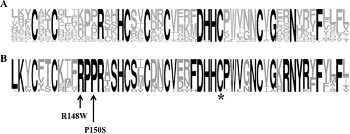
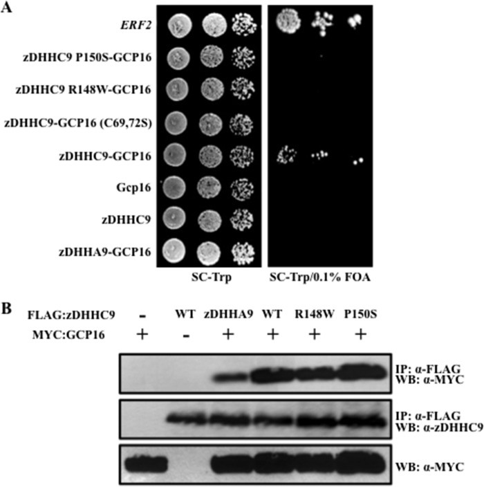
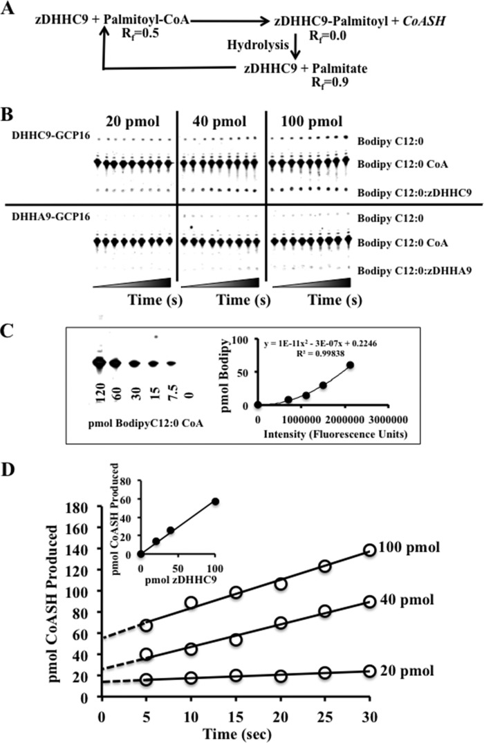
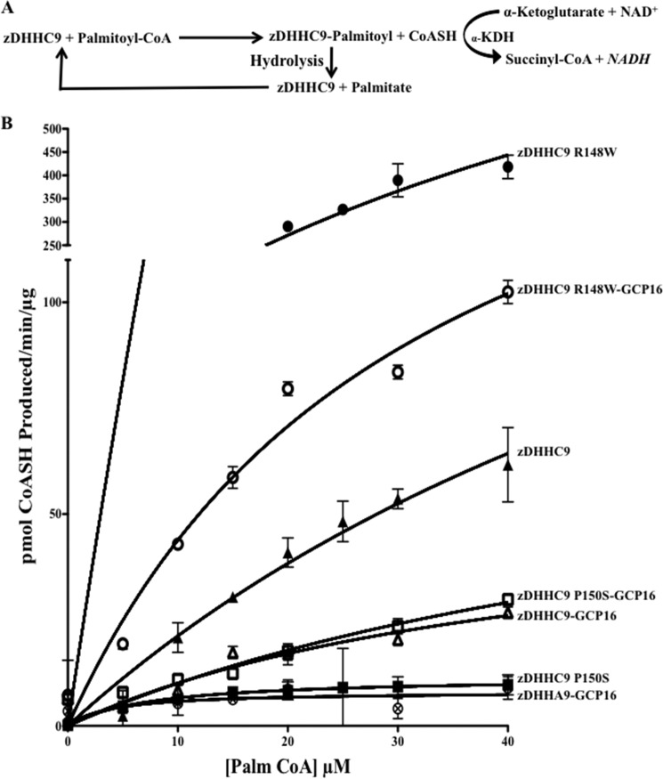
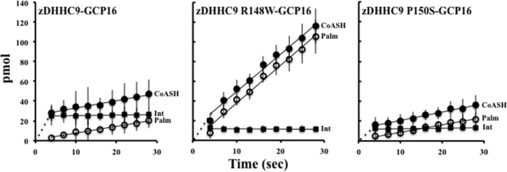
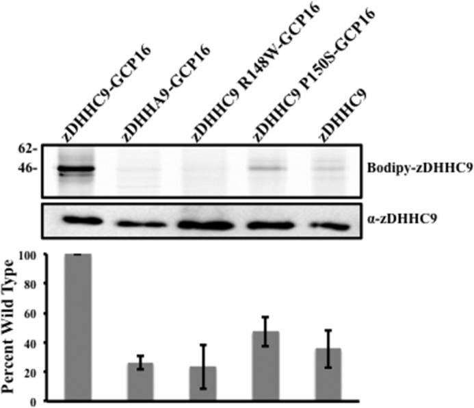
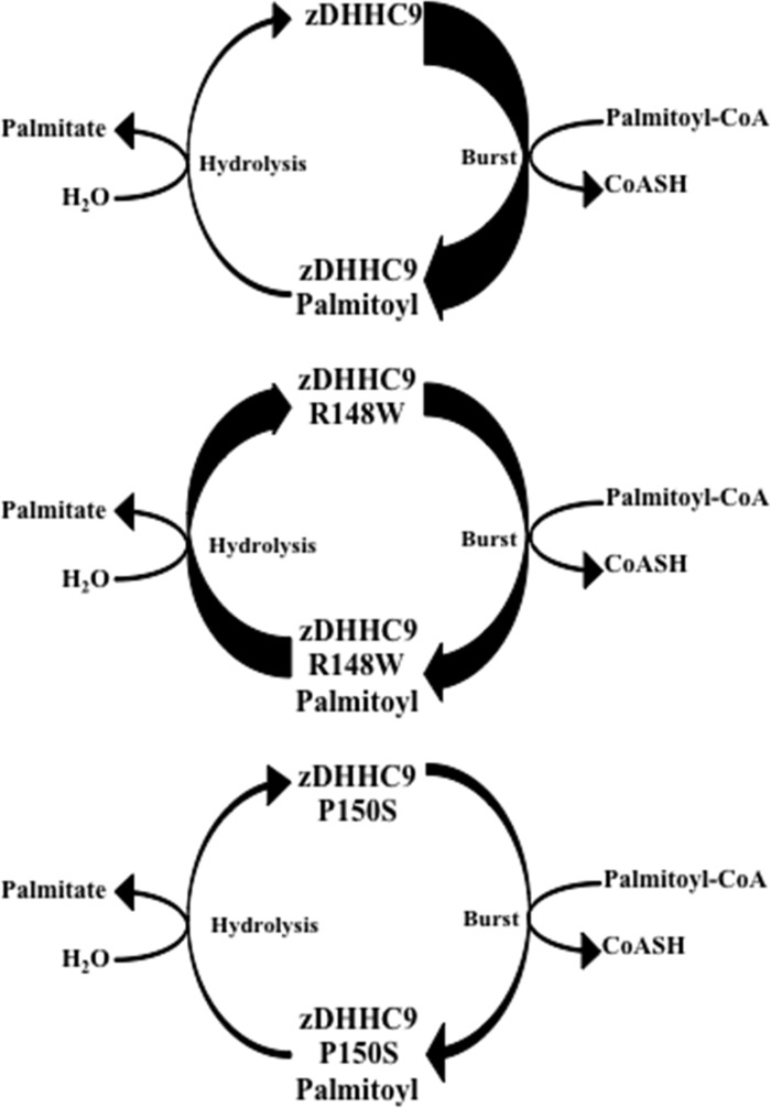
Early onset intellectual disabilities result in significant societal and economic costs and affect 1-3% of the population. The underlying genetic determinants are beginning to emerge and are interpreted in the context of years of work characterizing postsynaptic receptor and signaling functions of learning and memory. DNA sequence analysis of intellectual disability patients has revealed greater than 80 loci on the X-chromosome that are potentially linked to disease. One of the loci is zDHHC9, a gene encoding a Ras protein acyltransferase. Protein palmitoylation is a reversible modification that controls the subcellular localization and distribution of membrane receptors, scaffolds, and signaling proteins required for neuronal plasticity. Palmitoylation occurs in two steps. In the first step, autopalmitoylation, an enzyme-palmitoyl intermediate is formed. During the second step, the palmitoyl moiety is transferred to a protein substrate, or if no substrate is available, hydrolysis of the thioester linkage produces the enzyme and free palmitate. In this study, we demonstrate that two naturally occurring variants of zDHHC9, encoding R148W and P150S, affect the autopalmitoylation step of the reaction by lowering the steady state amount of the palmitoyl-zDHHC9 intermediate.

Keywords: Enzyme Mechanism; Post-translational Modification (PTM); Protein Acylation; Protein Palmitoylation; Ras Protein; X-linked Intellectual Disability; zDHHC Proteins.


Figures

FIGURE 1.
FIGURE 1.
Comparison of the consensus protein acyltransferase sequences with the amino acid sequence of zDHHC9. A, WebLogo sequence consensus representation of the 51-amino acid DHHC cysteine-rich domain using the 23 human protein acyltransferase protein sequences. The size of the amino acid letter code denotes the amount of conservation at that position; the boldface residues represent a minimum of 90% conservation. B, WebLogo sequence consensus representation of zDHHC9 and related cysteine-rich domains throughout evolution. The consensus is derived from the following: human zDHHC9, zDHHC14, and zDHHC18; S. cerevisiae Erf2; Ciona intestinalis zDHHC14 (GI: 198427890); Nematostella vectensis zDHHC9 (GI: 156377027); Drosophila melanogaster zDHHC9 (GI: 281366130); Strongylocentrotus purpuratis zDHHC14 (GI: 390334287), Amphimedon queenslandica zDHHC14 (GI: 340375495), Schistoma mansoni zDHHC9 (GI: 256084522), and Danio rerio zDHHC9 (GI: 158518002). An asterisk denotes the proposed catalytic cysteine residue of the DHHC motif (DHHC9 C169A). The arrows indicate the mutated positions of two naturally occurring isolates of human zDHHC9 (zDHHC9 R148W and zDHHC9 P150S).
FIGURE 2.
FIGURE 2.
zDHHC9-GCP16 interaction is required to suppress the loss of ERF2. A, to determine the ability of different zDHHC9 alleles and zDHHC9-GCP16 constructs to suppress the loss of ERF2 in S. cerevisiae, we performed a functional plasmid shuffle assay (22). Strain RJY1330 harboring plasmids expressing zDHHC9, zDHHC9-GCP16, zDHHC9 R148W-GCP16, zDHHC9 P150S-GCP16, zDHHC9-GCP16 (C69S,C72S), zDHHA9-GCP16, or ERF2 from the GAL1,10 promoter, were monitored for RAS2-independent growth on synthetic medium lacking tryptophan and supplemented with 5′-fluoroorotic acid (5′-FOA) to select for those strains capable of losing the URA3-linked RAS2 plasmid. Strains were initially grown in liquid synthetic medium lacking tryptophan and were spotted in 10-fold dilutions (50 × 103 initial cfu) on solid medium lacking tryptophan (top panel) and medium lacking tryptophan supplemented with 0.1% 5′-fluoroorotic acid (bottom panel). The plates were incubated for 4 days at 30 °C. B, mutations at zDHHC9 amino acid positions R148W and P150S do not interfere with the GCP16 interaction. Immunoprecipitates (IP) from whole cell extracts isolated from strain RJY1330 expressing zDHHC9-GCP16, zDHHC9, GCP16, zDHHA9-GCP16, zDHHC9 R148W-GCP16, and zDHHC9 P150S-GCP16 proteins were isolated using anti-FLAG-conjugated agarose. The immunoprecipitates were separated using SDS-PAGE (12%), transferred to nitrocellulose, and probed with antibodies to the c-Myc epitope (to identify myc-GCP16) or antibodies to the FLAG epitope (to identify zDHHC9). Western blot (WB) analysis of the whole cell extract utilizing antibodies to c-Myc was performed to demonstrate the presence of myc-GCP16 in the extracts.
FIGURE 3.
FIGURE 3.
Active site titration of zDHHC9-GCP16 complexes. A, schematic representation of the protein acyltransferase autopalmitoylation reaction. The reactants and products were separated using TLC as follows: 1) BODIPY® C12:0-CoA (Rf = 0.5); 2) zDHHC9-BODIPY® C12:0 intermediate (Rf = 0.0), and 3) BODIPY® C12:0 (Rf = 0.9). B, active site analysis using thin layer chromatography separation of autopalmitoylation reactants and products. Active site titration analysis measuring the amount of CoASH production was performed at room temperature (24 °C) using 20, 40, and 100 pmol of zDHHC9-GCP16 complex (or zDHHA9-GCP16 complex) and BODIPY® C12:0-CoA as the acyl donor. Time points were taken at 0, 5, 10, 15, 20, 25, 30, 45, and 60 s. The t0 point was taken prior to enzyme addition. As a control for nonspecific activity, 20, 40, and 100 pmol of the inactive zDHHA9-GCP16 enzyme were assayed, and the values were subtracted from the values determined for the wild type complexes (n = 2). C, amounts of the products were determined using a standard curve of known amounts of BODIPY® C12:0 CoA (n = 2). D, graphical representation of active site titration data. The amount of CoASH is calculated as the amount of BODIPY® C12:0 formed plus the amount of zDHHC9-BODIPY® C12:0 intermediate at the TLC origin (Rf = 0). There is a nearly 1 to 1 correlation between the amount of zDHHC9-GCP16 used and the amount of CoASH produced (slope = 0.58) (inset). The data represent the average of two (n = 2) experiments.
FIGURE 4.
FIGURE 4.
zDHHC9 undergoes autopalmitoylation in the absence of GCP16. A, schematic representation of the coupled in vitro autopalmitoylation reaction (see under “Experimental Procedures”). B, graphical representation of the rate of CoASH production (pmol/min/μg) over the first 10 min of the reaction versus the concentration (μm) of acyl donor (palmitoyl-CoA). The ordinate axis is broken to accommodate the activity measurement of zDHHC9 R148W. Complexes are denoted as follows: zDHHA9-GCP16 (circle with X); zDHHC9 P150S-GCP16 (open square); zDHHC9-GCP16 (open triangle); zDHHC9 R148W-GCP16 (open circle); zDHHC9 (closed triangle); zDHHC9 R148W (closed circle), and zDHHC9 P150S (closed square). Curve fit was performed using Prizm™ software.
FIGURE 5.
FIGURE 5.
In vitro measurement of zDHHC9-BODIPY® C12:0 intermediate hydrolysis rate. The reaction highlighted in Fig. 3A was used to determine the rate of CoASH production, rate of zDHHC9-BODIPY® C12:0 hydrolysis, and the amount of zDHHC9-BODIPY® C12:0 intermediate formed. The reactions were terminated by spotting onto the TLC plate. The plates were developed using a mobile phase of n-butyl alcohol/water/acetic acid (50:30:20) (v/v/v). BODIPY® fluorescence was visualized using excitation 488-nm/emission 520-nm filters (Typhoon, GE Healthcare). The amount (in pmol) of CoASH (CoASH, closed circles), BODIPY® C12:0 (Palm, open circles), and zDHHC9-BODIPY® C12:0 intermediate (Int, closed squares) produced are graphically represented for the zDHHC9-GCP16, zDHHC9 R148W-GCP16, and zDHHC9 P150S-GCP16 complexes. The data represent three independent experiments.
FIGURE 6.
FIGURE 6.
Mutations at positions Arg-148 and Pro-150 affect the autopalmitoylation activity of zDHHC9. In vitro autopalmitoylation reactions using BODIPY® C12:0-CoA as the acyl donor. Upper panel, in vitro steady state autopalmitoylation reactions were separated using SDS-PAGE under nonreducing conditions, and the fluorescence was visualized using excitation 488-nm/emission 520-nm filters (Typhoon, GE Healthcare). Lower panel, representative Western blot analysis used to quantify the amount of zDHHC9 and zDHHC9 mutants. The amount of BODIPY® C12:0 complexed to zDHHC9 was determined by using excitation 488-nm/emission 520-nm filters.
FIGURE 7.
FIGURE 7.
Model of autopalmitoylation reaction for mutant zDHHC9 complexes. The thickness of the arrow indicates the relative amount of “burst” or rate of hydrolysis.

References

    1. Zhang M. M., Tsou L. K., Charron G., Raghavan A. S., Hang H. C. (2010) Tandem fluorescence imaging of dynamic S-acylation and protein turnover. Proc. Natl. Acad. Sci. U.S.A. 107, 8627–8632 - PMC - PubMed
    1. Rocks O., Gerauer M., Vartak N., Koch S., Huang Z. P., Pechlivanis M., Kuhlmann J., Brunsveld L., Chandra A., Ellinger B., Waldmann H., Bastiaens P. I. (2010) The palmitoylation machinery is a spatially organizing system for peripheral membrane proteins. Cell 141, 458–471 - PubMed
    1. Fukata Y., Fukata M. (2010) Protein palmitoylation in neuronal development and synaptic plasticity. Nat. Rev. Neurosci. 11, 161–175 - PubMed
    1. Zhang W., Trible R. P., Samelson L. E. (1998) LAT palmitoylation: its essential role in membrane microdomain targeting and tyrosine phosphorylation during T cell activation. Immunity 9, 239–246 - PubMed
    1. Joseph M., Nagaraj R. (1995) Conformations of peptides corresponding to fatty acylation sites in proteins. A circular dichroism study. J. Biol. Chem. 270, 19439–19445 - PubMed

Publication types

LinkOut - more resources