. 2014 Dec;28(12):2376-87.
doi: 10.1038/leu.2014.126. Epub 2014 Apr 3.

FAS-antisense 1 lncRNA and production of soluble versus membrane Fas in B-cell lymphoma

Affiliations

FAS-antisense 1 lncRNA and production of soluble versus membrane Fas in B-cell lymphoma

L Sehgal et al. Leukemia. 2014 Dec.

Abstract

Impaired Fas-mediated apoptosis is associated with poor clinical outcomes and cancer chemoresistance. Soluble Fas receptor (sFas), produced by skipping of exon 6, inhibits apoptosis by sequestering Fas ligand. Serum sFas is associated with poor prognosis of non-Hodgkin's lymphomas. We found that the alternative splicing of Fas in lymphomas is tightly regulated by a long-noncoding RNA corresponding to an antisense transcript of Fas (FAS-AS1). Levels of FAS-AS1 correlate inversely with production of sFas, and FAS-AS1 binding to the RBM5 inhibits RBM5-mediated exon 6 skipping. EZH2, often mutated or overexpressed in lymphomas, hyper-methylates the FAS-AS1 promoter and represses the FAS-AS1 expression. EZH2-mediated repression of FAS-AS1 promoter can be released by DZNeP (3-Deazaneplanocin A) or overcome by ectopic expression of FAS-AS1, both of which increase levels of FAS-AS1 and correspondingly decrease expression of sFas. Treatment with Bruton's tyrosine kinase inhibitor or EZH2 knockdown decreases the levels of EZH2, RBM5 and sFas, thereby enhancing Fas-mediated apoptosis. This is the first report showing functional regulation of Fas repression by its antisense RNA. Our results reveal new therapeutic targets in lymphomas and provide a rationale for the use of EZH2 inhibitors or ibrutinib in combination with chemotherapeutic agents that recruit Fas for effective cell killing.


Conflict of interest statement

CONFLICT OF INTEREST

The authors declare no competing financial interests.

Figures
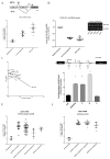
Figure 1
Figure 1. Expression of FAS-AS1 lncRNA inversely correlates with the levels of sFas mRNA in lymphomas
(A–B) The total cellular RNA isolated from healthy donors (n=4), primary lymphoma tissues (n=6), and B-cell lymphoma-derived cell lines (n=11) was analyzed for (A) the average sFas/total Fas RNA levels normalized to 1 in the healthy donors, and (B) FAS-AS1 lncRNA by qRT-PCR normalized to GAPDH. The inset image shows the expression of FAS-AS1 as determined by the RT-PCR from the total cellular RNA isolated from healthy donor (HD 35), primary lymphoma patient samples (P559; BL, P393; DLBCL, P225; MCL, P227;MCL). (C) The levels of FAS-AS1 lncRNA plotted against soluble Fas mRNA levels, to illustrate their relationship (n=10) in various cell lines with Pearson correlation coefficient of r2 = −0.58. (D) ChIP analysis of H3K27me3 on the promoter regions of FAS-AS1 using specific antibodies that recognize H3K27me3 and control IgG. The locations of primers used for the ChIP assay are indicated by arrows. The specific primer from the FAS-AS1 lncRNA (region C, +700) serves as the negative control, and the qPCR for α-satellite DNA served as a positive control for the H3K27me3 ChIP. Statistical significance was calculated by student’s T test(α satellite: P < 0.01, A: P < 0.01, B: P < 0.01, C: P < 0.11) (E–F) EZH2 expression levels in (E) 89 primary B cell lymphoma tissues and (F) 34 lymphoma-derived cell lines (Oncomine database; www.oncomine.org). Data sets in a single panel were from the same study. Gene expression profile of EZH2 was log transformed and normalized in different histopathological types of lymphomas.
Figure 2
Figure 2. High EZH2 expression and hyper-methylation of the FAS AS1 promoter is reversed upon treatment with DZNeP
(A) SP53 cells were treated with methyltransferase inhibitor DZNeP in different concentration in μM for 24 hours and analyzed for the levels of EZH2, H3K27me3, and Actin by IB (B) DZNeP 1uM was tested in vitro for the inhibition of EZH2-mediated methylation using microtiter plate precoated with histone H3 peptide substrate and H3K27me3-specific antibody. (P<0.01) (C–D) ChIP assays were performed to determine the extent of H3K27me3 on FAS-AS1 promoter in the presence or absence of the 1 uM DZNeP. The positions for the various primers are determined from the transcription start site (TSS, A;−400, B; −900) and the various control primers in Z-138 cells. The specific primer from the FAS-AS1 lncRNA (region C, +700) serves as the negative control, and the qPCR for α-satellite DNA served as a positive control for the H3K27me3 ChIP. The levels of methylation levels are reduced in b and c regions of FAS-AS1 lncRNA promoter. (E & F) Z-138 cells were treated with 1 uM DZNeP for 24 hours and total cellular RNA was isolated and subjected to qRT-PCR with primers specific for FAS-AS1 lncRNA(E) (P < 0.01) and sFas mRNA (F) (P < 0.01). (G) Lymphoma derived cell lines were incubated with and without 1 uM DZNeP for 24 hours and concentrations of sFas in the media were determined by ELISA (* denotes P < 0.01).
Figure 3
Figure 3. DZNeP stimulates the expression of lymphoma cell surface Fas and increases their sensitivity to FasL
(A) Surface levels of Fas in control and DZNeP-treated cells were analyzed by flow cytometry after staining of cells with anti-Fas antibody UB2 conjugated with PE. Representative results from one of least three independent experiments are shown (* denotes P < 0.01, ** denotes P < 0.01). (B) DZNeP-treated cells were incubated with FLAG-tagged FasL (100ng) for 20 minutes and the levels of bound ligand were analyzed by flow cytometry after staining with an anti-FLAG-PE secondary antibody or isotype control IgG-PE antibody. Representative data of 3 independent experiments are shown. The vertical dotted line intersecting the graph shows the increase in the Fas ligand binding to cells. (C) Cells were treated with DZNeP for 24hrs followed by incubation with FasL (100ng/mL) for 12 hours. Cells were analyzed by PI staining and flow cytometry for sub-G1 fraction. Mean value and SEM from at least 3 independent experiments with 3 replicates in each are shown. (D) The cell extracts from Z-138 cells treated with DZNeP, FasL or their combination as in (C) were analyzed by IB analysis for activation of indicated Fas signaling proteins.
Figure 4
Figure 4. Ectopic expression of FAS-AS1 inhibits alternative splicing of Fas and sensitizes lymphoma cells to FasL
(A–B) Z-138 cells were transfected with pcDNA3 expressing FAS-AS1 lncRNA or empty vector. The total cellular RNA was isolated 48 hrs post transfection and subjected to qRT-PCR with (A) FAS-AS1 to determine the efficiency of transfection (P < 0.01) or (B) sFas-specific primers to determine the sFas/mFas isoform ratio (P < 0.01). (C) Empty vector or pcDNA3-FAS-AS1 were co-transfected with pEGFP-N1. Surface levels of mFas in control and FAS-AS1 transfected cells (sorted based on GFP signal) were analyzed by flow cytometry after staining of cells with anti-Fas antibody UB2 conjugated with PE. Representative results from one of three independent experiments are shown. (D) Sorted cells from (C) were challenged with FasL (100ng/mL) for 12 hours and analyzed after PI staining and flow cytometry (Vector; P < 0.09, FAS-AS1; P < 0.01). (E) Z-138 cells were transfectd with increasing concentrations of FAS-AS siRNA and the level of FAS-AS1 lncRNA was analyzed by RT-PCR. (F & G) Z-138 cells transfectd with 25nM of scrambled- or FAS-AS1-specific siRNA were pre-treated with DZNeP, challenged with FasL (100ng/mL) for 12 hours, stained with PI, and analyzed for apoptosis by flow cytometry (DZNeP+sFas: Vector; P < 0.09, FAS-AS1 siRNA; P < 0.01).
Figure 5
Figure 5. Knockdown of EZH2 inhibits alternative splicing of Fas and sensitizes lymphoma cells to FasL
(A) Z-138 cells were transfected with either pLKO.1 vector control or pLKO.1 expressing shRNA targeting EZH2. 60 hrs post transfection the protein lysates were blotted with EZH2 antibody. Actin served as loading control. (B) The total cellular RNA was isolated 60 hrs post transfection and subjected to qRT-PCR with primers specific to EZH2,FAS-AS1 and sFas (vector control vs. shRNA EZH2.1; P < 0.01). (C) Empty vector- or pLKO.1carrying shRNA to EZH2 transfected cells were analyzed for the level of membrane Fas by flow cytometry. Representative results of three replicates are shown (vector control vs. shRNA EZH2.1; P < 0.01). (D) Empty vector- pLKO.1carrying shRNA to EZH2 transfected cells were challenged with FasL (100ng/mL) for 12 hours and analyzed after PI staining and flow cytometry (vector control vs. shRNA EZH2.1; P < 0.001).
Figure 6
Figure 6. FAS-AS1 binds RBM5 and interferes with RBM5-mediated exon 6 skipping of Fas pre-mRNA
(A) Prediction of the interaction map of FAS-AS1 lncRNA and RBM5 generated by use of the catRapid program (http://service.tartaglialab.com/page/catrapid_group). Interaction propensity is measured in procedure defined units (p.d.u.). The positive interaction propensity is shown for binding of FAS-AS1 lncRNA to the RNA Recognition Motifs in RBM5 [RRM1 98–178, RRM2 231–315]. A schematic representation of RBM5 protein is shown with location of RRM1 and RRM2 domains. (B) Whole-cell lysates from control or 1 uM DZNeP-treated Z-138 cells were immunoprecipitated with anti-RBM5 antibody or control IgG. Precipitates were analyzed by IB analysis for RMB5 and by qRT-PCR for the levels of co-precipitated FAS-AS1 lncRNA. The whole cell extracts from B were analyzed in immunoblots for EZH2, RBM5, H3K27me3, and H3 levels. (C–D) Real time-PCR analysis using primers for FAS-AS1 (RBM5 DZNeP, p<0.01) (C) and Fas pre-mRNA (RBM5 DZNeP, P < 0.01) (D) in immunoprecipitants of control antibody and RBM5 antibody on extracts of Z-138 cells treated with DZNeP.
Figure 7
Figure 7. Ibrutinib inhibits the expression of EZH2 and RBM5 and sensitizes lymphoma cells to Fas-mediated apoptosis
(A) Z-138 cells were treated with 10 uM ibrutinib (PCI) for 48 hours. Total cellular RNA was extracted and analyzed by qRT-PCR for the expression of mFas, sFas, EZH2, and RBM5 mRNAs and FAS-AS1 lncRNA. (P < 0.001) (B) Cell surface levels of Fas in control and ibrutinib-treated cells were stained with the PE-conjugated anti-Fas antibody UB2 and analyzed by flow cytometry. Representative results from at least three independent replicates are shown. (P < 0.01) (C) BJAB, SU-DHL-9 and Z-138 cells were treated with 10 μM Ibrutinib (24hrs) and then challenged with FasL (100ng/mL) for 12 hours, stained with PI and analyzed by flow cytometry. Representative results from at least three independent replicates are shown (P < 0.001).
Figure 8
Figure 8. Regulation of sFas expression and sensitization of lymphoma cells to Fas-mediated apoptosis by DZNeP and ibrutinib
(A) BTK promotes the expression of RBM5 and EZH2 in lymphoma cells. RBM5 promotes skipping of exon 6 from Fas pre-mRNA leading to production of soluble decoy sFas. EZH2 tri-methylates the FAS-AS1 promoter and represses transcription of FAS-AS1 lncRNA. (B) In DZNeP-treated cells, EZH2 activity is compromised, which promotes the expression of FAS-AS1 lncRNA. FAS-AS1 lncRNA binds and sequesters RBM5 protein preventing RBM5-mediated skipping of exon 6 and ultimately lower sFas, this leads to sensitization to Fas-mediated apoptosis. Because DZNeP affects EZH2, but not RBM5, the sFas production because of RBM5 still remains and the effect depends on the residual levels of RBM5 expression. (C) Ibrutinib treatment decreases expression of both EZH2 and RBM5 combined with enhanced expression of FAS-AS1 further which results in more pronounced decrease in the levels of sFas corresponding to more efficient interference with crosstalk between EZH2 and RBM5-mediated skipping of exon 6 and thus production of sFas. This translates into sensitization to Fas-mediated apoptosis.

References

    1. Nagata S. Fas and Fas ligand: a death factor and its receptor. Advances in Immunology. 1994;57:129–144. - PubMed
    1. Xerri L, Bouabdallah R, Devilard E, Hassoun J, Stoppa AM, Birg F. Sensitivity to Fas-mediated apoptosis is null or weak in B-cell non-Hodgkin's lymphomas and is moderately increased by CD40 ligation. British Journal of Cancer. 1998 Jul;78(2):225–232. - PMC - PubMed
    1. Thompson CB. Apoptosis in the pathogenesis and treatment of disease. Science. 1995 Mar 10;267(5203):1456–1462. - PubMed
    1. Lowe SW, Lin AW. Apoptosis in cancer. Carcinogenesis. 2000 Mar;21(3):485–495. - PubMed
    1. Rudin CM, Thompson CB. Apoptosis and disease: regulation and clinical relevance of programmed cell death. Annual Review of Medicine. 1997;48:267–281. - PubMed

Publication types

MeSH terms