. 2014 May;32(5):453-61.
doi: 10.1038/nbt.2890.

Detecting and characterizing circular RNAs

Affiliations

Detecting and characterizing circular RNAs

William R Jeck et al. Nat Biotechnol. 2014 May.

Abstract

Circular RNA transcripts were first identified in the early 1990s but knowledge of these species has remained limited, as their study through traditional methods of RNA analysis has been difficult. Now, novel bioinformatic approaches coupled with biochemical enrichment strategies and deep sequencing have allowed comprehensive studies of circular RNA species. Recent studies have revealed thousands of endogenous circular RNAs in mammalian cells, some of which are highly abundant and evolutionarily conserved. Evidence is emerging that some circRNAs might regulate microRNA (miRNA) function, and roles in transcriptional control have also been suggested. Therefore, study of this class of noncoding RNAs has potential implications for therapeutic and research applications. We believe the key future challenge for the field will be to understand the regulation and function of these unusual molecules.


Figures
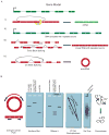
Figure 1
Figure 1. Splicing products and methods for detection
(A) Several mechanisms can form an apparent backsplice, illustrated here for a gene model in which exons are shown as rectangles, introns as thin lines and the transcription start site as a right-angled arrow. DNA is in green and RNA is in red. (i) Reverse transcriptase (RT) template switching, in which the RT enzyme transcribes another copy of an upstream exon. (ii) Tandem duplications in the DNA template resulting in repeated exons. (iii) Trans-backsplicing, in which one RNA molecule is spliced to another (shown by curved black line). Regular splicing events are shown as angled black lines. (iv) Exonic circRNAs can form by cis-backsplicing in which exons from the same RNA molecule are spliced together to form a circle (backsplicing shown by curved black line). (B) Molecular assays to distinguish exonic circRNA from other backsplice products, and diagrammatic representation of the expected results. (i) Divergent primers that would amplify in outward facing directions with respect to genomic sequence become properly inward facing an produce discreet amplicons when a backsplice connects outside sequences. (ii) The expected migration distance of a canonical linear RNA in a denaturing agarose gel, as well as the relative migration of exonic circRNA and RNAs resulting from trans-splicing or tandem duplication. (iii) Migration of RNA through an agarose gel before and after RNAse H treatment. Circular RNA, uniquely, results in a single band after being cut once. (iv) Two dimensional gel electrophoresis through two differently crosslinked polyacrylamide gels separates circular RNAs into an off-diagonal curve. (v) Gel-trapping holds circular RNAs in the well of an electrophoresis gel as linear RNAs migrate away. (vi) Exonuclease enrichment degrades linear RNAs while leaving a pool enriched for circular RNA.
Figure 2
Figure 2. Sequencing-based methods for identification of exonic circRNA
Several informatics methods have been used to identify the locations of backsplices using deep sequencing data from rRNA depleted RNA. (A) Paired-end reads may be mapped separately to the annotated transcriptome and the location of a backsplice inferred when the pairs have opposite orientations on either side of one or more splice sites. (B) Paired-end reads may be mapped directly to the genome and multiple reads that map out of genomic order suggest there might be a nearby backsplice. Stronger evidence is provided by the presence of multiple such reads accumulating in fixed “windows” of set length tiled across the genome. (C) A read can be segmented so that different parts of the read can be mapped to different parts of the genome; this allows backsplices to be mapped at nucleotide resolution without existing annotations.
Figure 3
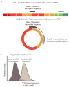
Figure 3. Informatic approach to identifying false positive backsplices
(A) Illustration of paired ends that would be inconsistent with underlying exonic circRNA. In the first example, the first read of the pair maps to a backsplice, but its paired read maps to an exon that is inconsistent with a circRNA because it lies in an exon beyond those involved in the backsplice. In the second example, the first read of the pair again maps to the backsplice, but its pair now maps to an exon between those two involved in the backsplice, consistent with a circRNA. The two sets of reads are then used to generate two distributions (B) that may be used to set empirical false discovery rates based on mapping quality features.
Figure 4
Figure 4. A combined biochemical and informatic approach to identify exonic circRNAs in mammalian cells, ‘CircleSeq’
Developed in part by our group. (A) Total RNA is depleted of rRNA, for example using RiboMinus or RiboZero methods rRNA depleted samples are split; one aliquot is treated with the RNase R exonuclease and the other is subjected to a mock treatment. Sequencing libraries are prepared from each and before deep sequencing and comparison of the samples, optimal RNase R digestion is confirmed by checking by quantitative RT-PCR that known exonic circRNAs are enriched in the RNase treated sample (not shown). Informatic approaches, such as that shown in Figure 2c can be applied to the data. (B) An example of data from CircleSeq in human tissues for the ANRIL noncoding RNA locus. Normalized mapped read depth for the mock treated sample (green, top) and for RNase R treated samples (orange, bottom) are shown (note different scales). Regions where there is enrichment of a group of exons in the rRNA-treated sample – suggestive of a circRNA – are shown in blue. Exons outside of these regions are depleted in the RNAase-treated sample.
Figure 5
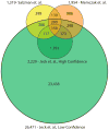
Figure 5. Comparison of circular RNAs identified by genomic studies
Numbers of circular RNAs identified by three genomic studies,, and the numbers of circular RNAs overlapping among these studies. Overlapping regions represent the number of species where both the splice donor and splice acceptor of the backsplice were identical in two or more works. Data from Memczak et. al. 2013 were taken from Supplementary Table 2 in that article. Two circles shown for Jeck et. al. show a high and low confidence set of circles reported in that work. High confidence circles were observed in two sequencing data sets from unenriched sequencing data, while low confidence circles were observed in only one. Sites taken from Salzman et. al. 2012 are from Supplementary Table S2 in that article, which required our inference of the genomic locations using RefSeq gene annotations.
Figure 6
Figure 6. Possible mechanisms for formation of exonic circRNAs
(A) direct backsplicing, in which two unspliced introns within a transcript pair and the intervening introns are spliced through the usual mechanism. Here, a branch point in the 5’ intron attacks the splice donor of the 3’ intron. The 3’ splice donor then completes the backsplice by attacking the 5’ splice acceptor. This forms a circular RNA. (B) Exon skipping resulting in circular RNA formation. In this case an exon skipping event creates a lariat containing an exon. This lariat is spliced internally, removing the intronic sequence and producing a circular RNA. (C) Design schema for an overexpression vector that produces circular RNA. This design was employed for CDR1as and SRY. Here the exon made to be circularized is included in an overexpression construct with upstream and downstream intron sequence. Additional sequence is placed 5’ in the transcript to produce pairing with downstream intron, as in the direct backsplicing mechanism (shown).
Figure 7
Figure 7. Genomic features of circular RNA
Circular RNAs are generated from exons that are longer than average, and are flanked by long introns containing inverted ALU repeats, as exemplified by the HIPK3 gene, shown with CircleSeq data set. In the HIPK3 gene, ribosomal RNA depleted sequencing (green) resulted in substantially more coverage of a single exon, which also demonstrated a large number of backsplice sequences. These sequences are also demonstrated to be true circles by their enrichment by RNAse R (brown), while all other exons are degraded. This circularized exon is the longest of the gene, is surrounded by the longest introns of the gene, and is flanked by ALU elements in a complementary orientation

Similar articles

Cited by

References

    1. Nigro JM, et al. Scrambled exons. Cell. 1991;64:607–613. - PubMed
    1. Cocquerelle C, Mascrez B, Hétuin D, Bailleul B. Mis-splicing yields circular RNA molecules. The FASEB Journal: Official Publication of the Federation of American Societies for Experimental Biology. 1993;7:155–160. - PubMed
    1. Kos A, Dijkema R, Arnberg AC, van der Meide PH, Schellekens H. The hepatitis delta (delta) virus possesses a circular RNA. Nature. 1986;323:558–560. - PubMed
    1. Sanger HL, GKDRHJGAKK Viroids are single-stranded covalently closed circular RNA molecules existing as highly base-paired rod-like structures. Proc Natl Acad Sci USA. 1976;73:3852. - PMC - PubMed
    1. Hansen TB, et al. Natural RNA circles function as efficient microRNA sponges. Nature. 2013 doi: 10.1038/nature11993. - DOI - PubMed

Publication types