. 2014 May 8;3(3):e000747.
doi: 10.1161/JAHA.113.000747.

Identification of Hic-5 as a novel scaffold for the MKK4/p54 JNK pathway in the development of abdominal aortic aneurysms

Affiliations

Identification of Hic-5 as a novel scaffold for the MKK4/p54 JNK pathway in the development of abdominal aortic aneurysms

Xiao-Feng Lei et al. J Am Heart Assoc. .

Abstract

Background: Although increased amounts of reactive oxygen species in the pathogenesis of abdominal aortic aneurysm (AAA) are well documented, the precise molecular mechanisms by which reactive oxygen species induce AAAs have not been fully elucidated. This study focused on the role of hydrogen peroxide-inducible clone 5 (Hic-5), which is induced by hydrogen peroxide and transforming growth factor-β, in the cellular signaling of AAA pathogenesis.

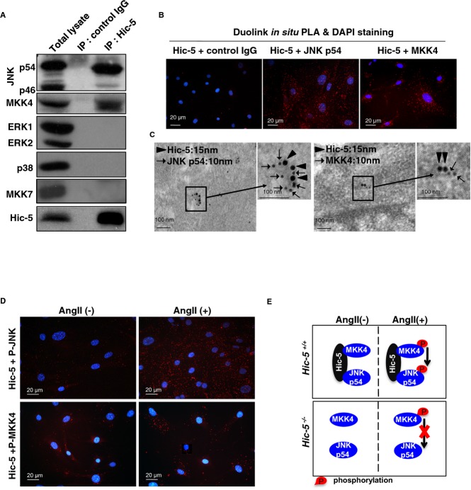
Methods and results: Using the angiotensin II-induced AAA model in Apoe(-/-) mice, we showed that Apoe(-/-)Hic-5(-/-) mice were completely protected from AAA formation and aortic rupture, whereas Apoe(-/-) mice were not. These features were similarly observed in smooth muscle cell-specific Hic-5-deficient mice. Furthermore, angiotensin II treatment induced Hic-5 expression in a reactive oxygen species-dependent manner in aortic smooth muscle cells in the early stage of AAA development. Mechanistic studies revealed that Hic-5 interacted specifically with c-Jun N-terminal kinase p54 and its upstream regulatory molecule mitogen-activated protein kinase kinase 4 as a novel scaffold protein, resulting in the expression of membrane type 1 matrix metalloproteinase and matrix metalloproteinase 2 activation in aortic smooth muscle cells.

Conclusion: Hic-5 serves as a novel scaffold protein that specifically activates the mitogen-activated protein kinase kinase 4/p54 c-Jun N-terminal kinase pathway, thereby leading to the induction and activation of matrix metalloproteinases in smooth muscle cells and subsequent AAA formation. Our study provided a novel therapeutic option aimed at inhibiting the mitogen-activated protein kinase kinase 4-Hic-5-p54 c-Jun N-terminal kinase pathway in the vessel wall, particularly through Hic-5 inhibition, which may be used to produce more precise and effective therapies.

Keywords: Hic‐5; JNK‐signaling scaffold protein; aneurysm; smooth muscle.


Figures

Figure 1.
Figure 1.
Hic‐5 deficiency blocked Ang II–induced AAA formation and AAA rupture in vivo. Apoe−/− and Apoe−/−Hic‐5−/− mice were treated with Ang II or saline for 4 weeks. A, Representative aortas from mice treated with saline or Ang II. The arrows point to typical AAAs in Apoe−/− mice. Scale bars, 1 mm. B, Hematoxylin and eosin (H&E [HE]) stains of aortic cross sections from Apoe−/− and Apoe−/−Hic‐5−/− mice after saline or Ang II infusion for 4 weeks. Scale bars, 500 μm. C, The incidence of Ang II–induced AAAs in Apoe−/− (n=15) and Apoe−/−Hic‐5−/− mice (n=9). No AAA formation was observed in the control groups (saline treatment) in both Apoe−/− (n=3) and Apoe−/−Hic‐5−/− mice (n=3). *P<0.05, significantly different from Ang II–infused Apoe−/− mice. D, Maximal diameters of the abdominal aortas of Apoe−/− and Apoe−/−Hic‐5−/− mice after the Ang II treatment for 4 weeks (n=9). Closed circles represent the means, and error bars denote SEM. *P<0.01, significantly different from Ang II–infused Apoe−/− mice. E, Survival curves of Apoe−/− and Apoe−/−Hic‐5−/− mice during the Ang II treatment. *P<0.05 significantly different from Apoe−/− mice. F, Typical AAA rupture in Ang II–infused Apoe−/− mice. The arrows point to a hematoma in the abdominal cavity. G, Victoria Blue H&E (VBHE) stains of aortic cross sections from Apoe−/− and Apoe−/−Hic‐5−/− mice after Ang II infusion. The arrows indicate ruptured elastic lamina in Apoe−/− mice. Scale bars, 100 μm. AAA indicates abdominal aortic aneurysm; Ang II, angiotensin II; Hic‐5, hydrogen peroxide–inducible clone 5.
Figure 2.
Figure 2.
The expression of Hic‐5 in the mouse aorta. A, Representative fluorescent immunostaining for Hic‐5 (red), α–smooth muscle actin (α‐SMA) (green), and DAPI for nucleus in aortic serial sections from Apoe−/−. B, Representative VBHE staining elastin and fluorescent immunostaining for Hic‐5 (green), α‐SMA (green), and monocyte/macrophage (MOMA) (red) in aortic serial sections from Apoe−/− and Apoe−/−Hic‐5−/− mice after saline or Ang II infusion for 7 days. Scale bars, 200 μm. C, Representative immunostaining for Hic‐5 and α‐SMA in aortic cross sections from Apoe−/− and Apoe−/−Hic‐5−/− mice after saline or Ang II infusion for 4 weeks. AAA indicates abdominal aortic aneurysm; Ang II, angiotensin II; Hic‐5, hydrogen peroxide–inducible clone 5; VBHE, Victoria Blue hematoxylin & eosin.
Figure 3.
Figure 3.
Smooth muscle–specific deletion of Hic‐5 inhibited Ang II–infused AAA formation in vivo. A, Time scheme for the generation of smooth muscle–specific knockout mice for Hic‐5 (SM‐Hic‐5KO). B, Immunoblots to assess the expression of Hic‐5 in the colon, lung, and aortic media from Hic‐5−/− and Hic‐5F/F/sCreY+ with or without the tamoxifen treatment. C, Representative aortas from Apoe−/−Hic‐5+/+ and Apoe−/−Hic‐5F/F/sCreY+ mice treated with Ang II after the tamoxifen injection. Scale bars, 1 mm. D, Immunoblots to analyze the expression of Hic‐5 in the aortic media. Quantitative analyses of Hic‐5 are shown in the right panel. *P<0.01 significantly different from Apoe−/−Hic‐5+/+ mice. E, The incidence of Ang II–induced AAAs in Apoe−/−Hic‐5+/+ (n=7) and Apoe−/−Hic‐5F/F/sCreY+ (n=8) after the tamoxifen injection. *P<0.05 significantly different from Apoe−/−Hic‐5+/+ mice. Saline infusion did not induce AAA formation in either group. F, Maximal diameters of abdominal aortas in both groups. Open circles represent Apoe−/−Hic‐5+/+ mice; triangles represent Apoe−/−Hic‐5F/F/sCreY+ mice. Closed circles represent the means, and error bars denote SEM. *P<0.01 significantly different from Ang II–infused Apoe−/− mice. G, Survival curve of both groups during the Ang II treatment after the tamoxifen injection. *P<0.05 significantly different from Apoe−/−Hic‐5+/+ mice. AAA indicates abdominal aortic aneurysm; Ang II, angiotensin II; Hic‐5, hydrogen peroxide–inducible clone 5.
Figure 4.
Figure 4.
Ang II upregulated the expression of Hic‐5 in the mouse aorta and cultured VSMCs. A, Immunoblot analysis of Hic‐5 expression in the aortic media from Ang II–infused Apoe−/− mice at the indicated times. The lower panel shows quantitative analyses of Hic‐5 expression after normalization with glyceraldehyde 3‐phosphate dehydrogenase (GAPDH). B, Immunoblots to analyze the expression of Hic‐5 in cultured Hic‐5+/+ VSMCs in response to Ang II with or without an ROS scavenger, 10 mmol/L N‐acetyl l‐cysteine (NAC). The lower panel shows the results of the densitometric analysis of immunoblots after normalization with GAPDH. Data are expressed as means±SEM of 3 independent experiments. *P<0.01 significantly different from untreated controls; **P<0.01 significantly different from the Ang II 10−8 mol/L treatment without ROS scavengers. AAA indicates abdominal aortic aneurysm; Ang II, angiotensin II; Hic‐5, hydrogen peroxide–inducible clone 5; ROS, reactive oxygen species; VSMCs, vascular smooth muscle cells.
Figure 5.
Figure 5.
Ang II–induced MMP expression and activation were inhibited in the Hic‐5−/− mouse aorta and cultured VSMCs. Representative immunoblots for proMMP2, MMP2, and MT1‐MMP in the aortic media after Ang II infusion for 7 days (A) and immunoblots for MMP2 and MT1‐MMP in cultured VSMCs after the Ang II treatment for 48 hours (F, upper 2 panels) in the indicated genotypes. (B, C, and G) Densitometric analyses of immunoblots for proMMP2, MMP2, and MT1‐MMP after normalization with GAPDH. Data are expressed as means±SEM of 3 independent experiments. *P<0.01 significantly different from Apoe−/− mice (B and C) or Hic‐5+/+ VSMCs (G). Gelatin zymography of conditioned medium from the whole aorta organ culture (D) or cultured VSMCs (F, lowest panel) in the indicated genotypes. E, Aortas from Apoe−/− and Apoe−/−Hic‐5−/− mice infused with saline or Ang II for 7 days were analyzed by in situ zymography for gelatinase activity. Active MMPs are indicated by the green color. Scale bars, 50 μm. H, VBHE staining of aortas from Apoe−/− and Apoe−/−Hic‐5−/− mice 4 weeks after Ang II infusion. The arrows indicate degradation of the elastic lamina in ApoE−/− mice. AAA indicates abdominal aortic aneurysm; Ang II, angiotensin II; Hic‐5, hydrogen peroxide–inducible clone 5; MMPs indicates matrix metalloproteinases; VBHE, Victoria Blue hematoxylin & eosin; VSMCs, vascular smooth muscle cells.
Figure 6.
Figure 6.
Hic‐5 deficiency inhibited the phosphorylation of c‐Jun N‐terminal kinase (JNK) in VSMCs. A, Representative immunoblots of VSMCs are shown for MAP kinases (ERK, p38, and JNK) and for their phosphorylated forms (P‐ERK, P‐p38, and P‐p54/p46 JNK, respectively) in the indicated genotypes after the Ang II (10−7 mol/L) or anisomycin (AN; 20 μg/mL) treatment. B, Quantitative analyses of P‐p54/p46 JNK are shown after normalization with GAPDH. C, Phosphorylation of p54/p46 JNK was detected using mouse aortas from Ang II–infused Apoe−/− and Apoe−/−Hic‐5−/− mice for 7 days. Quantitative analyses of P‐p54/p46 JNK are shown in the lower panel. D, Adenovirus‐mediated expression of flag‐tagged Hic‐5 (Ad‐Hic‐5/flag) in Hic‐5−/− VSMCs induced the phosphorylation of p54/p46 JNK. Hic‐5−/− VSMCs were infected with Ad‐Hic‐5/flag or the adenovirus‐mediated β‐galactosidase gene (Ad‐β‐gal) as a control. After 12 hours of infection, the expression of P‐JNK and Hic‐5 with or without the Ang II treatment was analyzed by immunoblots. E, Enhanced phosphorylation of MKK4 in Hic‐5−/− VSMCs. The levels of MKK4 phosphorylation induced by Ang II were detected at each of the indicated times. AN was used as a positive control. All results are means±SEM of 3 independent experiments. *P<0.01, P<0.05 significantly different from Hic‐5+/+ VSMCs (B and E) or Apoe−/−Hic‐5+/+ mice (C) or infected with Ad‐β‐gal gene (D). AAA indicates abdominal aortic aneurysm; Ang II, angiotensin II; Hic‐5, hydrogen peroxide–inducible clone 5; JNK, Jun N‐terminal kinase; MKK, mitogen‐activated protein kinase; VSMCs, vascular smooth muscle cells.
Figure 7.
Figure 7.
Hic‐5 interacted with p54 JNK and MKK4 in VSMCs. Coimmunoprecipitation of p54 JNK and MKK4 with the Hic‐5 antibody in the mouse aorta. Immunoblots for p54/p46 JNK, MKK4, ERK1/2, p38, MKK7, and Hic‐5 in the total lysates of the mouse aorta or that after immunoprecipitation with the Hic‐5–specific antibody or nonimmune mouse IgG as a negative control. IP, immunoprecipitation. B, Interaction of endogenous Hic‐5 and p54 JNK or MKK4 in cultured Hic‐5+/+ VSMCs. PLA was carried out to detect the proximal location of Hic‐5 and p54 JNK or that of Hic‐5 and MKK4 (shown as red dots) as described in “Methods.” All samples were stained with DAPI (blue) to visualize nuclei. C, Immunogold electron microscopy of Hic‐5+/+ mouse aortas was performed. Co‐localization of Hic‐5 with p54 JNK or that with MKK4 in mouse aortic SMCs was detected using secondary antibodies conjugated with larger (15‐nm) gold colloids for Hic‐5 (arrowheads) and smaller (10‐nm) gold colloids for p54 JNK or MKK4 (arrows), respectively. D, Interaction of Hic‐5 with P‐JNK or that with P‐MKK4 in cultured Hic‐5+/+ VSMCs with or without the Ang II treatment as demonstrated by the PLA method. E, Schematic diagram summarizing the interaction of Hic‐5 and p54 JNK or MKK4 in VSMCs. Red circles represent phosphorylation. Arrows indicate signal transduction for p54 JNK phosphorylation after the Ang II treatment. The red X‐mark represents inhibited signal transduction by Hic‐5 deficiency. AAA indicates abdominal aortic aneurysm; Ang II, angiotensin II; Hic‐5, hydrogen peroxide–inducible clone 5; JNK, Jun N‐terminal kinase; MKK, mitogen‐activated protein kinase; PLA, proximity ligation assay.
Figure 8.
Figure 8.
Ang II–induced AAA formation via Hic‐5 in mice. Ang II binds to its receptors and induces the production of ROS in VSMCs. Ang II–induced ROS promote the expression of Hic‐5 in VSMCs and Hic‐5 serves as a specific scaffold between JNK p54 and its upstream molecule, MKK4, which contributes to the transmission of the activated signal from MKK4 to JNKp54. The loss of Hic‐5 suppresses the JNK p54 pathway and results in the limited expression of proMMP2 and MT1‐MMP and activation of MMP2. AAA indicates abdominal aortic aneurysm; Ang II, angiotensin II; Hic‐5, hydrogen peroxide–inducible clone 5; JNK, Jun N‐terminal kinase; MMP, matrix metalloproteinase; MKK, mitogen‐activated protein kinase; ROS, reactive oxygen species.

Similar articles

Cited by

References

    1. Stanley JC, Barnes RW, Ernst CB, Hertzer NR, Mannick JA, Moore WS. Vascular surgery in the united states: workforce issues. Report of the society for vascular surgery and the international society for cardiovascular surgery, north american chapter, committee on workforce issues. J Vasc Surg. 1996; 23:172-181. - PubMed
    1. McCormick ML, Gavrila D, Weintraub NL. Role of oxidative stress in the pathogenesis of abdominal aortic aneurysms. Arterioscler Thromb Vasc Biol. 2007; 27:461-469. - PubMed
    1. Miller FJ, Jr, Sharp WJ, Fang X, Oberley LW, Oberley TD, Weintraub NL. Oxidative stress in human abdominal aortic aneurysms: a potential mediator of aneurysmal remodeling. Arterioscler Thromb Vasc Biol. 2002; 22:560-565. - PubMed
    1. Gavazzi G, Deffert C, Trocme C, Schappi M, Herrmann FR, Krause KH. NOX1 deficiency protects from aortic dissection in response to angiotensin II. Hypertension. 2007; 50:189-196. - PubMed
    1. Thomas M, Gavrila D, McCormick ML, Miller FJ, Jr, Daugherty A, Cassis LA, Dellsperger KC, Weintraub NL. Deletion of p47phox attenuates angiotensin ii‐induced abdominal aortic aneurysm formation in apolipoprotein E‐deficient mice. Circulation. 2006; 114:404-413. - PMC - PubMed

Publication types

MeSH terms

LinkOut - more resources