. 2014 Jun 3;53(21):3432-45.
doi: 10.1021/bi500346u. Epub 2014 May 20.

Insights into the architecture of the eIF2Bα/β/δ regulatory subcomplex

Affiliations

Insights into the architecture of the eIF2Bα/β/δ regulatory subcomplex

Andrew M Bogorad et al. Biochemistry. .

Abstract

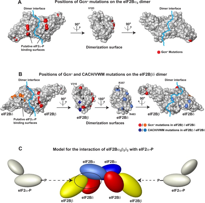
Eukaryotic translation initiation factor 2B (eIF2B), the guanine nucleotide exchange factor for the G-protein eIF2, is one of the main targets for the regulation of protein synthesis. The eIF2B activity is inhibited in response to a wide range of stress factors and diseases, including viral infections, hypoxia, nutrient starvation, and heme deficiency, collectively known as the integrated stress response. eIF2B has five subunits (α-ε). The α, β, and δ subunits are homologous to each other and form the eIF2B regulatory subcomplex, which is believed to be a trimer consisting of monomeric α, β, and δ subunits. Here we use a combination of biophysical methods, site-directed mutagenesis, and bioinformatics to show that the human eIF2Bα subunit is in fact a homodimer, at odds with the current trimeric model for the eIF2Bα/β/δ regulatory complex. eIF2Bα dimerizes using the same interface that is found in the homodimeric archaeal eIF2Bα/β/δ homolog aIF2B and related metabolic enzymes. We also present evidence that the eIF2Bβ/δ binding interface is similar to that in the eIF2Bα2 homodimer. Mutations at the predicted eIF2Bβ/δ dimer interface cause genetic neurological disorders in humans. We propose that the eIF2B regulatory subcomplex is an α2β2δ2 hexamer, composed of one α2 homodimer and two βδ heterodimers. Our results offer novel insights into the architecture of eIF2B and its interactions with the G-protein eIF2.


Figures

Figure 1
Figure 1
eIF2Bα dimerizes along the same interface as all its homologs with known structures. (A) Crystal structure of the archaeal eIF2Bα/β/δ homolog, aIF2B (PDB entry 1vb5), which is a homodimer. The two aIF2B subunits are colored red and yellow. The arm regions, which interact with the other subunit in the dimer, are labeled with arrows. (B) Crystal structure of eIF2Bα (PDB entry 3ecs) showing a dimer with a large buried surface. One subunit is colored cyan and the other blue. The arm regions, which interact with the other subunit in the dimer, are labeled with arrows. (C) eIF2Bα dimerizes using the same interface as aIF2B. Structure alignment of aIF2B and eIF2Bα, with the same orientation and coloring as in panels A and B, respectively. The CTDs of the eIF2Bα and aIF2B subunits were aligned [Cα rmsd of 1.38 Å (excluding the arm region)]. The interdomain orientations differ somewhat between eIF2Bα and aIF2B, as noted previously, and the NTDs of the proteins were not used in the alignment. Structure alignments were done in MOLMOL. (D) The eIF2Bα dimerization surface is highly conserved among eIF2Bα homologs. eIF2Bα is shown in surface representation. Amino acids are colored by sequence conservation from white (<30% conservation) to yellow (65% conservation) to green (100% conservation). The dimerization interface is marked with a light blue line. One subunit is omitted in the middle panel, to show the dimerization surface. (E) The eIF2Bα dimerization surface is highly hydrophobic. The display is as in panel D, except that the surface is colored by hydrophobicity and charge. Backbones are colored dark gray, hydrophobic side chains yellow, positively charged side chains blue, negatively charged side chains red, and the remaining side chains light gray. The three residues at the dimerization surface, whose mutation (designated eIF2Bα-TM) abolishes dimerization (see Figure 2), are labeled.
Figure 2
Figure 2
Size exclusion chromatography (SEC) of eIF2Bα and -β. (A) SEC traces of WT eIF2Bα (blue) and eIF2Bα-TM (green) at a concentration of 15 μM. The theoretical MWs for WT eIF2Bα and eIF2Bα-TM monomers are both ∼35 kDa. The apparent molecular weights (MWapp) from SEC are 60 kDa for WT eIF2Bα (dimer) and 33 kDa for eIF2Bα-TM (monomer). The positions of the markers used to calculate MWapp are shown with vertical dashed lines. (B) Calculated MWapp as a function of protein concentration. WT eIF2Bα (data from two independent sets of experiments are colored blue and light blue) is dimeric up to 3 μM, but its MWapp starts to gradually increase at higher concentrations, indicative of the formation of larger complexes. eIF2Bα-Δarm (red) is in equilibrium between the monomer and dimer, and possibly higher-order complexes, in the concentration range tested; eIF2Bα-TM (green) is clearly monomeric. (C) SEC trace of untagged eIF2Bβ at a concentration of 15 μM. The theoretical MW for an eIF2Bβ monomer is 39 kDa. The apparent molecular weight (MWapp) from SEC is 41 kDa (monomer). The positions of the markers used to calculate MWapp are shown with vertical dashed lines.
Figure 3
Figure 3
Models for the structure of eIF2Bβ and -δ. (A) Model for the proposed dimeric structure of eIF2Bβ (yellow) and eIF2Bδ (red), based on the structure of eIF2Bα2 and the sequence alignment shown in Figure S4 of the Supporting Information. The first eight residues of eIF2Bβ and the first 200 residues of eIF2Bδ were not modeled. Segments that have no counterpart in eIF2Bα or are not visible in the eIF2Bα crystal structure were modeled de novo and are colored black, because their real conformation is unknown. (B) The putative eIF2Bβ and -δ dimerization surfaces are highly conserved among eIF2Bβ and eIF2Bδ homologs. The eIF2Bβδ dimer model is shown in surface representation. Amino acids are colored by sequence conservation from white (<30% conservation) to yellow (65% conservation) to green (100% conservation). To show the dimerization surfaces, eIF2Bδ was omitted from the middle left panel and eIF2Bβ from the middle right panel. The dimerization interface is marked with a light blue line. The conserved surface unique to eIF2Bδ is circled in the right panel. (C) The putative eIF2Bβ and -δ dimerization surfaces are highly hydrophobic. The display is as in panel B, except that the surface is colored by hydrophobicity and charge. Backbones are colored dark gray, hydrophobic side chains yellow, positively charged side chains blue, negatively charged side chains red, and the remaining side chains light gray.
Figure 4
Figure 4
Sequence alignment of human eIF2Bα, -β, and -δ. Identical positions are shown as white letters on a red background; conserved positions are shown as red letters. The secondary structure and residue numbering above the alignment are for eIF2Bα. Amino acids located at the dimer interface are marked with a black asterisk below the alignment. The position of V183 in eIF2Bα is marked with a red asterisk. V183 is buried just under the dimerization surface and surrounded by residues that are part of the dimer interface (see also Figure 6A). The V183F mutation causes CACH/VWM, and was recently shown to affect eIF2Bα dimerization. The sequence alignment was obtained with HHpred from the HHsuite. This figure was generated with ESPript, using the BLOSUM62 homology scoring matrix.
Figure 5
Figure 5
Molecular docking indicates an eIF2Bβδ heterodimer utilizing the same interface as the eIF2Bα2 homodimer. (A) Highest-scoring model of the eIF2Bα2 dimer superimposed on the eIF2Bα2 dimer observed in the crystal structure of eIF2Bα (PDB entry 3ecs). The left subunits from each dimer (model and crystal structure) are aligned (colored gray). The second subunit in the modeled dimer is colored blue; the second subunit in the dimer found in the crystal structure is colored yellow. The interface rmsd between the two structures is 3.5 Å. None of the high-scoring models showed any similarity to any other crystal contacts observed in the eIF2Bα structure (PDB entry 3ecs). (B) Local energy landscape of the eIF2Bα2 models generated by docking, plotted with a model having the observed eIF2Bα2 crystal interface placed at the origin. The plot shows that such models are surrounded by a well-defined energy funnel. (C) Local energy landscape of the eIF2Bβδ models generated by docking, plotted with a model having the eIF2Bα2 crystal interface placed at the origin. The plot shows that, along the energy funnel, the models converge to ones with the interface seen in the eIF2Bα2 structure. (D) Local energy landscape of the eIF2Bβ2 models generated by docking, again plotted with a model having the eIF2Bα2 crystal interface placed at the origin. Unlike for the dimers shown in panels B and C, no funnel-shaped energy distribution is observed in the vicinity of such models, indicating that no stable eIF2Bβ2 homodimer is formed.
Figure 6
Figure 6
Model for the interaction of eIF2Bα2β2δ2 with eIF2α-P. (A) Positions of Gcn mutations (red) on the surface of the eIF2Bα2 dimer. The orientations are the same as in panels D and E of Figure 1. The dimerization interface is marked with a light blue line. In the central panel, only one eIF2Bα subunit is shown, to show the dimerization surface. Only mutations of surface-exposed residues that do not involve a glycine or a proline are shown, because these are least likely to affect the protein structure or stability. The approximate position of V183 (invisible because it is buried under the surface) is labeled in the central panel with a dashed arrow. (B) Positions of Gcn and CACH/VWM mutations on the surface of the eIF2Bβδ dimer, in the same orientation as the eIF2Bα2 dimer in panel A. The eIF2Bβδ dimer is in the same orientation as in Figure 3. The dimerization interface is marked with a light blue line. The two central panels show the dimerization surfaces of eIF2Bβ (left) and eIF2Bδ (right). Sites of Gcn mutations are colored red (eIF2Bβ) and orange (eIF2Bδ), and CACH/VWM mutations are colored navy (eIF2Bβ) and blue (eIF2Bδ). The clusters of CACH/VWM mutations at the eIF2Bβ and eIF2Bδ dimerization surfaces are visible in the two central panels. The cluster of CACH/VWM mutations in eIF2Bδ-NTD can be seen in the right panel (circled). Residues discussed in the text are labeled. The position of V316 (buried under the surface) is labeled in the left central panel with a dashed arrow. (C) Model for the interaction of eIF2Bα2β2δ2 with eIF2α-P. One eIF2Bα subunit is colored light blue, the other dark blue, eIF2Bβ yellow, and eIF2Bδ red. eIF2α-P (light gray) binds in a pocket (circled on the left) formed between one eIF2Bα subunit and one eIF2Bβδ dimer. The proteins are represented by solids, drawn approximately to scale with their respective sizes. Unphosphorylated eIF2α should bind to an overlapping surface on eIF2Bα2β2δ2.

Similar articles

Cited by

References

    1. Jackson R. J.; Hellen C. U.; Pestova T. V. (2010) The mechanism of eukaryotic translation initiation and principles of its regulation. Nat. Rev. Mol. Cell Biol. 11, 113–127. - PMC - PubMed
    1. Marintchev A.; Wagner G. (2004) Translation initiation: Structures, mechanisms and evolution. Q. Rev. Biophys. 37, 197–284. - PubMed
    1. Pestova T. V., Lorsch J. R., and Hellen C. U. T. (2007) The Mechanism of Translation Initiation in Eukaryotes. In Translational Control in Biology and Medicine (Mathews M. B., Sonenberg N., and Hershey J. W. B., Eds.) pp 87–128, Cold Spring Harbor Laboratory Press, Plainview, NY.
    1. Sonenberg N.; Hinnebusch A. G. (2009) Regulation of translation initiation in eukaryotes: Mechanisms and biological targets. Cell 136, 731–745. - PMC - PubMed
    1. Moreno J. A.; Halliday M.; Molloy C.; Radford H.; Verity N.; Axten J. M.; Ortori C. A.; Willis A. E.; Fischer P. M.; Barrett D. A.; Mallucci G. R. (2013) Oral treatment targeting the unfolded protein response prevents neurodegeneration and clinical disease in prion-infected mice. Sci. Transl. Med. 5, 206ra138. - PubMed

Publication types

LinkOut - more resources