. 2014 Jul 1;20(13):3446-57.
doi: 10.1158/1078-0432.CCR-13-2797. Epub 2014 May 8.

Effects of MAPK and PI3K pathways on PD-L1 expression in melanoma

Affiliations

Effects of MAPK and PI3K pathways on PD-L1 expression in melanoma

Mohammad Atefi et al. Clin Cancer Res. .

Abstract

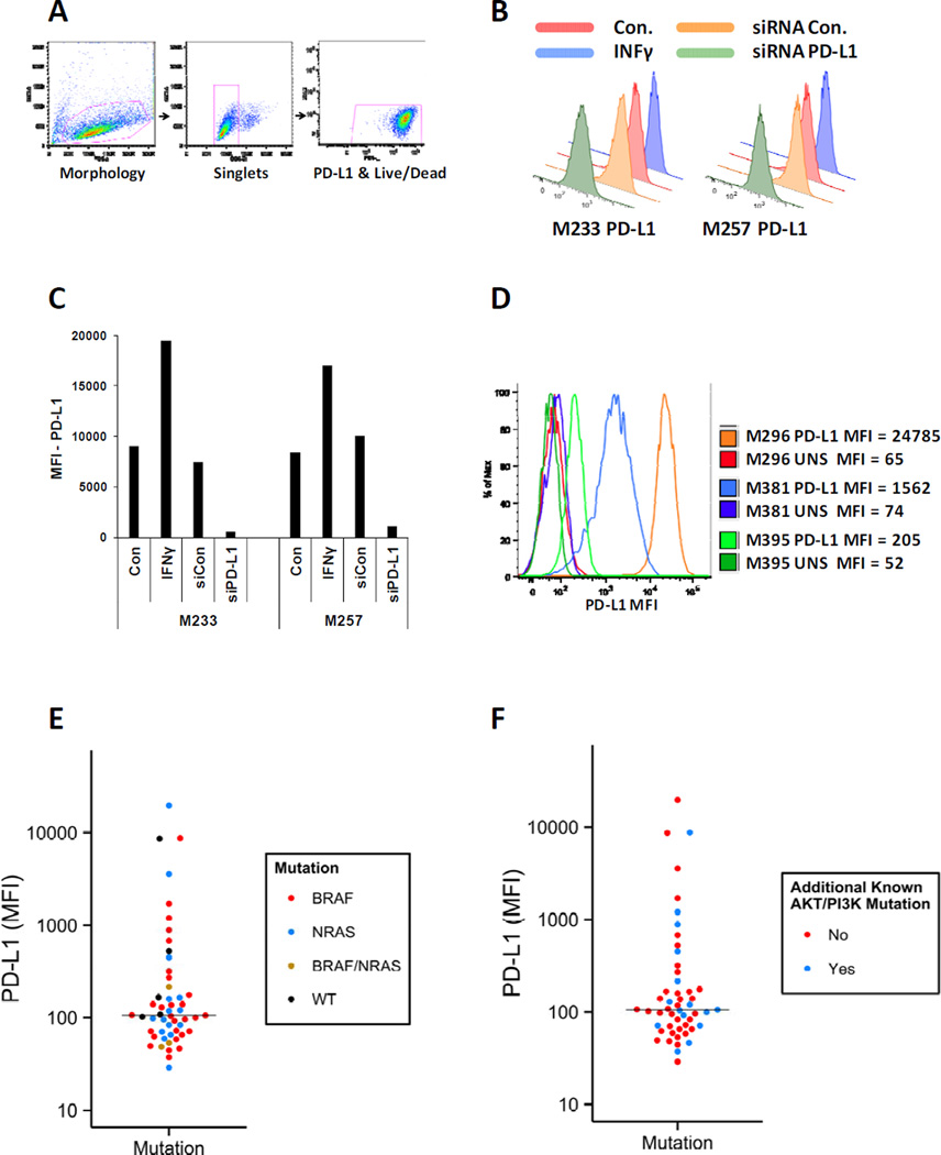
Purpose: PD-L1 is the main ligand for the immune inhibitory receptor PD-1. This ligand is frequently expressed by melanoma cells. In this study, we investigated whether PD-L1 expression is controlled by melanoma driver mutations and modified by oncogenic signaling inhibition.

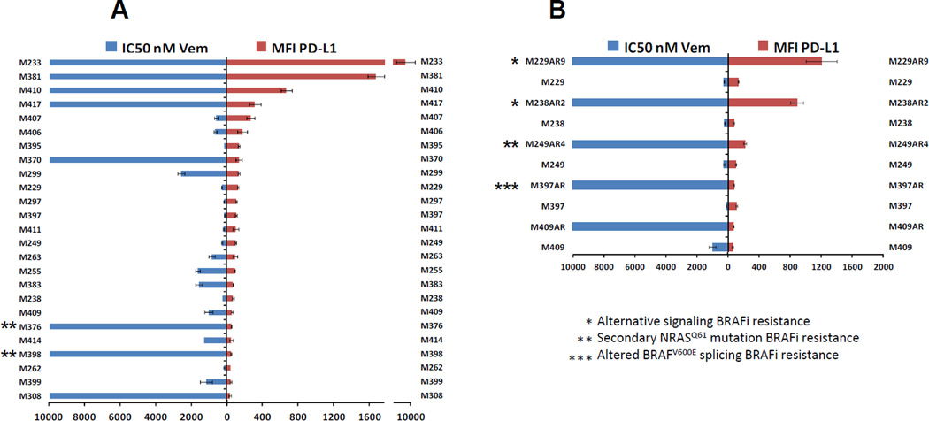
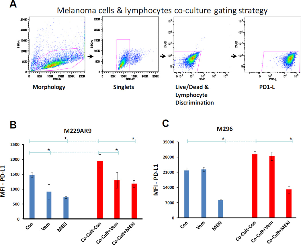
Experimental design: Expression of PD-L1 was investigated in a panel of 51 melanoma cell lines containing different oncogenic mutations, including cell lines with innate and acquired resistance to BRAF inhibitors (BRAFi). The effects of targeted therapy drugs on expression of PD-L1 by melanoma cells were investigated.

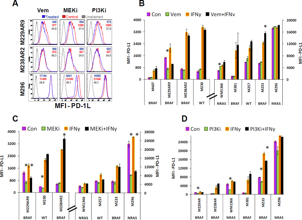
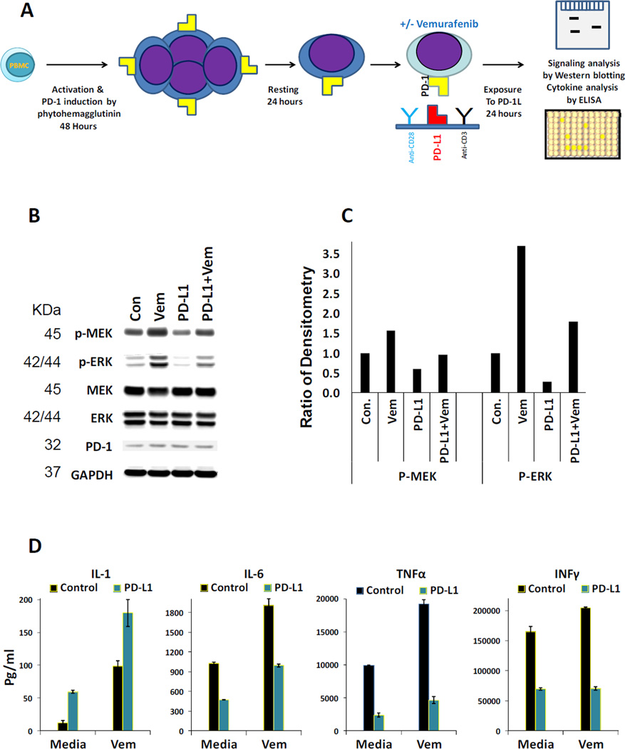
Results: No association was found between the level of PD-L1 expression and mutations in BRAF, NRAS, PTEN, or amplification of AKT. Resistance to vemurafenib due to the activation of alternative signaling pathways was accompanied with the induction of PD-L1 expression, whereas the resistance due to the reactivation of the MAPK pathway had no effect on PD-L1 expression. In melanoma cell lines, the effects of BRAF, MEK, and PI3K inhibitors on expression of PD-L1 were variable from reduction to induction, particularly in the presence of INFγ. In PD-L1-exposed lymphocytes, vemurafenib paradoxically restored activity of the MAPK pathway and increased the secretion of cytokines.

Conclusions: In melanoma cell lines, including BRAFi-resistant cells, PD-L1 expression is variably regulated by oncogenic signaling pathways. PD-L1-exposed lymphocytes decrease MAPK signaling, which is corrected by exposure to vemurafenib, providing potential benefits of combining this drug with immunotherapies.


Conflict of interest statement

No potential conflicts of interest were disclosed.

Figures

Figure 1
Figure 1. PD-L1 expression by melanoma cell lines according to their mutations in the MAPK or PI3K/AKT pathways
A) Gating strategy for detection of PD-L1 levels in melanoma cell lines stained with APC anti-PD-L1 antibody (catalog # 329708, Biolegend). For differentiation of live/dead, 7AAD exclusion staining was used. B) Validation of the anti-PD-L1 antibody by investigating its ability to detect induction of this ligand in the presence of 200u/ml of INFγ and reduction of this ligand after siRNA knockdown. Histograms show PD-L1 levels on the surface of two melanoma cell lines (M233 and M257) in the mentioned conditions. C) Bar graph of PD-L1 MFIs for the same cell lines in the mentioned conditions. D) Examples of flow cytometry histograms of PD-L1 levels in three melanoma cells lines with almost two log fold differences in expressions of this ligand. The PD-L1 levels were determined in the absence of INFγ. E) Expression levels of PD-L1 in a panel of melanoma cell lines containing 27 BRAF mutants, 15 RAS mutants, 3 BRAF/NRAS double mutant and 6 BRAF/NRAS wild types were measured by flow cytometry and graphed by using the MFI of this ligand for each cell line. The assays on each single sample were repeated at least twice. Each dot represents one cell line and is color coded according to the mutation status of BRAF and RAS genes. The median of PD-L1 expression for all the cell lines is shown by the horizontal line around MFI of 100. The MFI distributions of the different mutants are not significantly different (Kolmogorov–Smirnov test). F) The same cell lines in the panel are color coded according to the absence (red dots) or presence (blue dots) of mutations or amplification of genes involved in the PI3K/AKT pathway. Both MFI distributions are not significantly different (Kolmogorov–Smirnov test). The PD-L1 levels in figure E and F were determined in the absence of INFγ.
Figure 2
Figure 2. Comparison of PD-L1 expression in BRAF mutated melanoma cell lines with their responses to the BRAF inhibitor vemurafenib
A) Expressions of PD-L1 in BRAF mutated cell lines and their levels of sensitivities to the BRAF inhibitor vemurafenib were graphed side by side. Each assay was performed twice and resistance to vemurafenib was defined as an IC50 of higher than 1000nM. B) Side by side expression of PD-L1 and the IC50 levels of five pairs of sensitive parental cell lines and their in vitro developed vemurafenib resistant sublines indicated by the AR suffixes after the cell lines names. Mechanism of resistance due to the activation of alternative pathways, secondary NRAS mutation and truncated BRAF has been indicated by one, two or 3 asterisks, respectively. In both graph A and B, a bimodal pattern of PD-L1 expression among the highly resistant cell lines can be seen.
Figure 3
Figure 3. Effects of targeted therapy drugs in combination with INFγ on expression of PD-L1 in melanoma cell lines
A) Three examples of flow cytometry assays performed on melanoma cell lines treated with either the solvent (control), vemurafenib (1.5µM), MEKi (25nM), or PI3Ki (100nM) shown by histogram plots. In M229AR9 and M296 cell lines, MFI as the indication of PD-L1 expression level can be decreased significantly by some of the treatments, while in comparison with the unstained sample, the cell population is still in the positive range for expression of this ligand. B) The effect of vemurafenib (1.5µM) on expression of PD-L1 in melanoma cell lines in the presence and absence of INFγ (200u/ml). C) The effect of the MEK inhibitor, trametinib, at 25nM on expression of PD-L1 in melanoma cell lines in the presence and absence of INFγ (200u/ml). D) The effect of the PI3Ki at 100nM concentration on expression of PD-L1 in the presence and absence of INFγ (200u/ml). Graphs in panels B and C are separated between cell lines with low or high PD-L1 MFI to allow adequate evaluation of the different conditions. In panels B, C and D, the mutations in BRAF or NRAS genes of cell lines have been indicated under the name of each cell line. The assays on each single sample were performed at least twice and error bars are the standard errors. Significant effects of the signaling pathway inhibitors (Vem, MEKi, and PI3Ki) in the presence or absence of IFNγ are indicated (asterisk, t-test p-value<0.05). The effects of these targeted therapy drugs on expression of PD-L1 seem to be variable among the different cell lines causing decreases in some cases and induction of expression in some others particularly in the presence of INFγ.
Figure 4
Figure 4. Restoration of the MAPK pathway activity and enhancement of cytokine production by vemurafenib in lymphocytes exposed to PD-L1
A) PBMCs were primed by mitogen activation to express PD-1. This priming by mitogen activation leads to proliferation of activated T lymphocytes. Then in the presence and absence of vemurafenib, cells were exposed to anti-CD3/antiCD28 or anti-CD3/antiCD28 plus recombinant PD-L1 coated plates for 24 hours. B) Western blot analysis of the MAPK pathway activity in the lymphocytes exposed to the mentioned conditions described in the section A. C) Quantitation of the same Western blot analysis by densitometry. D) ELISA assays were performed in duplicates to detect the concentration of cytokines in the supernatants of lymphocytes primed and treated according to the same experimental settings described in the section A. Error bars are two standard deviations.
Figure 5
Figure 5. Inducing effect of lymphocytes and the reducing effect of MAPK pathway inhibitors on the expression of PD-L1 by melanoma cell lines
A) Gating strategy of the co-cultures. Melanoma cells were gated according to morphology (FSC-A vs SSC-A) to single cell discrimination (SSC-W vs SSC-A). The melanoma cells were then gated to perform live/dead and lymphocyte discrimination (CD45 vs 7AAD). These cells were then checked for PD-L1 positivity (PD-L1 vs 7AAD). The assays on each single sample were repeated at least twice. B) Evaluation of the expression of PD-L1 in the BRAF mutant melanoma cell line M229AR9 alone or co-cultured with lymphocytes in the absence and presence of the BRAF inhibitor, vemurafenib (1.5µM), or the MEKi, trametinib (25nM). M229AR9 control was off vemurafenib for 3 days before the harvest for flow cytometry (method and material section). Asterisks indicate significant statistical differences in PD-L1 expression upon treatment with vemurafenib and MEKi (Anova p-value<0.05). C) PD-L1 expression levels in the NRAS mutant melanoma cell line M296 alone or co-cultured with lymphocytes and the same treatments described in the section B. Asterisks indicate significant statistical differences in PD-L1 expression upon treatment with MEKi (Anova p-value<0.05).

References

    1. Alcalá AM, Flaherty KT. BRAF inhibitors for the treatment of metastatic melanoma: clinical trials and mechanisms of resistance. Clin Cancer Res. 2012 Jan 1;18(1):33–39. - PubMed
    1. Flaherty KT, Puzanov I, Kim KB, Ribas A, McArthur GA, Sosman JA, et al. Inhibition of mutated, activated BRAF in metastatic melanoma. N Engl J Med. 2010 Aug 26;363(9):809–819. - PMC - PubMed
    1. Flaherty KT, Infante JR, Daud A, Gonzalez R, Kefford RF, Sosman J, et al. Combined BRAF and MEK inhibition in melanoma with BRAF V600 mutations. N Engl J Med. 2012 Nov;367(18):1694–1703. - PMC - PubMed
    1. Hodi FS, O'Day SJ, McDermott DF, Weber RW, Sosman JA, Haanen JB, et al. Improved survival with ipilimumab in patients with metastatic melanoma. N Engl J Med. 2010 Aug 19;363(8):711–723. - PMC - PubMed
    1. Robert C, Thomas L, Bondarenko I, O'Day S, MD JW, Garbe C, et al. Ipilimumab plus dacarbazine for previously untreated metastatic melanoma. N Engl J Med. 2011 Jun 30;364(26):2517–2526. - PubMed

Publication types

MeSH terms