Review
. 2014 Jun;43(2):297-316.
doi: 10.1016/j.gtc.2014.02.015. Epub 2014 Apr 16.

Clinical implications and pathogenesis of esophageal remodeling in eosinophilic esophagitis

Affiliations
Review

Clinical implications and pathogenesis of esophageal remodeling in eosinophilic esophagitis

Ikuo Hirano et al. Gastroenterol Clin North Am. 2014 Jun.

Abstract

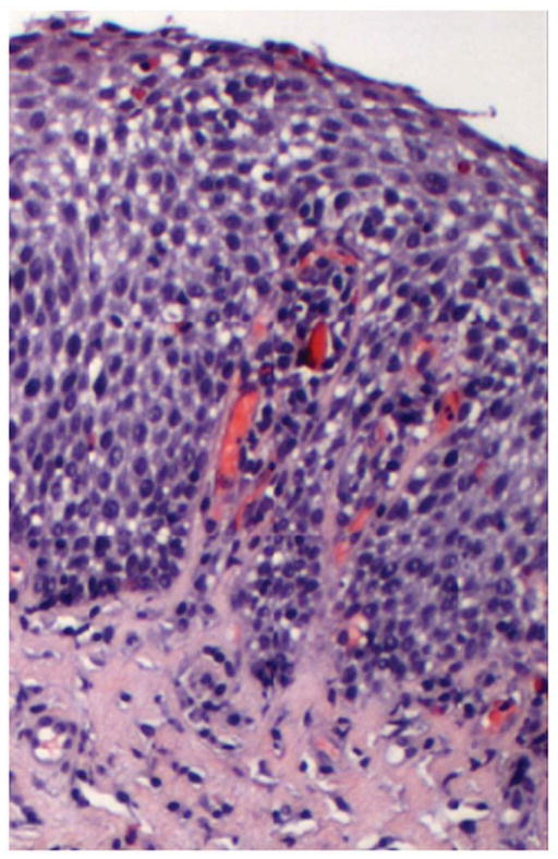
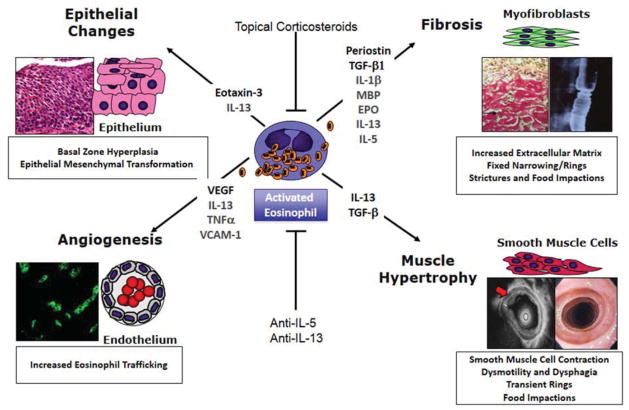
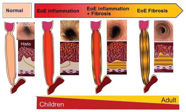
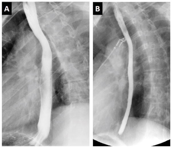
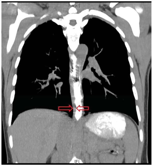
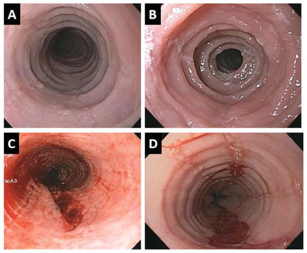
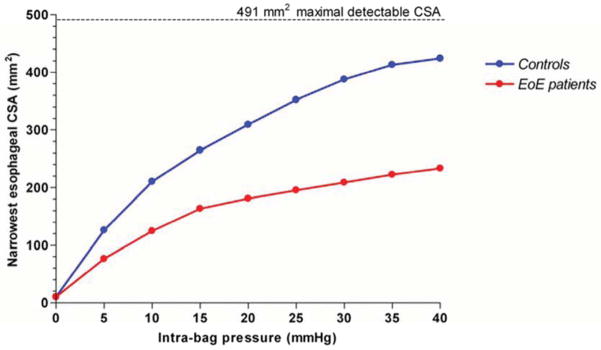
In eosinophilic esophagitis (EoE), remodeling changes are manifest histologically in the epithelium and subepithelium where lamina propria fibrosis, expansion of the muscularis propria, and increased vascularity occur. The clinical symptoms and complications of EoE are largely consequences of esophageal remodeling. Available therapies have demonstrated variable ability to reverse existing remodeling changes of the esophagus. Systemic therapies have the potential of addressing subepithelial remodeling. Esophageal dilation remains a useful, adjunctive therapeutic maneuver in symptomatic adults with esophageal stricture. As novel treatments emerge, it is essential that therapeutic end points account for the fundamental contributions of esophageal remodeling to overall disease activity.

Keywords: Dysphagia; Endoscopy; Eosinophilic esophagitis; Esophagitis; Fibrosis; Gastroesophageal reflux disease; Remodeling.


Figures

Figure 1
Figure 1
Histopathology of remodeling changes in eosinophilic esophagitis. The squamous epithelium shows basal zone hyperplasia and lamina propria shows increased collagen density in EoE.
Figure 2
Figure 2
Schematic representation of eosinophil induced esophageal remodeling, key interleukins and cytokines and its clinical consequences. Adapted from Aceves, S.S. and S.J. Ackerman, Relationships between eosinophilic inflammation, tissue remodeling, and fibrosis in eosinophilic esophagitis. Immunol Allergy Clin North Am, 2009. 29(1): p. 197–211, xiii–xiv; with permission.
Figure 3
Figure 3
Conceptual model of the consequences of esophageal remodeling. Esophageal remodeling over time leads to increasing subepithelial fibrosis that is associated with progressive esophageal structuring and narrow caliber esophagus. This model may explain phenotypic differences between pediatric and adult presentations of eosinophilic esophagitis.
Figure 4
Figure 4
Radiologic imaging in eosinophilic esophagitis. Barium esophagram in eosinophilic esophagitis. Panel A depicts a normal caliber esophagus in a patient with gastroesophageal reflux disease. Panel B shows an over 50% reduction in luminal diameter of the entire esophagus in an adult with eosinophilic esophagitis, a manifestation referred to as a narrow or small caliber esophagus.
Figure 5
Figure 5
Radiologic imaging in eosinophilic esophagitis. Coronal section of computed tomographic imaging illustrating the marked expansion of the esophageal wall in an adult with eosinophilic esophagitis. The imaging was obtained during ingestion of radiopaque contrast that clearly demarcates the inner lumen of the esophagus. The red arrows demarcate the mural thickness.
Figure 6
Figure 6
Endoscopic imaging of 4 adult patients with eosinophilic esophagitis. Panels A and B illustrate remodeling changes of esophageal rings and stricture. Panels C and D illustrate esophageal mural tears that occurred following esophageal dilation likely indicative of diffuse loss of esophageal elasticity.
Figure 7
Figure 7
Functional luminal imaging in eosinophilic esophagitis quantified remodeling effects of the esophagus. Esophageal distensibility plots in control subjects (blue) and eosinophilic esophagitis (red) demonstrating diminished distensibility for distension pressures above 5 mm Hg. The calculated value for constant cross sectional area in spite of increasing distension pressure is used to generate the distensibility plateau (DP). Data from Kwiatek, M.A., et al., Mechanical properties of the esophagus in eosinophilic esophagitis. Gastroenterology, 2011. 140(1): p. 82–90.

References

    1. Liacouras CA, et al. Eosinophilic esophagitis: updated consensus recommendations for children and adults. J Allergy Clin Immunol. 2011;128(1):3–20 e6. quiz 21–2. - PubMed
    1. Schoepfer AM, et al. Delay in Diagnosis of Eosinophilic Esophagitis Increases Risk for Stricture Formation, in a Time-Dependent Manner. Gastroenterology. 2013 - PubMed
    1. Dellon ES, et al. ACG clinical guideline: Evidenced based approach to the diagnosis and management of esophageal eosinophilia and eosinophilic esophagitis (EoE) Am J Gastroenterol. 2013;108(5):679–92. quiz 693. - PubMed
    1. Mishra A, et al. Esophageal remodeling develops as a consequence of tissue specific IL-5-induced eosinophilia. Gastroenterology. 2008;134(1):204–14. - PMC - PubMed
    1. Rubinstein E, et al. Siglec-F inhibition reduces esophageal eosinophilia and angiogenesis in a mouse model of eosinophilic esophagitis. J Pediatr Gastroenterol Nutr. 2011;53 (4):409–16. - PMC - PubMed