. 2014 May 8;157(4):869-81.
doi: 10.1016/j.cell.2014.03.040.

A chromatin-dependent role of the fragile X mental retardation protein FMRP in the DNA damage response

Affiliations

A chromatin-dependent role of the fragile X mental retardation protein FMRP in the DNA damage response

Roman Alpatov et al. Cell. .

Abstract

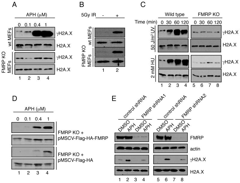
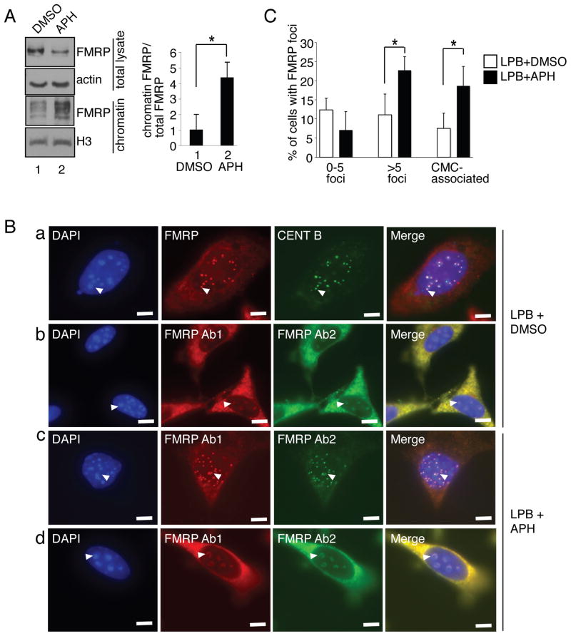
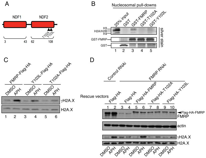
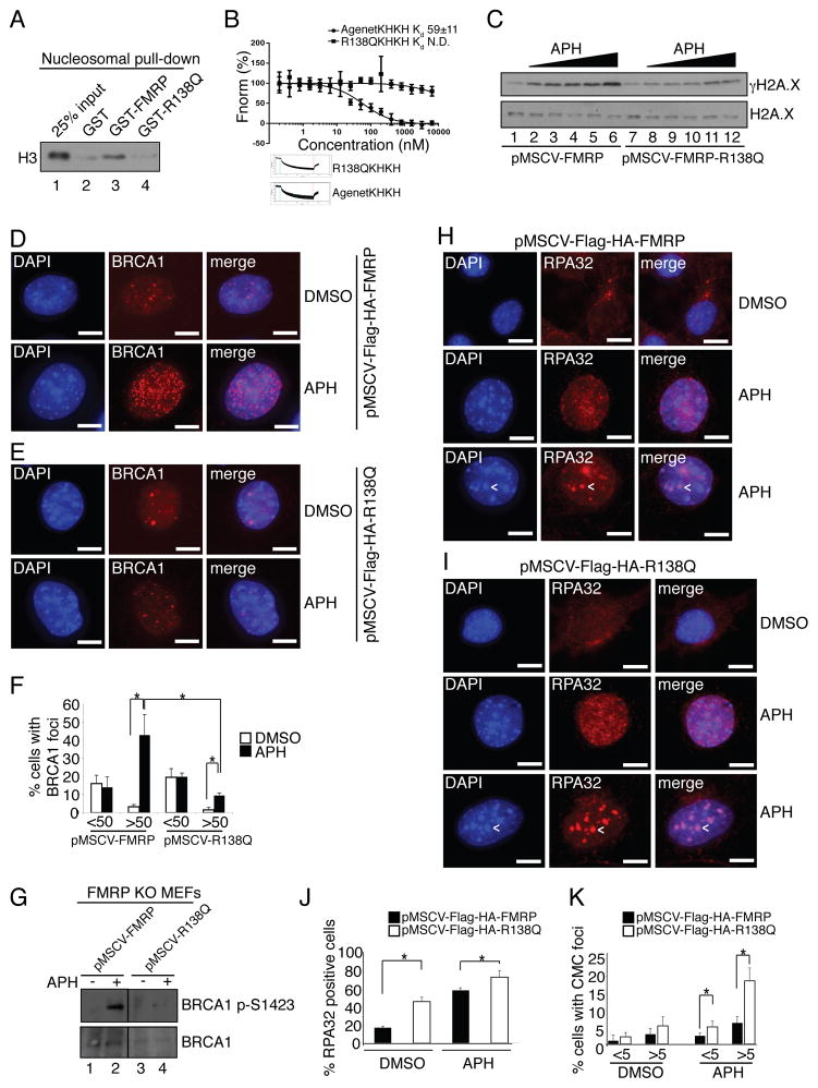
Fragile X syndrome, a common form of inherited intellectual disability, is caused by loss of the fragile X mental retardation protein FMRP. FMRP is present predominantly in the cytoplasm, where it regulates translation of proteins that are important for synaptic function. We identify FMRP as a chromatin-binding protein that functions in the DNA damage response (DDR). Specifically, we show that FMRP binds chromatin through its tandem Tudor (Agenet) domain in vitro and associates with chromatin in vivo. We also demonstrate that FMRP participates in the DDR in a chromatin-binding-dependent manner. The DDR machinery is known to play important roles in developmental processes such as gametogenesis. We show that FMRP occupies meiotic chromosomes and regulates the dynamics of the DDR machinery during mouse spermatogenesis. These findings suggest that nuclear FMRP regulates genomic stability at the chromatin interface and may impact gametogenesis and some developmental aspects of fragile X syndrome.


Conflict of interest statement

Conflict of Interest Statement

Y.S. is a co-founder of Constellation Pharmaceuticals and a member of its advisory board.

Figures

Fig. 1
Fig. 1. FMRP modulates histone H2A.X phosphorylation levels in response to replication stress
(A) Wild type but not FMRP KO MEFs exhibited dose-dependent γH2A.X induction in response to APH (lanes 1–4). See also Fig. S1A–C. (B) Wild type MEFs and FMRP KO MEFs exhibited similar degrees of γH2A.X induction (5-fold) in response to 5Gy of IR (lanes 1 and 2). (C) Wild type but not FMRP KO MEFs exhibited time-dependent γH2A.X induction in response to 50 J/m2 of UV irradiation or 2mM of HU (10-fold induction at 60 min post-treatment) (compare lanes 1–4 to lanes 5–8). (D) FMRP KO MEFs reconstituted with wild type Flag-HA-FMRP (pMSCV-Flag-HA-FMRP) or vector alone (pMSCV-Flag-HA) were exposed to various concentrations of APH. See also Fig. S1D. pMSCV-Flag-HA-FMRP MEFs exhibited more pronounced γH2A.X induction compared to pMSCV-Flag-HA cells (12-fold in Flag-HA-FMRP cells and 4-fold in Flag-HA cells (lanes 1–4). (E) FMRP RNAi HeLa cells but not control cells showed diminished γH2A.X induction in response to APH (3.4-fold and 8-fold, respectively, compare lanes 1,2 to 3,4 and 5,6 to 7,8). See also Fig. S1E,F and Fig. S2.
Fig. 2
Fig. 2. FMRP chromatin recruitment in response to replication stress
(A) MEFs were treated with DMSO (lane 1) or APH (lane 2). Chromatin fractions were isolated and Western blotted for FMRP. Bar graph, relative ratio of chromatin-associated FMRP to total FMRP. Asterisk, p<0.05, Student t-test. Data are an average of 3 independent experiments with standard deviation. (B) Immunostaining of nuclear FMRP in APH treated or DMSO treated MEFs in the presence of leptomycin B (LPB). Panel a, FMRP co-localized with CENT B next to chromocenters (CMCs). Arrowheads, representative co-localized FMRP (red) and CENT B (green) foci docked near CMCs. Panel b, Representative FMRP signal (Ab-1: anti-FMRP (Abcam) antibody (red), Ab-2: anti-FMRP (Calbiotech) antibody (green)) enveloping CMCs in LPB treated MEFs. Panel c, Representative FMRP foci in LPB+APH treated cells. Panel d, representative FMRP signals enveloping CMCs in LPB+APH treated MEFs. Arrowheads, selected FMRP foci wrapped around CMCs. Scale bar, 10μm. (C) APH treatment resulted in doubling of the number of cells with 5 or more FMRP foci (>5) or FMRP CMCs. Asterisks, p<0.05, Student t-test. Data are an average of 3 independent experiments with standard deviation. See also Fig. S3.
Fig. 3
Fig. 3. FMRP docking to chromatin is essential for FMRP-dependent modulation of γH2A.X levels in response to replication stress
(A) Diagram of AgenetFMRP. Mutations T102A and Y103L are demarcated by triangles. See also Fig. S4. (B) GST-FMRP or GST-FMRP carrying mutations in AgenetFMRP (GST-T102A and GST-Y103L) were incubated with isolated nucleosomes. Pull-down material was run on gradient gels followed by silver staining. A complete set of core nucleosomal histones including H3, H2A, H2B, and H4 were detected in wild type but not in mutant FMRP-mediated pull-downs (compare lanes 3–5). See also Fig. S5A. (C) Wild type FMRP (lanes 1 and 2) triggered more pronounced γH2A.X induction in FMRP KO MEFs in response to APH (12.8-fold) as compared to FMRP mutants (4-fold and 3-fold γH2A.X for Y103L and T102A mutants respectively) (lanes 3 and 4, 5 and 6). See also Fig. S1D. (D) FMRP RNAi in HeLa cells abolished γH2A.X induction in response to APH as compared to control RNAi (compare lanes 1,2 to lanes 3,4). Co-transfection with constructs expressing wild type but not mutant forms of FMRP restored the induction of γH2A.X in FMRP RNAi cells in response to APH (compare lanes 5,6 to lanes 7,8 and 9,10). The slower migrating band (in lanes 5–10) is Flag-HA-FMRP (indicated by an arrowhead).
Fig. 4
Fig. 4. Patient mutant R138Q is defective in γH2A.X induction and BRCA1 foci formation, and promotes excessive RPA retention on chromatin
(A) Unlike wild type FMRP, the R138Q FMRP mutant failed to bind nucleosomes in vitro (compare lanes 3 and 4). (B) Equilibrium binding analysis using recombinant MLA nucleosomes di-methylated at H3K79 and wild type AgenetKHKH (Kd=59 nM) or R138QKHKH (binding not detected). See also Fig. S5A and S6B. (C) FMRP KO MEFs rescued with wild type FMRP but not the R138Q FMRP patient mutant exhibited a dose-dependent γH2A.X response triggered by APH (0.05μM, 0.1μM, 0.3μM, 0.5μM, 1μM). See also Fig. S6C. (D,E) BRCA1 foci formation in FMRP KO MEFs rescued with wild type FMRP (D) in response to APH was more pronounced as compared to FMRP KO MEFs rescued with the R138Q FMRP patient mutant (E). See also Fig. S6D. (F) 40% of FMRP KO MEFs rescued with wild type FMRP exhibited >50 BRCA1 foci per cell upon APH treatment, compared to 10% in MEFs rescued with the R138Q patient mutant. (G) BRCA1 S1423 phosphorylation in FMRP KO MEFs rescued with wild type FMRP in response to APH was more pronounced as compared to rescue with the R138Q FMRP patient mutant (compare lanes 2 and 4). (H, I) RPA32 foci formation in FMRP KO MEFs rescued with wild type FMRP in response to APH was less pronounced as compared to FMRP KO MEFs rescued with the R138Q patient mutant (compare middle panels in H and I). Note the accumulation of a subset of RPA32 foci at CMCs (arrowheads, lower panels). (J, K) Quantification of total (J) and CMC-associated (K) RPA32 foci in FMRP KO MEFs rescued with wild type FMRP and R138Q patient mutant in response to APH. Percentage of cells positive for RPA32 increased from 10% to 50% upon APH treatment after rescue with wild type FMRP and from 40% to 70% after rescue with the R138Q mutant. Note increased numbers of RPA32 positive cells in the case of R138Q mutant rescue MEFs even in the absence of APH treatment. (K) 17 % of R138Q mutant rescue MEFs and 6% of wild type FMRP rescue MEFs had >5 CMC-associated RPA32 foci upon APH treatment. Asterisks, p<0.05, Student t-test. Data are an average of 3 independent experiments with standard deviation. Scale bars, 10 μm. See also Fig. S6D.
Fig. 5
Fig. 5. FMRP is present on meiotic chromosomes and regulates placement of γH2A
Immunofluorescence staining was performed on spread chromosomes from adult male primary spermatocytes, and cells were imaged by deconvolution microscopy. (A) Pachytene stage nucleus showing FMRP puncta along the chromosomes. SYCP1 marks the full length of the autosomes during the pachytene stage. Inset shows FMRP puncta (green) aligned along SYCP1-stained chromosomes (red). See also Fig. S7A. (B) γH2A.X and FMRP staining in wild type (left) and Fmr1 KO (right) primary spermatocyte nuclei at leptotene, zygotene, pachytene, and diplotene stages of meiotic prophase. SYCP3 accumulates on chromosomes beginning in leptotene and is present along their full length during pachytene. In Fmr1 KO cells, accumulation of γH2A.X is delayed in the leptotene stage. At the pachytene stage, γH2A.X is restricted to the sex chromosomes (arrowheads) in wild type cells, but remains at some autosomal locations in Fmr1 KO cells. Scale bars, 10 μm. (C) Percentage of cells retaining γH2A.X outside of the sex chromosomes in WT and KO pachytene spermatocytes. **P<0.01, Fisher’s exact test. See also Fig. S7B.
Fig. 6
Fig. 6. Fmr1 KO spermatocytes exhibit DNA repair defects and delayed resolution of single-strand intermediates at the pachytene stage
Staining of chromosome spreads was performed as in Fig. 5. (A) Co-staining of DMC1, MLH1, and the synaptonemal complex component SYCP3, showing retention of DMC1 and reduction of MLH1 in Fmr1 KO cells at mid-pachytene. (B) Numbers of WT and KO cells positive for DMC1 staining at mid-pachytene. ***P<0.0001, Fisher’s exact test. (C) MLH1 foci per mid-pachytene nucleus in WT and KO. **P<0.01, Mann-Whitney U test. (D) Number of chromosomes per mid-pachytene nucleus lacking MLH1 foci. In WT cells, there is at least one MLH1 focus per chromosome. ***P<0.0001, Mann-Whitney U test. Scale bars, 10 μm.
Fig. 7
Fig. 7. Abnormal BRCA1 and ATR loading and synapsis defects in Fmr1 KO spermatocytes
(A) Sample images of BRCA1 staining in pachytene spermatocytes in WT and KO animals. In wild type, BRCA1 staining is continuous and restricted to the sex chromosomes (arrowhead); in KO, it is discontinuous and frequently present on the autosomes. SYCP3 marks the chromosomes. (B) Sample images of ATR staining in pachytene spermatocytes in WT and KO animals. In wild type, ATR staining forms a cloud around the sex chromosomes (arrowhead) and is absent from the autosomes. In KO, ATR staining is retained in puncta on the autosomes and sometimes coats a complete autosome (bottom panels). SYCP3 marks the chromosomes. (C) Percentage of cells retaining BRCA1 or ATR outside of the sex chromosomes in WT and KO spermatocytes. *P<0.05; **P<0.01, Fisher’s exact test. (D) Co-staining of lateral (SYCP3) and central (SYCP1) elements of the synaptonemal complex shows discontinuous SYCP1 staining in Fmr1 KO cells, indicating defective synaptonemal complex formation. (E,F) Methylated H3K79 helps to recruit FMRP to chromatin. (E) Staining of FMRP in pachytene spermatocyte spreads from WT and Dot1L cKO mutants. Chromosome-associated FMRP signal is reduced in cKO cells, especially near the X and Y chromosomes. SYCP3 marks the chromosomes. (F) Quantitation of X- and Y-chromosome-associated FMRP foci. **P<0.01, unpaired t-test. Scale bars, 10 μm. See also Fig. S1C and S7C–E.

Comment in

  • FMRP: a new chapter with chromatin.
    He Q, Ge W. He Q, et al. Protein Cell. 2014 Dec;5(12):885-8. doi: 10.1007/s13238-014-0105-5. Protein Cell. 2014. PMID: 25327144 Free PMC article. No abstract available.

Similar articles

Cited by

References

    1. Adams-Cioaba MA, Guo Y, Bian C, Amaya MF, Lam R, Wasney GA, Vedadi M, Xu C, Min J. Structural studies of the tandem Tudor domains of fragile X mental retardation related proteins FXR1 and FXR2. PloS One. 2010;5:e13559. - PMC - PubMed
    1. Ascano M, Jr, Mukherjee N, Bandaru P, Miller JB, Nusbaum JD, Corcoran DL, Langlois C, Munschauer M, Dewell S, Hafner M, et al. FMRP targets distinct mRNA sequence elements to regulate protein expression. Nature. 2012;492:382–386. - PMC - PubMed
    1. Bear MF, Huber KM, Warren ST. The mGluR theory of fragile X mental retardation. Trends Neurosci. 2004;27:370–377. - PubMed
    1. Bernt KM, Zhu N, Sinha AU, Vempati S, Faber J, Krivtsov AV, Feng Z, Punt N, Daigle A, Bullinger L, et al. MLL-rearranged leukemia is dependent on aberrant H3K79 methylation by DOT1L. Cancer Cell. 2011;20:66–78. - PMC - PubMed
    1. Blonden L, van’t Padje S, Severijnen LA, Destree O, Oostra BA, Willemsen R. Two members of the Fxr gene family, Fmr1 and Fxr1, are differentially expressed in Xenopus tropicalis. Int J Dev Biol. 2005;49:437–441. - PubMed

Publication types